Case Report
High-Grade Chondrosarcoma of Nasal Septum-A Rare Case Report and Review of Literature
- Anju Khairwa, MD, PDCC 1*
1. Department of Pathology, University College of Medical Sciences, New Delhi, India.
*Corresponding Author: Anju Khairwa, MD, PDCC, Department of Pathology, University College of Medical Sciences, New Delhi, India.
Citation: Anju Khairwa. (2023). High-Grade Chondrosarcoma of Nasal Septum- A Rare Case Report and Review of Literature. International Journal of Clinical and Molecular Oncology, BioRes Scientia Publishers. 2(1):1-3. DOI: 10.59657/2993-0197.brs.23.006
Copyright: © 2023 Anju Khairwa, this is an open-access article distributed under the terms of the Creative Commons Attribution License, which permits unrestricted use, distribution, and reproduction in any medium, provided the original author and source are credited.
Received: July 30, 2023 | Accepted: August 25, 2023 | Published: December 22, 2023
Abstract
Chondrosarcoma of the nasal septum is a rare malignancy. Since 1927 to date, 69 cases of chondrosarcoma of nasal septum have been reported. In addition to the 70th case, we reported here. Mostly chondrosarcoma is present on the ends of long bones and 5-10% in the head-neck region. In our case, 50 years old female presented a bilateral nasal obstruction, and a biopsy was sent for nasal mass. Histomorphology suggested high-grade chondrosarcoma, which is positive for S-100 immunohistochemistry. Early intervention through radiology and biopsy will manage the patient with chondrosarcoma.
Keywords: chondrosarcoma; s100-; ihc; nasal septum
Introduction
Chondrosarcoma is a malignant cartilage tumor. It is a rare tumor, and 5-10% originate in the head and neck region.1 Chondrosarcoma mostly occurs in the long bone, ribs, pelvis, and laryngeal cartilage predominant site in the head & neck region.1,2 It rarely arises from the nasal septum.3 Usually, chondrosarcoma do not suspect clinically, radiologically, or histologically.4 Sometimes, it becomes difficult to diagnose, especially low-grade chondrosarcoma. Here, we are presenting a rare case of high-grade chondrosarcoma of nasal septum in elderly women.
Case Report
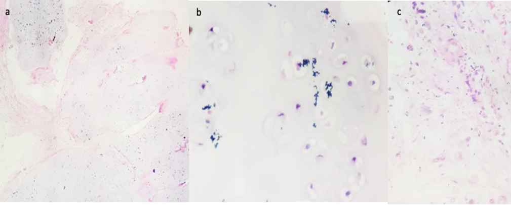
A 50-year-old female presented with bilateral nasal obstruction and swelling on the nose for two months. We received a biopsy from a nasal mass in our department. Grossly, we received two grey-white tissue bits altogether measuring 1.9x1.2x0.9 cm microscopic examination revealed a cartilaginous tumor arranged in a lobular fashion and increased cellularity (Figure 1a).
Figure1: A panel of microphotographs of Nasal Septum Chondrosarcoma; (a) Lobular arrangement of a cartilaginous tumor with increased cellularity (H&E, x40), (b) The tumor cells showed mild to moderate nuclear pleomorphism, vesicular nuclei, prominent nucleoli, few binucleated cells and 0-1 mitosis/hpf (H&E, x400), (c) The adjacent soft tissue revealed chronic inflammatory cells and invaded by the tumor (H&E, x400).
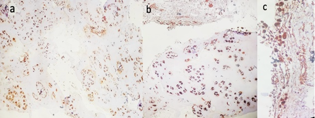
The tumor cells showed mild to moderate nuclear pleomorphism, vesicular nuclei, prominent nucleoli, few binucleated cells and 0-1 mitosis/hpf (Figur1b). The adjacent soft tissue of respiratory epithelium and subepithelial tissue with chronic inflammatory cells were invaded by the tumor (Figure 1c). Histomorphological features suggested chondrosarcoma of the nasal septum, Grad II. On immunohistochemical (IHC) examination showed, immunobiological (IHC) examination showed S-100 positivity in tumor cells and invasion in surrounding soft tissue (Figure 2a, b & c).
Figure 2: The tumor cells of chondrosarcoma of the Nasal Septum are positive for S-100, Immunohistochemistry (IHC) (a panel of microphotographs); (a) Scanner view (IHCx40), (b) Cartilage tumor and surrounding tissue positive for tumor cells (IHCx400), (c) Soft tissue showed invasion by tumor cells (IHCx400).
Discussion
Chondrosarcoma of the nasal septum is a rare malignancy. There were 69 cases of chondrosarcoma of nasal septum reported in the literature from 1927 to date [1,3,5,6-10]. In addition, 70th case chondrosarcoma of nasal septum we reported here within this century. The rare entity of chondrosarcoma of the nasal septum changed now and needs an update of it. The age of chondrosarcoma presentation is reported to be 40-50 years and commonly seen in males [11]. Our case is also 50 years but a female patient.
Most of the chondrosarcoma from nasal septum reported low grade in nature in literature [8,10-11]. In our case, we found high-grade (grade-2) chondrosarcoma of the nasal septum, which corroborated with the findings of Kainuma et al. and Sahu et al.5-6 Histologically, three grades of chondrosarcoma were reported based on cellularity, nuclear atypia, and mitotic activity [12]. In grade1- there is an abundant chondroid matrix with low cellularity, mild nuclear atypia, occasional binucleated cells and no mitosis; in grade 2- increased cellularity, decreased chondroid matrix, moderate nuclear pleomorphism, vesicular nuclei, few binucleations, and mitosis present; in grade 3 more cellular, myxoid matrix, highly pleomorphic irregular nuclei, prominent nucleoli, and increased mitosis [12].
Conclusion
The treatment of choice for chondrosarcoma of the nasal septum is surgery. Most of the studies reported an endoscopic approach under general anaesthesia [6,8-9,11]. The surgical approach should clear the margins from the tumor. On follow-up, our case expired due to a secondary infection during chemo-radiotherapy. We recommended early radiology and biopsy intervention to help in the detection and management of patients with chondrosarcoma.
References
- Downey TJ, Clark SK, Moore DW. (2001) Chondrosarcoma of the nasal septum. Otolaryngol Head Neck Surg 125:98–100.
Publisher | Google Scholor - Thompson LD, Gannon FH. (2002) Chondrosarcoma of the larynx: a clinicopathologic study of 111 cases with a review of the literature. Am J Surg Pathol. 26 (7):836–51.
Publisher | Google Scholor - Rassekh CH, Nuss DW, Kapadia SB, et al. (1996) Chondrosarcoma of the nasal septum: skull base imaging and clinicopathologic correlation. Otolaryngol Head Neck Surg 115:29–37.
Publisher | Google Scholor - Mirra JM, Gold R, Downs J, Eckardt JJ. (1985) A new histologic approach to the differentiation of enchondroma and chondrosarcoma of the bones. A clinicopathologic analysis of 51 cases. Clin Orthop Relat Res. 214–237.
Publisher | Google Scholor - Sahu PK, Goyal L, Bothra J, Vadhanan S, Sharma P. (2020) Chondrosarcoma of Nasal Cavity: a Rare Entity. Indian Journal of Surgical Oncology 11:288-292.
Publisher | Google Scholor - Kainuma K, Netsu K, Asamura K et al. (2009) Chondrosarcoma of the nasal septum: A case report. Auris Nasus Larynx 36, 601–605.
Publisher | Google Scholor - Gupta N, Pandey AK, Dimri K. (2021) Chondrosarcoma of the Nasal Septum—A Rare Subsite: Case Report with Review of Literature. Ind J Med Paediatr Oncol 42:506–509.
Publisher | Google Scholor - Bahgat M, Bahgat Y, Bahgat A, Elwany Y. (2012) Chondrosarcoma of the nasal septum. BMJ Case Rep. bcr2012006266.
Publisher | Google Scholor - Belgioia L, Vaccara EM, Bacigalupo A, Corvò R. (2017) Management of Nasal Septum Chondrosarcoma Occurring in Elderly: A Case Report. Cureus. 9(7):e1497.
Publisher | Google Scholor - Dass AN, Wilfred CG, Shek WHT, Ho WK. (2008) Case 139: Nasal Septum Low-Grade Chondrosarcoma. Radiology 249:714–717.
Publisher | Google Scholor - Maingi S, Padiyar BV, Sharma N. (2019) Chondrosarcoma of the Nasal Septum: A Case Report. Dubai Med J 2 (3): 107–110.
Publisher | Google Scholor - Magnano M, Boffano P, Machetta G, et al. (2015) Chondrosarcoma of the nasal septum. Eur Arch Otorhinolaryngol. 272:765-772.
Publisher | Google Scholor


