

Short Report
Comparison of A Histopathologic Grading Between the Xenografte Mouse Model of Lung Cancer Adenocarcinoma, and Colloid Gold with Camostat Mesilate Powder Intaking Group by Hematoxylin and Eosine Stain
- Chur Chin *
Department of Emergency Medicine, New life Hospital Bokhyun-dong, Bukgu, Daegu, Korea.
*Corresponding Author: Chur Chin, Department of Emergency Medicine, New life Hospital Bokhyun-dong, Bukgu, Daegu, Korea.
Citation: Chin C. (2024). Comparison of A Histopathologic Grading Between the Xenografte Mouse Model of Lung Cancer Adenocarcinoma, and Colloid Gold with Camostat Mesilate Powder Intaking Group by Hematoxylin and Eosine Stain. Clinical Case Reports and Studies, BioRes Scientia Publishers. 5(2):1-3. DOI: 10.59657/2837-2565.brs.24.117
Copyright: © 2024 Chur Chin, this is an open-access article distributed under the terms of the Creative Commons Attribution License, which permits unrestricted use, distribution, and reproduction in any medium, provided the original author and source are credited.
Received: March 02, 2024 | Accepted: March 15, 2024 | Published: March 18, 2024
Abstract
Previously, we developed nontoxic, broad-spectrum virucidal gold nanoparticles, less than 10nm sized, modified with sulfonic acids with camostat, a serine protease inhibitor can introduce gold nanoparticles to the influenza virus via ionic bonds. Moreover, this nontoxic drug irreversibly removes gene fragments inhibit insertion into the host genome. Lewis’s lung cancer cell line derived xenografts are generated by transplanting into immune-deficient mice.
Keywords: histopathologic grading; lung cancer; eosine stain
Description
Previously, we developed nontoxic, broad-spectrum virucidal gold nanoparticles, less than 10nm sized, modified with sulfonic acids with camostat, a serine protease inhibitor can introduce gold nanoparticles to the influenza virus via ionic bonds [1]. Moreover, this nontoxic drug irreversibly removes gene fragments inhibit insertion into the host genome [2]. Lewis’s lung cancer cell line derived xenografts are generated by transplanting into immune-deficient mice. We have identified the response of the lung adenocarcinoma to colloid gold with camostat mesilate powder intaking. Comparative analysis of the histology by formalin-fixation and paraffin-embedding were done from 4 control mice and 5 intaking mice. All tumors were collected and engrafted within 2h post resection. Each case was reviewed by pathologists to confirm the diagnosis. Mice were sacrificed by intraperitoneal injection of ketamine/xylazine (90/8mg/kg). 3 weeks after implantation.
Lewis lung carcinoma is a hypermutated Kras/Nras–mutant cancer with extensive regional mutation clusters in its genome. A tumor that spontaneously developed as an epidermoid carcinoma in the lung of a C57BL mouse. Syngeneic models have proven to be useful in predicting clinical benefit of therapy in preclinical experiments. The cells were anaplastic, varying in size and shape; and they appeared to have little cytoplasm. The nuclei of the cells were highly distorted and prominent.
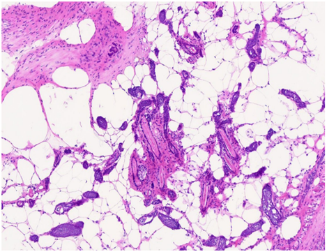
Figure 1(A): Control: high-grade components: micropapillary and solid patterns by the International Association for the Study of Lung Cancer.
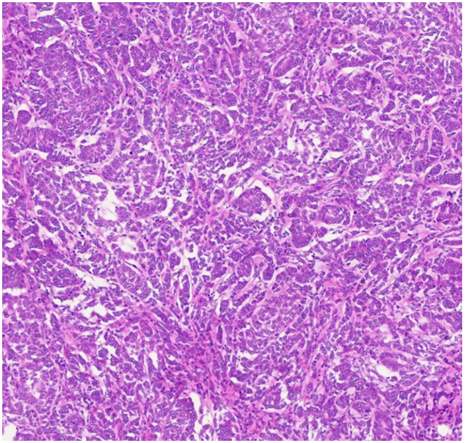
Figure 1(B): Intake group: lepidic and papillary patterns
Figure 1: Histologic analysis of colloid gold with camostat mesilate powder intaking group and Xenograft mouse model: (A) control group: cribriform pattern characterized by nests of neoplastic cells with perforated sieve; (B) intake group: lepidic patterns.
Table 1: Comparative analysis of the histology by hematoxylin-eosine stain: (A) Control group: grade 3; (B) Intake group: grade 1
| Predominant histologic pattern, n (%) | Control | Intake group |
| Lepidic | 5 | |
| acinar | 2 | |
| papillary | 1 | |
| Solid | 4 | |
| Complex glands (cribriform and fused glands) | 3 |
References
- Chin C. (2021). Cell entry inhibitor with sulfonated colloid gold as new potent broad spectrum virucides. J Infect Dis Ther., 9:1-4.
Publisher | Google Scholor - Chin C. (2024). Collloidal Gold with Camostat Mesylate Powder Remove DNA and RNA from Living Object, International journal of clinical case report and review. 16(2):1-5.
Publisher | Google Scholor - Andrea L Moreira, Paolo S Ocampo, Yuhe Xia, Hua Zhong, Prudence A Russell, et al. (2020). A Grading System for Invasive Pulmonary Adenocarcinoma: A Proposal From the International Association for the Study of Lung Cancer Pathology Committee. J Thorac Oncol., 15(10):1599-1610.
Publisher | Google Scholor


