

Review Article
Virtopsy: A New Perspective of Postmortem to Determine the Cause of Death
- Roshini K *
- Sweta Bisht
- Amit Chauhan
- Fr. Jobi Xavier
Department of Life Sciences, School of Sciences, CHRIST (Deemed to be University), Bengaluru, Karnataka, India.
*Corresponding Author: Amit Chauhan,Department of Life Sciences, School of Sciences, CHRIST (Deemed to be University), Bengaluru, Karnataka, India.
Citation: K. Roshini, S. Bisht, A. Chauhan, Fr. J. Xavier. (2024). VIRTOPSY: A New Perspective of Postmortem to Determine the Cause of Death. International Journal of Medical Case Reports and Reviews, BioRes Scientia Publishers. 3(4):1-8. DOI: 10.59657/2837-8172.brs.24.057
Copyright: © 2024 Amit Chauhan, this is an open-access article distributed under the terms of the Creative Commons Attribution License, which permits unrestricted use, distribution, and reproduction in any medium, provided the original author and source are credited.
Received: April 15, 2024 | Accepted: May 07, 2024 | Published: June 11, 2024
Abstract
Virtopsy is known as a non-invasive method which uses various imaging techniques to analyse, examine and evaluate a corpse and find the cause of death. This emerging technique plays a key role in answering the cause of death in unnatural and suspected deaths leading to the conclusion of the case. This study was initiated to review the advancement in the technologies used in virtual autopsy as a prominent emerging technique in the field of forensic pathology. During the SARS-COV-2 pandemic this autopsy was used as it is sensitive and avoids any kind of contaminants and toxins to be spread. One of prominent benefit is that during the conduction of virtopsy, the corpse is not contaminated by any contaminants, toxins or other external factors which may disrupt the examination further leading to a false report or statement. As beliefs and cultures forbid traditional autopsies because it multiplies the wounds and harms the corpse, but by implementing virtopsy it offers an alternative to this concern and help to find the cause of death without sacrificing their culture and beliefs. Therefore, virtopsy stands as an enhanced, useful technique and better documentation method for gathering forensic pathology findings from head to toe of the corpse.
Keywords: virtual autopsy; death; postmortem; computed tomography; multi slice-computed tomography; magnetic resonance imaging
Introduction
Autopsy is performed to determine the cause of death in unnatural and suspected death cases. Along with the advancement in the field of forensic science, virtual autopsy has taken over the traditional method autopsy to find the cause of death. It involves imaging techniques that is used in clinical medicine such as Computed Tomography (CT), Magnetic Resonance Imaging (MRI), etc., to determine the cause of the death. In the year of 1895, Roentgen discovered X-rays that was used for the examination specifically in concern to the Computed Tomography (CT) in the year of 1971 followed in the year 1989, Spiral CT technology which provided 3D representation of body structures and in 1990, Magnetic Resonance (MR) technology by Ros et al., [2][5]. A new generation of CT scanners that could create many cross-sections of an entire body in less than a minute was launched in 1998 [2]. Traditional autopsy is the standard and the best method for postmortem examination, also CT scanning alone could be used to determine the cause of death. In February 2006, the Institute of Forensic Medicine at the University of Southern Denmark made CT scanning a standard part of every autopsy [3]. The concept of using contemporary digital cross-sectioning methods, such as Magnetic Resonance Imaging (MRI) and Multi Slice-Computed Tomography (MSCT), as an additional tool in the examination of fatal diving occurrences is not new and has been shown to be quite valuable. The head, abdomen, thorax, and lower extremities were all scanned using MRI in the axial, sagittal, and coronal planes. For example- MSCT and MRI can be used as an invasive procedure to determine the diver drowned but passed away because of gas embolism [4]. Virtual autopsy has various advantages over traditional autopsy such as it a non-destructive method of examination of the corpse, it allows for the acquisition of data that is not dependent upon single examination or examiner and can involve many experts for the examination of the corpse [23]. Compared to traditional autopsy, MSCT and MRI have numerous advantages such as, they mainly non-invasive techniques, and the data collected can be visualized in situ, stored, and can be reinterpreted at any time. The 3D scanning technique helps to determine key features that are critical to find in a traditional autopsy in case of entry angles of a knife or a bullet, or cases related to medical malpractices [19]. The goal of virtopsy is to objectively record and examine physical characteristics and injuries which caused the death. Both MSCT and MRI technologies have made significant advancement. Except for the decayed body, which will be discovered after almost three weeks, MSCT and MRI procedures can be carried out on corpse on average 32 hours after death [1].
Emerging Techniques of Vir topsy
Computed Tomography (CT) scanner
The CT scan has a dual-slice scanners with a two parallel detector in row and the air-cooled X-ray tube is required for a brief cooling in between each scan. Leth, P. M. (2007) discussed 100 cases performed in 2006 at the Institute of Forensic Medicine, University of Southern Denmark of sequential autopsies. An oil-cooled X-ray tube was also used in setting up more autopsies. Each time, the head, neck, thorax, and abdomen are scanned separately for four times. Further scans might be required in some circumstances, such as when examining the lower extremities of car accident victims. Before the organs are returned to the body, the CT scan results are communicated to the forensic pathologist. The final forensic report includes CT scans. For the further research and analysis, the double-blind CT scan and autopsy data are entered into a SPSS database. At present, the CT scan is done right before the autopsy, but it might be better to do it before the external medicolegal examination, or before deciding whether an autopsy is necessary. After that, it might turn out that some autopsies are not necessary at all, or it might turn out that an unexpected CT finding makes it necessary to perform an autopsy in cases where it would not have been requested otherwise [3]. Significant information from the CT scan was not visible during the autopsy in 11 of the cases. These were two cases of bone metastases, two cases of pneumothorax, one pelvic fracture, four extremities fractures, and two cases of severe heart compression due to hydrothorax. In 27% of cases in external examination the cause of death can be identified by CT scan and forensic chemistry by 32% in external examination [3]. Figure 1 represents the traditional autopsy and the virtual autopsy images during the examination of the corpse to determine the cause of death.
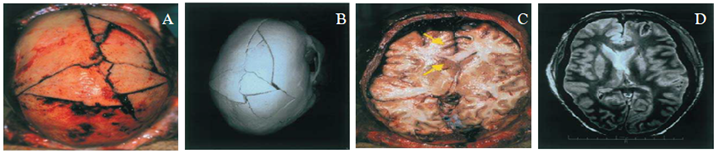
Figure 1: Burst fracture of the calvaria after blunt injury from a falling tree (Case 007): (a) at autopsy; (b) corresponding 3D reconstruction from CT data obtained before autopsy; (c) cranial view of brain; (d) corresponding axial fast SE proton density MR image, caudal view [1].
Poulsen and Simonsen (2007) employed spiral CT scanner. At the Institute of Forensic Medicine in Copenhagen, 525 medico-legal cases were routinely CT-scanned before the post-mortem examination in 2003. The results of the two assessments were contrasted. According to preliminary findings, the CT scanner was unparalleled in its ability to detect abrasions of the extremity bones, particularly multiple fractures, and crushing fractures. As a resultant scanner shows the fractures in-situ, which provides a much more complete picture. For example- using CT scanning the moderate fluid accumulation in the thoracic cavity can be seen more clearly and the pelvic region was found to benefit greatly from CT scanning. All fractures were visible, and the CT scan pictures aided the pathologist in identifying small fracture lines in the pelvic area and other localizations. The drawback of CT scan imaging technique is observed in cases of hypostasis and putrefaction due to the density of tissue which are frequently mistaken as pneumonia through the imaging methods. In 2010, Filo grana and colleagues conducted study on the implementation of the imaging techniques to support in the field of forensics has seemingly increased. The advanced CT imaging technology have led some authors to propose that postmortem radiological techniques and virtual autopsies are suitable alternatives to traditional autopsies [8]. In one of the case studies where three patients in the group of other pathologies passed away from malignant illness, in two of these cases, the cause of death was correctly identified both before and after PMCT; the causes were pleural carcinomatosis in a breast cancer patient and respiratory failure brought on by cachexia in a patient with metastatic esophageal cancer. This demonstrates that, even when the precise cause of death cannot be determined, PMCT can still point to a cardiovascular or perfusive cause of death [17]. In cases of drowning and blood aspiration in thoracic trauma, PMCT imaging of the lungs is frequently used as a forensic tool and as a post mortem method in addition to or in place of autopsy. In case of covid-19, virtual autopsy helped in the significant examination of spread of infection between the patients and operators [18]. As a non-invasive autopsy technique CT, MRI, photogrammetry, 3D surfaces scanning, PMCT angiography, and percutaneous post-mortem biopsy are utilized to enhance the visibility of the anatomic regions during forensic analysis. PMCT is considered one of the best technologies for the visualization of skeletal traumatic lesions, intravascular gas present in foreign bodies. At the same time, it has very low sensitivity in the differentiation of soft tissue that interfere in the investigation of it [18].
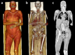
Figure 2: Virtual autopsy of PMCT images in a fatal case of natural death (myocardial infarction) [18].
CT scan gives an accurate result in the case of gunshot injury, where the projectile, angle of shot and bullet tract can be visualized and provides digital image and which makes it easy to preset the report in the court as compared to the traditional autopsy [3].
Multi Slice-Computed Tomography (MSCT)
In 2004, Jackowski conducted a study based on the utilization of MSCT to find the injuries from head and neck in case of venous air embolism (VAE). This protocol is based on the density differences to create a 3D model of all gas-tissue borderlines within the scanned volume. The use of MSCT has made it easier to evaluate cases of suspected VAE after the facts. As a resultant of this study, he could find how much air was embolized. The MSCT test was carried out on a scanner with four or eight detector rows. A collimation of 4/8 1.25 mm was used to scan important forensic areas. The volume data was used to calculate up to 1200 axial cross-sections with customized slice thickness and reconstruction intervals. In this process, the image acquisition takes approximate ten minutes time. A volume rendering protocol for isolating air structures was used to process the acquired MSCT data on a workstation. VAE will probably be found more frequently during routine post mortem MSCT examinations than at routine autopsies (9). The main advantages of MSCT are its rapid isotropic documentation and, in turn, its enormous potential for postprocessing in two dimensions and three dimensions [12].
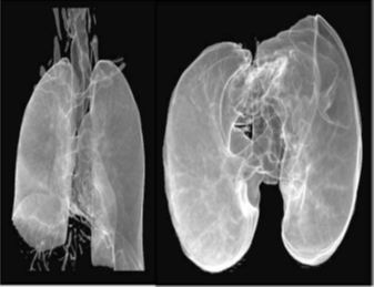
Figure 3: 3D air structure reconstruction using MSCT of the air-filled spaces within the chest (gunshot to the head); left: antero-posterior, right: caudo-cranial view [9].
In 2011 Gudda and his colleagues examined the bodies of four newborns over the course of six months using MSCT scanner which was connected to 16 detectors to scan each body. Four forensic infant cases were examined using pmMSCT scans, while two cases involved neonaticide, one involved an unknown cause of death, and one involved a stillbirth.
Multi Slice-Computed Tomography (MSCT) and Magnetic Resonance Imaging (MRI)
The standardized protocol and additional sequences for MRI was studied in 2012. In this study T1 and T2 weighted fast spin-echo sequences were systematically utilized. As a resultant of to T1-weighted images in the axial plane with a slice thickness of 4 mm and gaps of 1 mm, T2-weighted images of the head were obtained in each of the three planes. The classic pattern of blood aspiration to the lungs, signs of vital reaction to the gunshot, and air embolism to the heart and vessels were also revealed by MSCT or MRI. In gunshot victims, MSCT and MRI prove its significance for cross-sectional imaging data, three-dimensional reconstruction of the face and body surface or the skeleton along with visualization of fracture patterns from inward and outward beveling at the bone defects entrances and exits. It successfully helps to determine the fragmentation of bones and missiles, laceration, elevated intracerebral pressure, and a brain contusion, wound tract analysis and bullet track documentation [12]. All gunshot-caused complex skull fractures and brain injuries, including wound channels and deeply driven bone splinters, were documented by MSCT, MRI, 2D MPR, and 3D SSD reconstruction. The characteristic pattern of blood aspiration to the lungs, air embolism to the heart and arteries, and vital response to the gunshot were also shown on the MSCT and/or MRI and residue from gunshots left beneath and inside the skin were also visualized clearly.
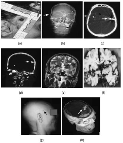
Figure 4: CT and MRI data of gunshot victim. (a) Photo of entrance wound. (b) CT view. (c) air and bleeding in the entrance area. (d) showing projectile from another view. (e) MRI cross-section correspond to (d). (f) autopsy finding. (g) 3D CT surface reconstruction of the face. (h) 3D CT reconstruction of the skull after removal of the top of the skull [12].
By following his earlier study, he continued to employ the utilization of MSCT and MRI in the analysis of the other body organs including brain, cardiovascular system, lung, skin, subcutaneous fat, muscle, respiratory system, circulatory system [1]. In case of putrefied corpse, it is difficult to perform traditional autopsy due liquification of the body and its organs, but postmortem imaging can recognize massive gas accumulation within the vascular system, body cavities, and soft tissues [22].
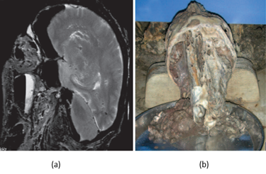
Figure 5: Putrefaction at postmortem MR imaging in a body that had been underwater for more than 1 year. (a) Sagittal T2-weighted MR image depicts intracerebral structures in the putrefied brain, thereby allowing cross-sectional exclusion of gross pathologic cerebral findings. (b) Autopsy photograph fails to allow cerebral assessment, since the liquefied intracranial structures became indistinguishable when the skull was opened [22].
In case of mummified body, MSCT is the most appropriate technique to be used as it was of great benefit even when the time lapse from death to imaging process was significant, also when the body was mummified [23]. By using the MSCT technique on the mummified body the remains of the internal organs, muscles and ligaments which were preserved, and the pathological conditions of the mummified body was able to be studied.
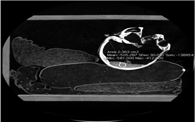
Figure 6: The position of the head and remains of soft tissues [23].
Feasibility Of Virtual Autopsy
In forensic pathology, we could establish the cause of death, determination of time since death as well as the tool which have caused the injuries. At the same time, it has proven its significance by estimating the gender, height, age, race, facial imaging reconstruction, etc. Motive of imaging equipment in virtopsy viz CT and MRI, is to obtain image data that cannot be acquired using traditional anatomy studies, thus solving many problems in autopsy, and providing a high social application value. Since CT is a high-speed examination technique which completes the scanning of the whole body in minutes, it is mainly applied to deceased persons with bullet trauma, especially in brain trauma. The new MSCT technology, as opposed to the older generation of CT scanners, enables quick documentation of the entire body. When compared to MRI, which requires a longer examination time, CT picture contrast is only dependent on the variability of X-ray penetration [12]. Thayyil et al. mentioned that the minimally invasive autopsy has accuracy like that of conventional autopsy for detection of cause of death or any pathological abnormality post death in case of foetuses, newborns, and infants, but was less accurate in older children [19].
In drowning case, the CT scan can provide the information about volume, density, size of the lungs and the amount of liquid observed in organs to determine the cause of death. In traffic accident reconstruction, bones can be examined by MR images with the aim to find bone bruises which is a sign of impact caused during the accident and bone bruises also can be detected from the bones of decomposed bodies. In smuggling cases, CT scan can be used, as X-rays in many cases does not reveal about the presence of heroin and gold rings in different containers or capsule in the body.
However, this new emerging technique cannot be used in toxicological cases where imaging techniques cannot provide the information about the odor and texture of organs, as well as the volume measurements [23].
Table 1: Diagnosis of pathological cases and accuracy percentage in various cases performed by virtual autopsy.
| Type of technique used | Type of pathological identification | Type of examination (internal/ external/ organs) | Percentage(Accuracy rate) | References |
| Computed Tomography (CT) scanning | Drowning cases | Internal & organs | 70% | Malizia, A., Filograna, L., Ryan, C. P., & Manenti, G. (2020). |
| Firearm projectile injuries | External & internal | 72.1–100% | Rosário Junior, A. F. D., Souza, P. H. C., Coudyzer, W., Thevissen, P., Willems, G., & Jacobs, R. (2012). | |
| Smuggling cases | Organs | 95.6 – 100% | Kružić, I., Jerković, I., Mihanović, F., Marušić, A., Anđelinović, Š., & Bašić, Ž. (2018). | |
| Magnetic Resonance Imaging (MRI) | Traffic accident reconstruction | External & internal | 94.81% | Buck, U., Christe, A., Naether, S., Ross, S., & Thali, M. J. (2009). |
| Diagnosis of burnt and charred body | Internal & organs | 90% | Haque, M. A. (2021). | |
| Multi Slice-Computed Tomography (MSCT) | In case of mummified body. | Internal & organs | 90% | Kružić, I., Jerković, I., Mihanović, F., Marušić, A., Anđelinović, Š., & Bašić, Ž. (2018). |
Conclusion
According to the research protocols, Computed Tomography is mainly used for hard tissues, Magnetic Resonance is used for soft tissues and combination of both for differentiation between adjacent structures. In traditional autopsy the main difficulty is to predict the depth of the foreign body where the virtual autopsy can answer this difficulty with accurate and clear information Performing virtual autopsies on corpse to establish the cause of death is less time consuming as it takes less time to scan the whole body as compared to the time consumed in opening the body cavities [21]. Virtual autopsy involving Postmortem Multidetector Computed Tomography (MDTC) or MRI in combination with software for 3-dimensional visualization is a new alternative to medical autopsy [20]. This also gives preferable diagnosis than traditional autopsy. The regions of the body which are not approachable by routine traditional autopsy can be scanned while performing virtual autopsy. Samples can be collected from least approachable area like spine using minimally invasive technique [21].
Declarations
Ethical Consideration: NA
Conflict of Interest: NA
Source of Funding: NA
References
- Thali, M. J., Yen, K., Schweitzer, W., Vock, P., Boesch, C., Ozdoba, C., ... & Dirnhofer, R. (2003). Virtopsy, a new imaging horizon in forensic pathology: virtual autopsy by postmortem multislice computed tomography (MSCT) and magnetic resonance imaging (MRI)-a feasibility study. Journal of forensic sciences, 48(2):386-403.
Publisher | Google Scholor - Filograna, L., Pugliese, L., Muto, M., Tatulli, D., Guglielmi, G., Thali, M. J., & Floris, R. (2019, February). A practical guide to virtual autopsy: why, when and how. In Seminars in Ultrasound, CT and MRI. 40(1):56-66.
Publisher | Google Scholor - Leth, P. M. (2007). The use of CT scanning in forensic autopsy. Forensic science. medicine and pathology, 3(1):65-69.
Publisher | Google Scholor - Plattner, T., Thali, M. J., Yen, K., Sonnenschein, M., Stoupis, C., Vock, P., ... & Dirnhofer, R. (2003). Virtopsy-postmortem multislice computed tomography (MSCT) and magnetic resonance imaging (MRI) in a fatal scuba diving incident. Journal of forensic sciences, 48(6):1347-1355.
Publisher | Google Scholor - Dirnhofer, R., Jackowski, C., Vock, P., Potter, K., & Thali, M. J. (2006). VIRTOPSY: minimally invasive. imaging-guided virtual autopsy. Radiographics, 26(5):1305-1334.
Publisher | Google Scholor - Bolliger, S. A., & Thali, M. J. (2015). Imaging and virtual autopsy: looking back and forward. Philosophical Transactions of the Royal Society B: Biological Sciences. 370(1674): 20140253.
Publisher | Google Scholor - Poulsen, K., & Simonsen, J. (2007). Computed tomography as routine in connection with medico-legal autopsies. Forensic science international, 171(2-3):190-197.
Publisher | Google Scholor - Filograna, L., Tartaglione, T., Filograna, E., Cittadini, F., Oliva, A., & Pascali, V. L. (2010). Computed tomography (CT) virtual autopsy and classical autopsy discrepancies: radiologist's error or a demonstration of post-mortem multi-detector computed tomography (MDCT) limitation? Forensic science international, 195(1-3):e13-e17.
Publisher | Google Scholor - Jackowski, C., Thali, M., Sonnenschein, M., Aghayev, E., Yen, K., Dirnhofer, R., & Vock, P. (2004). Visualization and quantification of air embolism structure by processing postmortem MSCT data. Journal of Forensic Science, 49(6):JFS2004047-4.
Publisher | Google Scholor - Kauczor, H. U., Riepert, T., Wolcke, B., Lasczkowski, G., & Mildenberger, P. (1995). Fatal venous air embolism: proof and volumetry by helical CT. European journal of radiology, 21(2):155-157.
Publisher | Google Scholor - Guddat, S. S., Gapert, R., Tsokos, M., & Oesterhelweg, L. (2013). Proof of live birth using postmortem multislice computed tomography (pmMSCT) in cases of suspected neonaticide: advantages of diagnostic imaging compared to conventional autopsy. Forensic science, medicine. and pathology, 9(1):3-12.
Publisher | Google Scholor - Thali, M. J., Yen, K., Vock, P., Ozdoba, C., Kneubuehl, B. P., Sonnenschein, M., & Dirnhofer, R. (2003). Image-guided virtual autopsy findings of gunshot victims performed with multi-slice computed tomography (MSCT) and magnetic resonance imaging (MRI) and subsequent correlation between radiology and autopsy findings. Forensic science international, 138(1-3):8-16.
Publisher | Google Scholor - Rosário Junior, A. F. D., Souza, P. H. C., Coudyzer, W., Thevissen, P., Willems, G., & Jacobs, R. (2012). Virtual autopsy in forensic sciences and its applications in the forensic odontology. Revista Odonto Ciência, 27:5-9.
Publisher | Google Scholor - Buck, U., Christe, A., Naether, S., Ross, S., & Thali, M. J. (2009). Virtopsy—noninvasive detection of occult bone lesions in postmortem MRI: additional information for traffic accident reconstruction. International journal of legal medicine, 123(3):221-226.
Publisher | Google Scholor - Wan, L., Song, Y. X., Li, Z. D., Liu, N. G., Wang, Y. H., Wang, M. W., ... & Chen, Y. J. (2020). The approach of virtual autopsy (VIRTOPSY) by postmortem multi-slice computed tomography (PMCT) in China for forensic pathology. Forensic Imaging, 20: 200361.
Publisher | Google Scholor - Donchin, Y., Rivkind, A. I., Bar-Ziv, J., Hiss, J., Almog, J., & Drescher, M. (1994). Utility of postmortem computed tomography in trauma victims. The Journal of trauma, 37(4): 552-555.
Publisher | Google Scholor - Sonnemans, L. J., Kubat, B., Prokop, M., & Klein, W. M. (2018). Can virtual autopsy with postmortem CT improve clinical diagnosis of cause of death? A retrospective observational cohort study in a Dutch tertiary referral centre. BMJ open, 8(3):e018834.
Publisher | Google Scholor - Malizia, A., Filograna, L., Ryan, C. P., & Manenti, G. (2020). Post-mortem investigation through virtual autopsy techniques: proposal of a new diagnostic approach to reduce the risks of operators during emergencies. International Journal of Safety and Security Engineering, 10(4):535-541.
Publisher | Google Scholor - Timonov, P., Novakov, S., Sivkov, S., Fasova, A., Novakov, I., & Spasov, S. (2019). The advantage of the virtual forensic autopsy-a new approach which could benefit forensic expertise. Journal of Forensic and Legal Medicine, 62:69-71.
Publisher | Google Scholor - Wichmann, D., Obbelode, F., Vogel, H., Hoepker, W. W., Nierhaus, A., Braune, S., ... & Kluge, S. (2012). Virtual autopsy as an alternative to traditional medical autopsy in the intensive care unit: a prospective cohort study. Annals of internal medicine, 156(2): 123-130.
Publisher | Google Scholor - Haque, M. A. (2021). Medical Professionals' Perspective on Virtual Autopsy: Comprehensive Analysis and Validation Study with Respect to Traditional Autopsy. Medico-Legal Update, 21(4).
Publisher | Google Scholor - Dirnhofer, R., Jackowski, C., Vock, P., Potter, K., & Thali, M. J. (2006). VIRTOPSY: minimally invasive, imaging-guided virtual autopsy. Radiographics, 26(5):1305-1333.
Publisher | Google Scholor - Kružić, I., Jerković, I., Mihanović, F., Marušić, A., Anđelinović, Š., & Bašić, Ž. (2018). Virtual autopsy in legal medicine: literature review and example of application on the mummified remains. Medicine. Law & Society, 11(2)67-90.
Publisher | Google Scholor


