Case Report
Type IV Flexor Digitorum Profundus Avulsion: A Case Report
- Mohammed Alwatari 1*
- Ahmed Khalaf 2
- Mohammed Alghannami 3
- Fatima Ghalib Alnajar 4
- Maryam Alwattary 5
1University of Baghdad College of Medicine, Iraq.
2Plastic surgeon at medical city plastic surgery department Baghadad, Iraq.
3Medical student at University of Baghdad College of medicine, Iraq.
4Medical student at University of Wasit College of medicine, Iraq.
5Family medicine board student at medical city department Baghdad, Iraq.
*Corresponding Author: Mohammed Alwatari, University of Baghdad College of Medicine, Iraq.
Citation: Alwatari M, Khalaf A, Alghannami M, Alnajar G. F, Alwattary M. (2023). Type IV Flexor Digitorum Profundus Avulsion: A Case Report. Clinical Case Reports and Studies, BioRes Scientia Publishers. 3(6):1-4. DOI: 10.59657/2837-2565.brs.23.090
Copyright: © 2023 Mohammed Alwatari, this is an open-access article distributed under the terms of the Creative Commons Attribution License, which permits unrestricted use, distribution, and reproduction in any medium, provided the original author and source are credited.
Received: November 27, 2023 | Accepted: December 11, 2023 | Published: December 18, 2023
Abstract
The avulsion of the flexor digitorum profundus, also known as the jersey finger, is a well-known injury that can be treated surgically. It has been classified into four types, among them type IV which involves tendon avulsion from an associated bony fragment with subsequent retraction to the palm or proximal interphalangeal joint is very rare. We present a case of type IV injury in a 45-year-old man with a pulling-on injury. The repair followed a pull-out technique (reinserting the tendon into the avulsed fragment); the entire reduction was tied over a button on the dorsal aspect of the nail and was augmented with a volar plate. The case was reported due to its rarity. We found that early surgical management of this problem greatly facilitates optimum surgical results and that early rehabilitation is essential for good long-term results.
Keywords: jersey finger; flexor digitorum profundus; interphalangeal joint
Introduction
Avulsion of the flexor digitorum profundus, also known as jersey finger, is a well-known injury that can be treated surgically, the mechanism of injury is hyperextension of the distal phalanx with the FDP muscle in a state of maximal contraction; the ring finger is the most commonly involved digit [1, 2]. It has been classified into three types by Leddy and Packer based primarily on the level of retraction of the injured tendon [3]. followed by a fourth type suggested by Smith [4] (Table 1). Type II is the most common variety while type IV is very rare [5]. Among these types, type IV has the most challenging surgical management because you need to both ensure good fixation of the fracture with the tendon and to allow early rehabilitation [6].
Table 1: Types of jersey finger injury [1,2].
| Type | Description |
| I | Tendon is retracted to palm due to ruptured vincula |
| II | Tendon is retracted to PIP due to intact vincula |
| III | Tendon is attached to large bony fragment with minimal retraction |
| IV | Tendon is avulsed from an associated bony fragment with subsequent retraction to the palm or PIP |
Case Report
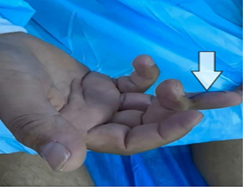
A 45-year-old man presented with an inability to flex the DIP joint of the right ring finger for 10 days precipitated by pulling on injury of a starter cord when he was trying to turn on his generator. On examination, there were no palmar findings and the digit was neurovascularly intact. However, there was loss of active flexion of the D.
Figure 1: Loss of active flexion of the DIP joint of the right ring finger.
IP joint of the right ring finger (Figure 1). Imaging was ordered and showed fracture avulsion of the distal phalanx (Figure 2), the diagnosis was made on the basis of history, examination, and imaging of the affected finger.
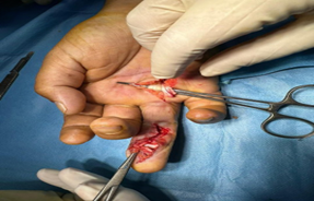
Figure 3: Surgical exploration showing the profundus tendon retracted to the palm level with a large bone avulsion at the base of the phalanx.
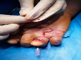
The repair followed the pull-out technique consisting of reinserting the tendon into the avulsed fragment, which was in turn reduced into the defect in the distal phalanx. The entire reduction was held with a suture weaved through the tendon, passed through the fragment and the distal phalanx, and tied over a button on the dorsal aspect of the nail (Figure 4), the repair was augmented with a volar plate.
Figure 4: The entire reduction was held by a suture passed through the tendon, fragment and the distal phalanx.
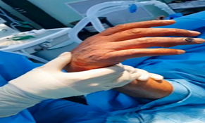
Immediately after surgery, passive tenodesis was normal indicating a good prognosis (Figure 5). Three weeks later the sutures are removed and the patient is undergoing vigorous physiotherapy right now with excellent regain of function and range of motion.
Figure 5: Normal passive tenodesis immediately after surgery.
Discussion
Jersey finger is almost always caused by an injury and the most common mechanism is pulling on a jersey or a starter cord. The most reliable physical finding is a complete lack of active flexion at the distal IPJ [1,2]. Careful physical examination of the injury is very important to prevent a delay in diagnosis. If a loss of active flexion at the DIP joint is demonstrated, surgical exploration is highly encouraged. Early management of this problem also greatly facilitates optimum surgical results. It is recommended that patients with FDP avulsion of any type to have exploration and repair as soon as possible, because of the difficulty of accurately predicting the level of flexor tendon retraction preoperatively on the basis of x-ray films [1,7,8]. The surgical approach reported here (Pull-Out) is the most frequently reported technique for the tendon repair. It is recommended doing independent repairs of the bone and tendon, as rigid fixation of the fracture is the most important factor for good results. Early rehabilitation is mandatory for good results [2,6,9]. When the diagnosis is delayed, treatment depends on many factors including the age of the patient, the functional disability, and the particular occupational and recreational requirements of the patient. Stabilization of the distal IPJ by tenodesis or arthrodesis is usually advisable, but in selected cases, tendon reconstruction can be considered [5].
Declarations
Conflict of interest statement
None declared.
Funding
None declared.
Data availability
The author confirms that all data generated during this study are included in this article and are available for open access.
Authors’ contributions
All authors contributed to the paper conception and design, collection and assembly of data, manuscript writing and final approval of manuscript
Disclaimers
None
Ethics approval and consent to participate
Informed consent was taken from the patient to publish his photos and information.
References
- Buscemi MJ, Jr., Page BJ, (1987). 2nd. Flexor digitorum profundus avulsions with associated distal phalanx fractures. A report of four cases and review of the literature. Am J Sports Med, 15(4):366-370.
Publisher | Google Scholor - Henry SL, Katz MA, Green DP. (2009). Type IV FDP avulsion: lessons learned clinically and through review of the literature. Hand (N Y), 2009;4(4):357-361.
Publisher | Google Scholor - Leddy JP, Packer JW. (1977). Avulsion of the profundus tendon insertion in athletes. J Hand Surg Am., 1977;2(1):66-69.
Publisher | Google Scholor - Smith JH, Jr. (1981). Avulsion of a profundus tendon with simultaneous intraarticular fracture of the distal phalanx--case report. J Hand Surg Am., 1981;6(6):600-601.
Publisher | Google Scholor - Langa V, Posner MA. (1986). Unusual rupture of a flexor profundus tendon. J Hand Surg Am. 11(2):227-229.
Publisher | Google Scholor - Allard R, Gras M. (2023). Jersey finger type IV: a case report. Hand Surg Rehabil., 42(4):369-373.
Publisher | Google Scholor - Trumble TE, Vedder NB, Benirschke SK. (1992). Misleading fractures after profundus tendon avulsions: a report of six cases. J Hand Surg Am, 17(5):902-906.
Publisher | Google Scholor - Takami H, Takahashi S, Ando M, Masuda A. (1997). Flexor digitorum profundus avulsion with associated fracture of the distal phalanx. Arch Orthop Trauma Surg., 116(8):504-506.
Publisher | Google Scholor - Eglseder WA, Russell JM. (1990). Type IV flexor digitorum profundus avulsion. J Hand Surg Am., 15(5):735-739.
Publisher | Google Scholor


