Case Report
The Micro abrasion Associated with External Bleaching in The Management of Patients with Dental Fluorosis-A Case Report
1Public health dentist at Jebeniana regional hospital, Tunisia.
2Dentist specialist in public health, at the regional hospital of Houmet Souk Tunisia.
*Corresponding Author: I. Cherni,2Dentist specialist in public health, at the regional hospital of Houmet Souk Tunisia.
Citation: M. Karray, I. Cherni. (2024). Ossified inflammatory epulis: A case report. Dentistry and Oral Health Care, BioRes Scientia Publishers, 3(2): 1-4. DOI: 10.59657/2993-0863.brs.23.028
Copyright: © 2024 I. Cherni, this is an open-access article distributed under the terms of the Creative Commons Attribution License, which permits unrestricted use, distribution, and reproduction in any medium, provided the original author and source are credited.
Received: January 18, 2024 | Accepted: February 08, 2024 | Published: February 24, 2024
Abstract
Dental fluorosis is a developmental anomaly which affects hard tissues because of fluoride overloading during dental organogenesis. It impacts teeth appearance which impairs patients’ quality of life and their sociocultural integration. Many therapeutics have been applied to treat this anomaly like dental bleaching, resin infiltration for mild cases and prosthetic solutions for advanced ones. The combination of some technics was indicated to reach optimal results. The aim of this presentation is to focus on the benefit of the association of micro abrasion and external bleaching. Thereby, the treatment protocol is described and illustrated through the case of a 27-year-old female who has had white and few brownish spots. In the first time, dental scaling was done, and a sensitivity toothpaste was prescribed 15 days before teeth bleaching. Afterward, micro abrasion was applied, then, in the next appointment, external dental bleaching was done. The result was very satisfactory, and no sensitivity was reported. The association of micro abrasion and external bleaching might be then a reasonable compromise in the case of mild or moderate fluorosis. It gives an acceptable esthetic aspect, a minimal substance loss, transient secondary effects, and affordable cost.
Keywords: micro-abrasion; lightning; fluorosis
Introduction
Fluorosis is a developmental anomaly that affects hard tissues following fluoride overload during the organogenesis of dental crowns. Medical authorities recommend, for the purpose of care protection, a total daily intake of fluorinated agents equal to 0.05 mg/Kg, without exceeding 1mg [1]. It affects the aesthetic appearance of teeth which may present stains of variable colors (chalky white, brown, brown) associated or not with loss of substances [2]. This aesthetic damage constitutes a real problem with a significant impact on the quality of life of patients and their sociocultural integration [3]. Several therapies have been proposed to overcome this anomaly such as different lightening techniques (micro abrasion, chairside whitening, ambulatory whitening) and more recently the use of infiltrating resin [4]. In this work, we will describe the stages of treating dental fluorosis using the chairside whitening technique by illustrating it with a clinical case.
Presentation of the clinical case
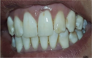
This is a 27-year-old patient, in good general condition, who presented to our consultation because of the presence of stains on her teeth. Upon questioning, the patient did not report any dentin sensitivity. The clinical examination showed the existence of opaque spots and other slightly brownish ones related to cracks on the 11, 12, 21, 22 and 23. The patient also presented tartar especially in the anterior sector. -lower (figure 1).
Figure 1: preoperative photo
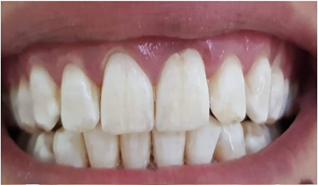
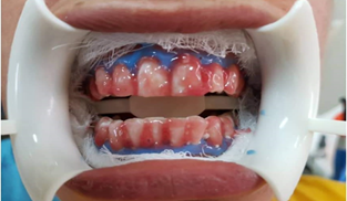
Her treatment began with a good scaling-root planing, the motivation of the patient and the prescription of a sensitivity toothpaste that she had to use for 15 days before starting whitening to prevent the appearance of possible sensitivity. In the following session, 4 cycles of micro abrasion were done using the opalustre® (figure 2). After one week, the patient was seen again, an application of a fluoride gel (Floropal ®) took place 10 minutes before the application of the lightening product. The cracks were filled with fluid resin and the gums were protected by the photopolymerizable dam. Opalescence extra-boost ® was subsequently applied to the teeth for 30 minutes without light activation. (Figure 3).
Figure 2: Photo after my microabrasion
Figure 3: Application of Extraboost ®
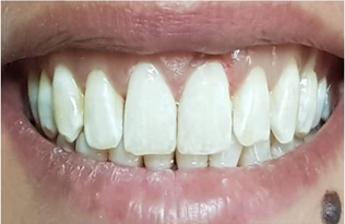
The patient continued brushing with sensitivity toothpaste in the following days, and she was seen again after 2 weeks to replace the composite resin at the cracks (figure 4).
Figure 4: Final result
Discussion
The different lightening techniques have been applied, each separately, to treat dental fluorosis. But the principle of using treatment using combined techniques has brought many advantages. In our case, we combined micro abrasion and external lightening on a chair. Indeed, micro abrasion, which is a chemo-mechanical treatment using an abrasive agent and an acid, is intended to eliminate or improve superficial enamel dyschromia [5]. The thickness removed varies from 20 to 200 µm depending on the acid concentration and the duration of the application [5]. Brown spots, which are generally more superficial than white spots, disappear after micro abrasion enamel in approximately 100% of cases compared to a success rate of 75% for opaque ones [5]. During the micro abrasion procedure, the erosive action of the acid causes the disorganization of the prismatic structure of the enamel. A mineral matrix is produced at the periphery during its reorganization, which allows the formation of a highly compressed aprismatic enamel layer, reinforced with particles from the micro abrasion material (such as silica) and/or polishing pastes (like fluorides) which will gradually remineralize upon contact with saliva [6,7]. Several microabrasion materials exist on the market, the most available and used are: Prema ®, Premier Dental Company (Philadelphia, PA, United States) containing 10% hydrochloric acid and abrasive particles of silicon carbide whose particle size is 30 to 60 µm and Opalustre ® (Ultradent, South Jordan, Utah, United States), containing 6.6% hydrochloric acid and silicon carbide microparticles with a particle size of 20 to 160 µm [8]. Microabrasion is carried out using special rubber cups, mounted on a contra-angle at a low speed of 300 rpm, at a rate of 10 seconds per tooth and a standardized force of 100 grams, equivalent to 2 Bars [9]. It remains reserved for dyschromia limited to the outer layer of enamel and constitutes a solution for treating mild to moderate fluorosis [10-12]. The micro abrasion /external lightening association is of particular interest because the loss of enamel thickness after micro abrasion can allow the underlying dentin to show through, giving a yellowish appearance. Lightening will then make it possible to reduce the saturation of the color and harmonize it, especially since in the case of fluorosis, the teeth have a chalky and cloudy appearance. This aspect, even reduced by micro abrasion, remains visible, hence the interest in completing the treatment with external lightening which will optimize the aesthetic result by reducing the contrast between healthy enamel and stained enamel [13,14]. In an in vitro study, Franco et al. (2016) reported that the combination of micro abrasion and external whitening had no influence on micro-hardness or surface roughness of teeth, whether with immediate or delayed combination [15]. . Castro et al. (2014) reported in a randomized clinical trial that there was no statistically significant difference between the two techniques (micro abrasion alone and micro abrasion associated with external lightening) because in both treatment groups there was a improvement of the aesthetic appearance without the occurrence of side effects (dentin sensitivity or gingival irritation). However, patients who underwent external whitening after micro abrasion were more satisfied with the appearance of their teeth [16].
Conclusion
The micro abrasion /external lightening combination may be a reasonable compromise in the case of mild to moderate fluorosis. It provides an acceptable aesthetic appearance, minimal loss of substance, transient side effects and an affordable cost [17,18]. This is the case of the clinical situation illustrated in this work where the result of the combination of the two techniques in addition to bonding at the level of the cracks perfectly responded to the aesthetic request of the patient.
References
- Chirani R., Foray H. (2004). Dental fluorosis: etiological diagnosis. EM- consults, 12(3).
Publisher | Google Scholor - Sundfeld D, Pavani CC, Pini N, Machado LS, Schott TC, Sundfeld RH. (2019). Enamel Microabrasion and Dental Bleaching on Teeth Presenting Severe-pitted Enamel Fluorosis: A case Report. Oper Dent.
Publisher | Google Scholor - Romero MF, Babb CS, Delash J, Brackett WW. (2018). Minimally invasive esthetic improvement in a patient with dental fluorosis by using microabrasion and bleaching: a clinical report. J Prosthet Dent. 120(3) :323-326.
Publisher | Google Scholor - Giovanni T., Eliades T., Papageorgiou S. (2018). Interventions for dental fluorosis: a systematic review. J Esthet Restoring Dent, 30(6) :502-505.
Publisher | Google Scholor - Croll TP. (1989). Enamel microabrasion: the technique. Quintessence Int. 1989 June; 20(6):395-396.
Publisher | Google Scholor - Pini NIP, Lima DANL, Sundfeld RH, Ambrosano GMB, Aguiar FHB, Lovadino JR. (2017). Tooth enamel properties and morphology after microabrasion: an in-situ study. J Investigate Clin Dent.
Publisher | Google Scholor - Pini NI, Costa R, Bertoldo CE, Aguiar FH, Lovadino JR, Lima DA. Enamel morphology after microabrasion with experimental compounds. Contemp Clin Dent. 2015 Apr- Jun; 6(2):1.
Publisher | Google Scholor - Pini NI, Sundfeld -Neto D, Aguiar FH, Sundfeld RH, Martins LR, Lovadino JR et al. (2015). Enamel microabrasion: An overview of clinical and scientific considerations. World J Clin Cases, 3(1):34-35.
Publisher | Google Scholor - Deshpande AN, Joshi NH, Pradhan NR, Raol RY. (2017). Microabrasion-remineralization: An innovative approach for dental fluorosis. J Indian Soc Pedod Prev Dent, 35 (4):384-387.
Publisher | Google Scholor - Sundfeld RH, Franco LM, Gonçallves RS, de Alexandre RS, Machado LS, Neto DS. (2014). Accomplishing esthetics using enamel microabrasion and bleaching- a case report. Oper Dent. 39 (3):223-227.
Publisher | Google Scholor - Sunfeld RH, Sunfeld-Neto D, Machado LS, Franco LM, Fagundes TC, Briso AL. (2014). Microabrasion in tooth enamel discoloration defects: three cases with long-term follow-ups. J Appl Oral Sci. 22(4):347-354.
Publisher | Google Scholor - Pandey P, Ansari AA, Moda P, Yadav M. (2013). Enamel microabrasion for aesthetic management of dental fluorosis. BMJ Case Rep.
Publisher | Google Scholor - Gençer MDG, Kirzioglu Z. (2019). A comparison of effectiveness of resin infiltration and microabrasion treatments applied to developmental enamel defects in color masking. Dent Mater J, 38(2):295-302.
Publisher | Google Scholor - Sundfeld D, Pavani CC, Schott TC, Machado LS, Pini NIP, Bertoz APM et al. (2019). Dental bleaching on teeth submitted to enamel microabrasion 30 years ago-a case report of patients’ compliance during bleaching treatment. Clin Oral Investig. (1):321-326.
Publisher | Google Scholor - Franco LM, Machado LS, Salomao FM, Dos Santos PH, Briso AL, Sundfeld RH. (2016). Surface effects after a combination of dental bleaching and enamel microabrasion : an in vitro and in situ study. Dent Mater J. 35(1):13-20.
Publisher | Google Scholor - Castro KS, Ferreira AC, Duarte RM, Sampaio FC, Meireles SS. (2014). Acceptability, efficacy and safety of two treatments protocols for dental fluorosis: a randomized clinical trial. J Dent. 42(8):938-944.
Publisher | Google Scholor - Perete-de-Freitas CE, Silva PD, Faria-E-Silva AL. (2017). Impact of Microabrasion on the Effectiveness of tooth bleaching. Braz Dent J. 28(5):612-617.
Publisher | Google Scholor - Gupta A, Dhingra R, Chaudhuri P, Gupta A. (2017). A comparison of various minimally invasive techniques for the removal of dental fluorosis stains in children. J Indian Soc Pedod Prev Dent. 35(3):260-268.
Publisher | Google Scholor


