

Research Article
The Magseed Marker in Impalpable Breast Cancer: A Single Centre Review of Efficacy and Safety
Craigavon Area Hospital, Northern Ireland.
*Corresponding Author: Norah Scally, Craigavon Area Hospital, Northern Ireland.
Citation: L Armstrong, N Scally, J Lockhart, H Mathers. (2022). The Magseed Marker in Impalpable Breast Cancer: A Single Centre Review of Efficacy and Safety. Journal of Clinical Surgery and Surgical Research. 1(1); DOI: 10.59657/2992-9989.brs.22.002
Copyright: © 2022 Norah Scally, this is an open-access article distributed under the terms of the Creative Commons Attribution License, which permits unrestricted use, distribution, and reproduction in any medium, provided the original author and source are credited.
Received: September 30, 2022 | Accepted: October 18, 2022 | Published: October 26, 2022
Abstract
Aim: A cohort observational study was conducted, aiming to review the overall efficacy and safety of the Magseed localisation technique in non-palpable breast cancer, in comparison to wire guided excisions (WGE).
Methods: Data was collected in a single centre, on all patients undergoing image guided breast conserving surgery between April 2018 and August 2019, with a planned accumulation of 100 patients undergoing Magseed localisation. During the same period, data was also collected for 100 consecutive patients undergoing WGE. Primary end points observed included excision volumes and re-excision rates.
Results: Re-excision rates due to positive margins were significantly higher in the WGE group (WGE 26% vs. Magseed 8%, p<0.05). Specimen sizes by weight were also larger for WGE group (WGE 50.4g vs. Magseed 31.2g p=<0.05). 41% of WGE cases were delayed (average 62mins) when compared to 0% of Magseed cases.
Conclusions: The previously recognised gold standard method of WGE has documented limitations. Our study illustrates the Magseed marker as an effective alternative method of lesion localisation, which reduces re-excision rates, improves theatre scheduling, and reduces volume of excision tissue, improving overall cosmetic outcome.
Keywords: magnetic seed; breast cancer; localization; wire; lumpectomy
Introduction
In the advent of screening and with increasing awareness, impalpable breast lesions or those measuring <2cm>
Traditionally guide wires are used to intra-operatively identify impalpable breast lesions. This is a method which has been utilised since the 1960’s and is the most commonly used technique in centres worldwide. However, many limitations of the guide wire technique have been recognised, most notably migration of the wire tip itself which in turn compromises its accuracy. Furthermore, there have been reports of high positive margin rates, leading to additional surgery for re-excision [4]. Significant patient discomfort has also been reported during the insertion of the wire and during the transport of the patient to and from the radiology department. A desire to improve the patient experience and comfort has led to the development of alternative localisation techniques such as the Magseed and Radioactive seed. Implementation of the radioactive seed technique is often limited due to radiation safety requirements [2].
The Magseed marker is a small metallic seed, deployed by a needle delivery system [3], that can be visualised on mammography and ultrasound. It received FDA 510(k) clearance in March 2016 and is approved for placement at any time prior to surgey [5]. Crucially for radiologists and surgeons, the Magseed marker cannot be broken on deployment, following implantation, or damaged with diathermy during surgery.
Materials and Methods
This observational study was carried out in a single centre between April 2018 and August 2019. Data was collected on all patients undergoing image guided breast conserving surgery during this period, with a planned accumulation of 100 patients undergoing Magseed localisation. Data was retrieved from an institutional database search and patient follow-up obtained from up-to-date regional electronic care records. During the same period, data was also collected for 100 consecutive patients undergoing WGE. Data collected included: patient age, insertion technique, histological subtype, tumour size, total specimen excision size and volume, and any secondary surgical procedure performed due to requirement of cavity shaves.
Clinical decisions and management plans, included proposed localisation techniques, were devised upon a consensus at a weekly multidisciplinary meeting, which involved the surgical, oncological and radiology team members. Only patients with clinically non-palpable breast cancer and histologically proven carcinoma of the breast were included. Patients who had a pacemaker in situ, or those whose lesion was greater than 6cm from skin were not assigned to the Magseed group. To avoid bias, those undergoing procedures involving multiple wires or therapeutic mammoplasties were excluded.
Our primary end points in our study included excision volumes and re-excision rates. Specimen weight and volumes alongside tumour size were all recorded. Any margin <1mm>
The Magseed marker was introduced to Craigavon Area Hospital in April 2018 and was the first unit in Ireland to complete 100 cases using this technique. This localisation method was adopted by all three consultants within the unit. Patients were allocated to a localisation technique at a weekly multidisciplinary meeting. Patients who had a pacemaker or those whose lesion was greater than 6cm from skin were not assigned to the Magseed group.
The Magseed device consists of a 5 mm x 1 mm cylindrical nonradioactive paramagnetic steel and iron oxide seed.6 The seed is deployed via a preloaded 18 gauge, under ultrasonographic or mammographic guidance by radiologists. Intra-operatively the Magseed is identified and located by the Sentimag probe. This has a numerical display and an audio tone, the frequency of which correlates to the distance of the probe from the Magseed, thus allowing the surgeon to accurately gauge the distance of the seed and precisely locate the lesion. Non-ferromagnetic retractors are used so as not to cause interference with the Magseed detection.
Results
100 patients were collected prospectively for each localisation group, with a mean follow up time of 187 ± 64 days. Following multidisciplinary discussion, 2 patients were assigned to the WGE group, due to the presence of a pacemaker. Demographic and disease characteristics were well balanced between the Magseed and wire-guided group (See table 1). All patients within the study population were female. The vast majority of patients underwent ultrasound guided insertions in both groups (87% and 89%) (see table 1). The median length of time from insertion to procedure in the Magseed group was 9 days (1-33).
Table 1
| Magseed | Wire guided | P Value | |
| Total | 100 | 100 | - |
| Age (Median) | 59 (35-82) | 68(35-82) | - |
| Histology | |||
| Ductal | 71 | 71 | |
| Lobular | 6 | 6 | |
| DCIS | 6 | 13 | |
| Other | 17 | 10 | |
| Margins | |||
| Negative | 92 | 74 | |
| Positive | 8 | 26 | <0> |
| Lumpectomy (g) (Median) | 31.2 (8-92) | 50.4 (13-268) | <0> |
| Tumour Volume (mm3) (Median) | 11(1-28) | 19 (1-110) | 0.022 |
| Guidance Modality | |||
| USS | 89 | 87 | |
| Stereotactic | 11 | 13 | |
| Timing | |||
| Insertion (days pre op) | Median 9 days (1-33) | - | |
| Neoadjuvant Chemotherapy (n) | 60 | 11 |
Re-excision rates, due to positive margins, were significantly higher in the WGE group (WGE 26% vs. Magseed 8%, p<0 xss=removed xss=removed>
The start time of each procedure was reviewed via the theatre management system, and a delayed start time was considered >15 minutes. 41% of WGE cases had a delayed start time, with an average time of 62mins. Delays were in relation to the insertion of the wire, transport of the patient to the radiology department and co-ordination with the radiology team. In contrast, none of the Magseed localisation patients experienced a delay in operating start time.
Thirty-one patients returned the state trait questionnaires, 3 excluded due to non-completion. The population included 14 wire guided and 14 Magseed localisation patients. Scale based average scores for each localisation population were 41 and 36 for WGE and Magseed localisation, respectively. Higher scores positively correlated higher anxiety with wire guided localisation.
Discussion
The Magseed marker is a small, non-radioactive, para-magnetic seed, (Fig.1) which similarly to the guide wire, can be inserted under stereotactic or ultrasound guidance. However, in contrast, it can be inserted at any time in the pre-operative period, which can be instrumental in improving patient flow and theatre scheduling. It is also derived from low nickel steel; therefore, there is no concern for a nickel allergy. It is accompanied by the Senti-Mag probe (Fig 2), which allows accurate transcutaneous detection intra-operatively. The Sentimag system was originally launched at the end of 2012 for sentinel lymph node biopsies (SLNB) [8].
Breast conserving surgery is the preferred surgical method for small breast lumps, aiding overall cosmetic results. Previous studies have reported larger specimen volumes in wire guided procedures compared to other techniques [4,6,9]. This may be due to the migration of the wire tip itself, or due to the substantial distance from the tip of the wire to the transcutaneous entry site. This distance can make it difficult to maintain the wires accuracy, thus leading to a more extensive dissection intra-operatively. Our results illustrated a statistically significant difference in excision weight between the groups, which can impact overall cosmetic outcome. There was also a statistical difference between the tumour volumes in each group, perhaps suggesting smaller tumours were assigned to the Magseed group. This may be also illustrated in the larger number of DCIS patients with the Magseed group (n=13 vs. n=6). It is also important to note the larger volume of patients who underwent neoadjuvant chemotherapy within the Magseed group. This may have led to a significant or complete histological response, thus leading to a smaller tumour size.
WGE has historically been associated with high positive margin rates requiring additional surgery [3]. A recent NHS England review showed reoperation was required in approximately 20% of breast conserving cases; with significant variation amongst Trusts [3]. Again, this can be due in part to migration of the wire, from its original position, between placement and surgery. In contrast to the guidewire, the Magseed provides a point source which enables continuous reorientation during surgery using the Sentimag probe. It also allows for extracorporeal confirmation of the Magseed within the excision specimen. Plain radiographs can also be performed of the specimen to confirm its location. Our results demonstrated a higher rate of positive margins and therefore a higher rate in return to theatre in the WGE group (WGE 26% vs. Magseed 8%, p<0>Powell et al. also reviewed the use of the Magseed marker, with a similar population size. When compared to published figures of WGE, they also concluded favourable re-excision rates withing the Magseed group [10].
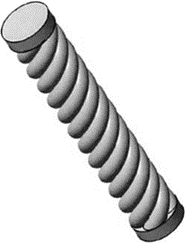
Figure 1: The Magseed Marker structure- 5 × 1 mm paramagnetic steel seed
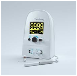
Figure 2: The Sentimag Probe- allows for accurate transcutaneous detection of the Magseed
According to the literature, delays in theatre start times for WGE cases average 40 minutes [11]. The typical journey of a patient undergoing a wire guided procedure begins on the elective admission ward, where they await the insertion of the wire. For this reason, it is not feasible to place this patient first on the operating list, nor is it possible to add more than 4 patients to any one list. The journey time to and from the nuclear medicine department for sentinel node localisation, along with wire insertion, can take up to 1 hour. These factors lead to issues in theatre scheduling, create delays and cancellations, and have negative impacts on both patients and staff. In comparison, a patient undergoing Magseed localisation can have in inserted at any point prior to the procedure. This allows the patient to be placed first on the operating list, without any expected delays. Furthermore, this theatre streamlining and reduction in delays can potentially increase list capacity.
The mental health burden amongst women with breast cancer extends beyond their initial diagnosis throughout the breast cancer journey [12]. Previous studies have commented on the overall patient dissatisfaction and discomfort experienced with guide wires [14]. The protrusion of a foreign body can be unsettling for a patient. In addition to this, they are informed that their movement may hinder the wire’s accuracy. Our results suggest that Magseed localisation may result in lower patient anxiety pre-operatively when compared to guide wire localisation.
The radiology team responsible for the insertion of the Magseed required minimal additional training upon its introduction, as the deployment of the seed is similar to other radiological marker placement. With careful co-ordination between the breast and radiology team, the majority of Magseed patients were able to have their seed inserted on the day of their pre-operative assessment, reducing their attendances to the department.
One limitation of this study was the exclusion of lesions >6cm from the skin edge in the Magseed group. This is turn may have influenced the selection criteria to each localisation group, preferentially selecting breast lumps that were surgically more easily accessible to the Magseed group. We also recognise the inclusion of neoadjuvant therapy cases have influenced the tumour size in each group, with a larger volume of neoadjuvant cases in the Magseed group resulting in a statistical significant difference in tumour size overall.
Conclusion
The previously recognised gold standard method of wire guided excisions has limitations for both patients and staff alike. Our study has illustrated the Magseed marker as a safe and reliable alternative localisation technique to the guide wire. Magseed localisation has been proven to reduce re-excision rates due to positive margins and facilitate lower excision volumes resulting in both improved cosmetic outcomes and overall patient satisfaction.
Acknowledgements
Ethical Statement
The authors are accountable for all aspects of the work in ensuring that questions related to the accuracy or integrity of any part of the work are appropriately investigated and resolved.
Funding
No funding was received for this study.
Conflicts of Interest
No conflicts of interest
We can confirm that we have been actively involved in the preparation of the paper.
We agree to publication.
Author Contributions
(I) Conception and design: Ms Lara Armstrong, Ms Helen Mathers
(II) Administrative support: Ms Helen Mathers, Ms Norah Scally
(III) Provision of study materials or patients: Ms Lara Armstrong, Ms Jessica Lockhart
(IV) Collection and assembly of data: Ms Lara Armstrong, Ms Jessica Lockhart
(V) Data analysis and interpretation: Ms Lara Armstrong, Ms Jessica Lockhart
(VI) Manuscript writing: All authors
(VII) Final approval of manuscript: All authors
References
- Welch HG, et al. (2016). Breast-cancer tumour size, overdiagnosis, and mammography screening effectiveness. N Engl J Med. 375(15):1438e47.
Publisher | Google Scholor - Fisher B, Anderson S, Bryant J, Margolese RG, Deutsch M, Fisher ER, et al. (2002). Twenty-year follow-up of a randomized trial comparing total mastectomy, lumpectomy, and lumpectomy plus irradiation for the treatment of invasive breast cancer. N Engl J Med. 347:1233-1241.
Publisher | Google Scholor - Cheang, Ellen et al. (2018). Innovations in image-guided preoperative breast lesion localization. The British journal of radiology. 91(1085):20170740.
Publisher | Google Scholor - Jeevan R et al. (2012). Reoperation rates after breast conserving surgery for breast cancer among women in England: Retrospective study of hospital episode statistics. BMJ.
Publisher | Google Scholor - Harvey JR, et al. (2018). Safety and feasibility of breast lesion localization using magnetic seeds (Magseed): a multi-centre, open-label cohort study. Breast Canc Res Treat. 169(3):531e6.
Publisher | Google Scholor - Hayes MK. (2017). Update on preoperative breast localization. Radiol Clin North Am. 55:591-603.
Publisher | Google Scholor - Zacharioudakis K, Down S, Bholah Z, Lee S, Khan T, Maxwell AJ, Howe M, Harvey J. (2019). Is the future magnetic? Magseed localisation for non-palpable breast cancer. A multi-centre non randomised control study. Eur J Surg Oncol.
Publisher | Google Scholor - Available at: https://www.endomag.com/sentimag/
Publisher | Google Scholor - Karakatsanis A, et al. (2017). Superparamagnetic iron oxide nanoparticles as the sole method for sentinel node biopsy detection in patients with breast cancer, BJS.
Publisher | Google Scholor - Powell M, Gate T, Kalake O, Ranjith C, Pennick MO. (2021). Magnetic Seed Localization (Magseed) for excision of impalpable breast lesions-The North Wales experience. The Breast Journal.
Publisher | Google Scholor - Dauer LT. (2013). Radioactive seed localisation with 125I for non-palpable lesions prior to breast lumpectomy and/or excisional biopsy methodology, safety, and experience of initial year, Health Physics. 105(4):356-365.
Publisher | Google Scholor - Hayes MK, et al. (2017). SAVI scout improves operating room start times compared with wire localization.
Publisher | Google Scholor - Small S, McAdam A and Mathers H. (2019). Analysis of patient anxiety related to magseed and guide-wire localisation techniques. EurJ Surg Oncol. 45(5):925.
Publisher | Google Scholor - Bloomquist EV, et al. (2016). A Randomised Prospective Comparison of Patient-Assessed Satisfaction Clinical Outcomes with Radioactive Seed, Breast Journal, March. 22(2):151-157.
Publisher | Google Scholor


