

Case report
The Aesthetic Restoration of Missing Anterior Teeth with A Dmls Prosthesis-A Case Report
- Nikhil Vitthal Dayama *
- Mariyam Ali
- Pratibha Katiyar
- Neeranshi Swaroop
- Asra Ahmed
- Mirza Hamza Baig
Department of Prosthodontics, Crown and Bridge, Career Post Graduate Institute of Dental Sciences and Hospital, Lucknow, UP.
*Corresponding Author: Nikhil Vitthal Dayama.Department of Prosthodontics, Crown and Bridge, Career Post Graduate Institute of Dental Sciences and Hospital, Lucknow, UP.
Citation: Nikhil V Dayama, Ali M, Katiyar P, Swaroop N, Ahmed A, et al. (2023). The Aesthetic Restoration of Missing Anterior Teeth with A Dmls Prosthesis-A Case Report. International Journal of Medical Case Reports and Reviews, BRS Publishers. 2(2); DOI: 10.59657/2837-8172.brs.23.020
Copyright: © 2023 Nikhil Vitthal Dayama, this is an open-accsess article distributed under the terms of the Creative Commons Attribution License, which permits unrestricted use, distribution, and reproduction in any medium, provided the original author and source are credited.
Received: May 22, 2023 | Accepted: June 02, 2023 | Published: June 16, 2023
Abstract
The most significant social connecting gesture and vital to professional advancement is a confident, appealing smile. Patients may experience social and psychological discomfort as a result of the loss of their anterior teeth. The contemporary dental situation provides numerous treatment options for the aesthetic and functional replacement of a congenitally or traumatically lost anterior tooth in young and adult patients, including implant crown, conventional FDP, Resin Bonded FDP, or RPD. The most important aspect of an aesthetic anterior restoration is patient satisfaction with the cosmetic and functional outcome of therapy. This case report discusses the aesthetic correction of missing maxillary and mandibular anterior teeth using intra-oral scanning and DMLS PFM crowns.
Keywords: aesthetic restoration; intra-oral scanning; dmls pfm crowns
Introduction
A beautiful confident smile is desired by all. Over time, evolving dental technology and rising aesthetic expectations have produced predictable, more aesthetically pleasing, and more useful outcomes. Pierre Fauchard (1678–1761) of France, worked with a number of his colleagues to modernize, advance, and support dentistry as well as aesthetic procedures1,2. An aesthetic makeover aims to create a calm and stable masticatory system where the teeth, tissues, muscles, skeletal structures, and joints all work together harmoniously [3]. According to reports (Mange and Besler, 2010; McLaren and Culp, 2013) [4]. face, dent gingival, and dental aesthetics are the fundamental ideas for aesthetic analysis. To plan a proper aesthetic rehabilitation, it is necessary to meet the expectations of the patients at the end of the treatment considering
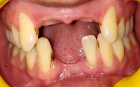
Figure 1: Intra-oral view.
all these parameters (Meres et al., 2016) [5]. This case report discusses the restoration of missing maxillary and mandibular anterior teeth (12-22, 31-42) using DMLS PFM crowns.
Case report
A 28-year-old male patient reported to the Department of Prosthodontics and Crown & Bridge, Career Postgraduate Institute of Dental Science and Hospital, Lucknow with a chief complaint of unpleasant appearance due to missing anterior teeth. Missing teeth IRT 12, 11, 21, 22, 31, 41, and 42 were revealed on intra-oral examination (Figure 1). Due to the financial constraints of the patient, the options of the implant-supported prosthesis and CAD-CAM zirconia crown were ruled out and DMLS PFM crowns were suggested for better marginal adaptation and aesthetics.
Diagnostic impressions were made with irreversible hydrocolloid and diagnostic models were poured followed by diagnostic mounting. Tooth preparation was done it 12-22 and 31-42 (Figure 2) and an intra-oral scanning was carried out (Figure 3) followed by previsualization. Scanned models with the design of the prosthesis were obtained (Figure 4). Bisque try-in was done (Figure 5) followed by cementation of the final DMLS PFM prosthesis (Figure 6). Patient education and motivation about the maintenance of the prosthesis and hygiene were carried out.
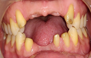
Figure 2: Tooth Preparation.
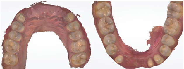
Figure 3: Intra Oral Scanning.
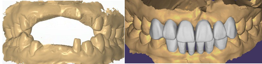
Figure 4: Designing of DMLS Prosthesis.
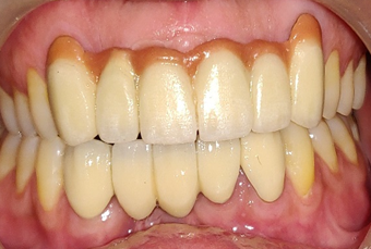
Figure 5: Bisque Tria.
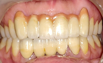
Figure 6: Final Prosthesis.
Discussion
The creation of an ideal smile necessitates an examination and appraisal of the face, lips, gingival tissues, and teeth, as well as an understanding of how they appear combined. Davis (2007) defines smile design as a collection of aesthetic factors that make face aesthetics compatible with dent gingival structures [1]. Alternatively, it is frequently referred to as the aesthetic treatment of anterior teeth in the visible aesthetic zone (Zimmermann and Mewl, 2015) [6]. CAD/CAM was first used in dentistry for ceramic inlays and on lays two decades ago. In 1991, the Procura system (Nobel BioCare, Goteborg, Sweden) was introduced for individualized dental restorations. After that, a quick production strategy utilizing 3D printing (DMLS) and metal powders in CAD models was implemented [7]. The particle size used in DMLS crowns and bridges is 3–14 µm. This results in a density of 99.9%, which leads to stronger copings, when paired with a very fine tip laser (0.1 mm). Restorations produced by the method are incredibly precise and well-detailed. Clinically acceptable variation is present as long as the specimen has a constant level of strength and quality [8,9].
The present case report describes the rehabilitation of the anterior edentulous area using DMLS PFM crowns which is a cost-efficient alternative for CAD-CAM zirconia and implant crowns resulting in acceptable aesthetic and good marginal fit. The teeth were prepared and temporization is done to assess the aesthetics. Once, the patient was satisfied with the temporary crowns, an Intraoral scan was used for final impressions. Compared to traditional impression methods, digital impressions have been demonstrated to be more accurate. With the use of digitization, it is simpler to transport the scanned information and accurately build the prosthesis [10]. The designing of DMLS copings was done, followed by porcelain build-up. Gingival porcelain was also added to make the prosthesis more aesthetic and natural. The final prosthesis was cemented using GIC cement. The anterior guidance was verified in centric and eccentric movements. The patient was satisfied with the outcome of the prosthesis.
Conclusion
Happiness is a state of mind. It is brought about by a feeling of well-being, security and confidence in one’s self. It is thus essential for the dentist to give a pleasing smile to the patient when working in the anterior zone. The treatment plan has to be thoroughly discussed with the patient and if needed, interdisciplinary approach can be executed.
References
- Sharma A, Bhardwaj V, Gupta J, Kadina R. Smile Design: A Modern Approach.
Publisher | Google Scholor - US YÖ, YÜZBAŞIOĞLU E, ALBAYRAK B, ÖZDEMİR G. (2021). Digital Smile Design: Predictable Results. Journal of Experimental and Clinical Medicine, 38(3): 123-128.
Publisher | Google Scholor - Bhuvaneswar an M. (2010). Principles of smile design. Journal of conservative dentistry, 13(4):225-232.
Publisher | Google Scholor - McLaren, E.A., Culp, L. (2013). Smile analysis: The photoshop smile design technique part 1. J. Cos met. Dent, 1:98-108.
Publisher | Google Scholor - Meres, C., De Souza, G., Albino, L., Oliar, F., Pia, E., Lima, G. (2016). Digital smile design for computer-assisted esthetic rehabilitation: Two-year follow-up. Opera. Dent, 41:13-22.
Publisher | Google Scholor - Zimmermann, M., Mewl, A. (2015). Virtual Smile Design Systems: A Current Review, 18:303-317.
Publisher | Google Scholor - Venkatesh KV, Nandini VV. D. (2013). direct metal laser sintering: a digitized metal casting technology. The Journal of Indian Prosthodontic Society, 13(4):389-392.
Publisher | Google Scholor - Orator A, Jonsson D, Mohsen A, von Steyer PV. (2011). The fit of cobalt–chromium three-unit fixed dental prostheses fabricated with four different techniques: A comparative in vitro study. Dental Materials, 27(4):356-363.
Publisher | Google Scholor - Quante K, Ludwig K, Kern M. (2008). Marginal and internal fit of metal-ceramic crowns fabricated with a new laser melting technology. Dental Materials, 24(10):1311-1715.
Publisher | Google Scholor - Aswani K, Wankhade S, Khalikar A, Deogade S. Accuracy of an intraoral digital impression: A review. The Journal of the Indian Prosthodontic Society, 20(1):27.
Publisher | Google Scholor


