Clinical Image
Textbook Presentation of Cannonball Pulmonary Metastases
1Associate professor in Medicine Department at SMS Medical College, Jaipur, Rajasthan, India.
2Final year MBBS student at SMS Medical College, Jaipur, Rajasthan, India.
*Corresponding Author: Vansh Bagrodia, Final year MBBS student at SMS Medical College, Jaipur, Rajasthan, India.
Citation: Sharma V, Bagrodia V. (2024). Textbook Presentation of Cannonball Pulmonary Metastases, Clinical Research and Reports, BioRes Scientia Publishers. 2(1):1-2. DOI: 10.59657/2995-6064.brs.24.008
Copyright: © 2024 Vansh Bagrodia, this is an open-access article distributed under the terms of the Creative Commons Attribution License, which permits unrestricted use, distribution, and reproduction in any medium, provided the original author and source are credited.
Received: October 03, 2023 | Accepted: October 17, 2023 | Published: January 06, 2024
Abstract
This case report presents a late sixties male patient who arrived with massive hemoptysis and breathing difficulties. Shock symptoms were evident, alongside distinct cannonball-like lung lesions on X-ray, prompting consideration of metastases. Clinical evaluation and attendant history suggested renal cell carcinoma (RCC) as an underlying cause. Despite swift intervention, the patient deteriorated rapidly, succumbing to recurring hemoptysis within hours. This report underscores the challenging diagnosis and management of aggressive metastatic RCC, particularly underscored by the unique X-ray presentation, emphasizing the significance of acute medical presentations.
Keywords: male patient; metastatic RCC; X-ray; cannonball pulmonary
Case
A male patient in his late sixties presented to our tertiary specialist unit with massive hemoptysis and progressive dyspnea [mMRC (Modified Medical Research Council) grade 4]. The patient was alert, conscious, and oriented to time, place, and person. On examination, the patient had tachycardia (pulse rate - 125 beats/minute) and low blood pressure (87/56 mmHg), indicating that he was in shock [modified shock index = 1.9]. Room air oxygen saturation was 87%. To stabilize the patient, he was put on oxygen via a mask at a flow rate of 6 L/min, and transfusion of 2 units of packed red blood cells was started after cross-matching.
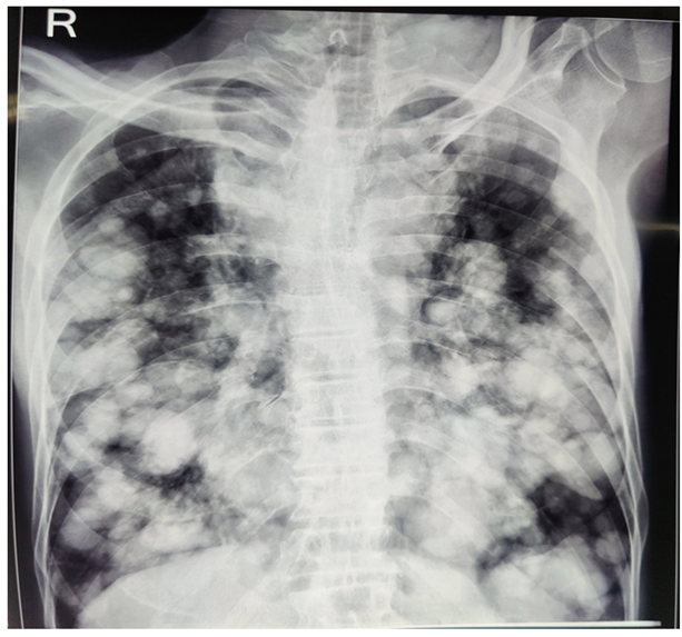
An emergency room chest X-ray showed remarkable cannonball-like lesions scattered all over both lung fields (figure 1). Preliminary history taken from attendants revealed that the patient had complaints of vague discomfort in the left lumbar region, painless intermittent hematuria, and persistent low-grade fever for the past 2 months. During this time period, the patient experienced a significant weight loss of 7 kilograms [12% of the initial body weight]. Attendants also reported the presence of a lump in the left lumbar region of the patient.
Based on the history given and the presence of a ballotable mass in the left lumbar region upon bimanual palpation, a diagnosis of renal cell carcinoma (RCC) was suspected. The cannonball-like lesions scattered all over both lung fields on the chest X-ray were suspected to be metastases from a likely renal cell carcinoma. However, before the patient could be sent for further imaging and biopsy to confirm the presence of a malignant renal mass, he had another bout of massive hemoptysis and succumbed to shock within just 4 hours of the initial presentation to the emergency room.
RCC commonly leads to metastases with a cannonball appearance and was the most likely primary pathology of our patient, based on the history given by attendants. Unfortunately, it could not be confirmed with any imaging or histopathology due to the rapid deterioration of the patient in a short span of time [1]. The patient's age also supported the suspicion of RCC, as most patients are diagnosed with RCC between ages 65 and 74 [2]. The lung is the most frequent site of metastasis and recurrence of RCC, with more than 50–60% of patients developing lung metastases [3]. Massive hemoptysis is associated with a high mortality rate of 17.8 Percentage according to studies and was the reason for the mortality of our patient as well [4].
Figure 1: Remarkable cannonball-like lesions scattered all over both lung fields.
References
- Kshatriya R, Patel V, Chaudhari S, Patel P, Prajapati D, Khara N, Paliwal R, Patel S. (2016). Cannon ball appearance on radiology in a middle-aged diabetic female. Lung India, 33(5):562-568.
Publisher | Google Scholor - Padala SA, Barsouk A, Thandra KC, Saginala K, Mohammed A, Vakiti A, Rawla P, Barsouk A. (2020). Epidemiology of Renal Cell Carcinoma. World J Oncol, 11(3):79-87.
Publisher | Google Scholor - Gonnet, A., Salabert, L., Roubaud, G. et al. (2019). Renal cell carcinoma lung metastases treated by radiofrequency ablation integrated with systemic treatments: over 10 years of experience. BMC Cancer, 19:1182.
Publisher | Google Scholor - Reechaipichitkul W, Latong S. (2005). Etiology and treatment outcomes of massive hemoptysis. Southeast Asian J Trop Med Public Health, 36(2):474-480.
Publisher | Google Scholor


