Research Article
Radiation Doses Associated with Some Computed Tomography Examinations
- T. M. Taha *
- Talaat Salah Ahmed
- Mohamed Aboalez
Nuclear Research Center, Atomic Energy Authority, Cairo, Egypt.
*Corresponding Author: T. M. Taha, Nuclear Research Center, Atomic Energy Authority, Cairo, Egypt.
Citation: Taha T. M., Ahmed T. S., Aboalez M. (2024). Radiation Doses Associated with Some Computed Tomography Examinations, Clinical Case Reports and Studies, BioRes Scientia Publishers. 7(1):1-11. DOI: 10.59657/2837-2565.brs.24.173
Copyright: © 2024 T. M. Taha, this is an open-access article distributed under the terms of the Creative Commons Attribution License, which permits unrestricted use, distribution, and reproduction in any medium, provided the original author and source are credited.
Received: September 05, 2024 | Accepted: September 12, 2024 | Published: September 23, 2024
Abstract
Radiation doses associated with Computed Tomography is higher than the corresponding doses associated with x-ray examinations by more than one hundred. The research study aims to evaluate the radiation doses associated with CT examinations in examinations in Hospital A in one of the radiology departments. This hospital participated in a dose survey for the three most commonly used CT protocols. CT dose indices are calculated and shown by CT machine via Picture archiving communication system. The volume CT dose index, dose-length product, and effective dose for the head were 48.7 mGy, 600 mGy.cm, and 1.38 mSv; for the chest, 15.5 mGy, 349 mGy.cm and 4.89 mSv; for the abdomen- pelvic, 14 mGy, 551 mGy.cm, and 5.3 mSv. The diagnostic reference dose levels were lower than those of international organizations. The study concluded that the effective doses for head, chest and abdomen pelvic are calculated and can be used as additional tools to verify the quality control of CT scanners.
Keywords: head CT; chest CT; abdomen-pelvic CT; PACs
Introduction
Medical x-rays are the largest man -made source of public exposure to ionizing radiation. The contribution of CT to the collective dose in 1991-1996 was 34% [1,2]. Medical radiation protection principles should include justification and optimization by applying the ALARA principle, as low as reasonably achievable. There are few studies about the evaluation a radiation doses associated with computed tomography examinations in Cairo that means Egypt needs to evaluate the effective doses for all CT examinations for all the hospitals of ministry of health. The effective dose for CT examinations plays an important role in generating a diagnostic dose baseline for the CT scanners. The paper is aimed to calculate the radiation doses associated with CT examinations in Hospital A in Cairo.
Materials and Methods
CT scanner specifications
The study was performed using CT scanner. It includes a Siemens CT machine with 64 multislice as described in Table 1. The demographic data are presented Table 2.
Table 1: Specification of CT scanner.
| Number | CT Manufacture | Scanner Type | Detector type |
| 2 | Siemens | MDCT 64 | Scintillation detectors |
Table 2: Biometric parameters and main CT acquisition parameters for Hospital A.
| Biometric Parameters | Main CT Acquisition Parameters | ||||||
| CT Protocol | No. of Patients | Age (year) | Weight (kg) | Kilo Voltage, kV | Effective mAs | Section Thickness (mm) | Number of Slices |
| Head | 50 | 33 ± 14 | 76 ± 17 | 120 ± 0 | 172 ± 0 | 0.5 ± 0 | 188 |
| Chest | 50 | 52 ± 15 | 85 ± 25 | 120 ± 0 | 250 ± 0 | 2.0 ± 0 | 142 |
| Abdomen-Pelvic | 50 | 45 ± 15 | 86 ± 19 | 120 ± 0 | 134 ± 0 | 3.0 ± 0 | 124 |
CT Dosimetric Unit
CTDIw: Weighted Computed Tomography Dose Index [6].

CTDIvol: Volume Computed Tomography Dose Index [6].

and l = the table increment per axial scan (mm). Since pitch is defined as the ratio of the table travel per rotation (I) to the total nominal beam width (N x T) [3].
The Dose-Length product (DLP) is calculated as presented in equation 6.

Effective dose was estimated multiplying CT dose length product CT, mGy.cm by a corresponding normalized conversion coefficient that is defined as specific only to the anatomic region (k), mSv/mGy. cm for multislice CT scans [4-7].
Results
The mean values of CTDIvol and DLP for fifty CT examinations for each case for different weight intervals presented as shown in Table 3. The Hospital A in Cairo country participated in a dose survey for the three most commonly used CT protocols. CTDIvol, DLP for head/sinus, chest was recorded by (PACS) from Hospital A as shown in Table 4.
Table 3: CTDIvol (mGy) and DLP (mGy.cm) at CT scanner for Hospital A.
| CT protocol | Dosimetric data | 41-60 kg | 61-80 kg | 81-100 kg | 101-120 kg |
| Head | CTDIvol l(mGy) | 48.85±-5.0 | 48.80±1.6 | 48.95±12 | 48.36±8 |
| DLP (mGy.cm) | 602.38±148 | 601.37±208 | 602.75±141 | 593.85±234 | |
| Chest | CTDIvol (mGy) | 14.40±2.5 | 14.10±6 | 15.20±3.5 | 17.00±2.5 |
| (DLP) (mGy.cm) | 107.60±-46 | 312.80 | 342.40±149 | 342.50±135 | |
| Abdomen-Pelvic | CTDIvol (mGy) | 6.47±2.5 | 10.06 | 14.91±2.9 | 21.83±2.9 |
| (DLP) (mGy.cm) | 279.80±102 | 379.31 | 420.20±88 | 921.23±220 |
Table 4: Mean values of CTDIvol, DLP and effective dose for CT protocols.
| CT Protocol | Dosimetric Data/CT Examination | CT Scanner A | ACR-2018 |
| Head | CTDIvol (mGy) | 61.80± 1.8 | 56.00 |
| DLP (mGy.cm) | 598±29 | 962.00 | |
| E (mSv) | 2.55 ± 0.13 | 2.21 | |
| Chest | CTDIvol (mGy) | 15.50 ± 0.62 | 10.00 |
| DLP (mGy.cm) | 349±67 | 400.00 | |
| E (mSv) | 9.21 ± 0.06 | 6.80 | |
| Abdomen-Pelvic | CTDIvol (mGy) | 10±0.1 | 16.00 |
| DLP (mGy.cm) | 379±25 | 781.00 | |
| E (mSv) | 8 ± 0.8 | 11.72 |
Dose Optimization of Abdomen-Pelvic CT Protocol
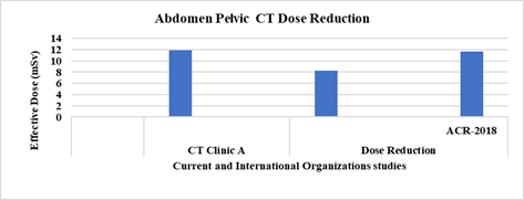
Twenty cases for abdomen- pelvic CT examinations were classified into two groups. The physical parameters for group one was examined for of 120 kV, 224 mAs and group two was examined for 120 kV, 182 mAs respectively. Effective for doses the abdomen-pelvic CT protocol is reduced to 55% due to decrease the tube current-product time, 224 mAs to 182 mAs that effect on the dose out [6] as presented in Figure 1.
Figure 1: Dose Reduction for Abdomen Pelvic CT Examination in CT scanner Versus the American College of Radiology.
Discussion
The CTDIvol and dose length product, DLP corresponding CT protocols in CT clinic were recorded for about 150 patient sizes of selected CT scanner. The demographic data was collected for many patient weight intervals as shown in Table 3. The variation in Cvol and dose length product, DLP for head CT scan for different weights is not significance because of little difference in the number of slices.
The CTDIvol and DLP for chest CT scan for patient’s weights greater than 100 kg increase by a about 10% than patient’s weights lower than 100 kg may be due to difference patient’s chest size.
CTDIvol and DLP for lower limb CT examinations for different patient’s weight ranges increased gradually due to decreasing pitch factors. Table 4 present the volume CT dose index (CTDIvol), dose-length product (DLP), and effective dose) for adult’s head, chest and abdomen- pelvic CT Examinations for Hospital A in Cairo and comparison with international organizations. In the CT clinic A, CTDIvol, DLP, and effective dose for the head were 48.7 mGy, 600 mGy.cm and 1.38 mSv; for the chest, 15.5 mGy, 349 mGy.cm and 4.89 mSv; for the abdomen, 14 mGy, 551 m4Gy.cm and 5.3 mSv. The effective dose for head CT, chest CT and abdomen CT was presented as shown in Table 4.
Conclusion
The results show that the effective doses for head, chest and abdomen pelvic are calculated. Effective doses for the -abdomen-pelvic CT protocol is reduced to 55% due to optimize the tube current product time.
Abbreviations
CTDIvol: Computed Tomography volume dose index,
DLP: dose length product,
ACR: American College of Radiology,
CT: Computed Tomography,
kVp: kilovoltage, peak kilovoltage (Kvp),
milli-Ampere-second: mAs,
Pitch factor: P,
slice-width: T,
length of scan: L,
Picture archiving communication system: PACs,
CTDIvol: Volume Computed Tomography Dose Index.
References
- Foley SJ, McEntee MF, Rainford LA. (2012). Establishment of CT Diagnostic Reference Levels in Ireland. Br J Radiol. 85(1018):1390-1397.
Publisher | Google Scholor - Saravanakumar A, Vaideki K, Govindarajan KN, Jayakumar S. (2014). Establishment of Diagnostic Reference Levels in Computed Tomography for Select Procedures in Pudhuchery, India. J Med Phys. 39(1):50-55.
Publisher | Google Scholor - Heyer CM, Mohr PS, Lemburg SP, Peters SA, Nicolas V. (2007). Image Quality and Radiation Exposure at Pulmonary CT Angiography With 100- Or 120-Kvp Protocol: Prospective Randomized Study. Radiology. 245(2):577-583.
Publisher | Google Scholor - M. A. S Sherer, P. J. Visconti, R. E. Russell, K. W. Haynes. (2018). Radiation Protection in Medical Radiography, 8th Edition book, Elsevier.
Publisher | Google Scholor - General Diagnostic Radiology Practice Parameter/American College of Radiology, (2018).
Publisher | Google Scholor - Patient dosimetry, in Diagnostic Radiology Physics: A handbook for teacher and students, (2014). International Atomic Energy Agency, 555.
Publisher | Google Scholor - Chu PW, Kofler C, Haas B, Lee C, Wang Y, et al. (2024). Dose Length Product to Effective Dose Coefficients in Adults. Eur Radiol. 34(4):2416-2425.
Publisher | Google Scholor


