Caser Report
Post-Traumatic Persistent Complex Regional Pain Syndrome in Ankle: Recurrence or No-Healing?
- Salvatore Gioitta Iachino *
- Festini Capello Michele Paolo
- Schaller Christian Anton Ernst
Department of Orthopedics and Traumatology, Bressanone Hospital, Italy.
*Corresponding Author: Salvatore Gioitta Iachino, Department of Orthopedics and Traumatology, Bressanone Hospital, Italy.
Citation: Salvatore G Iachino, F.C.M Paolo, S.C.A. Ernst. (2023). Post-Traumatic Persistent Complex Regional Pain Syndrome in Ankle: Recurrence or No-Healing?, Journal of Clinical Rheumatology and Arthritis, BRS Publishers. 1(2); DOI: 10.59657/jcra.brs.23.004
Copyright: © 2023 Salvatore Gioitta Iachino, this is an open-access article distributed under the terms of the Creative Commons Attribution License, which permits unrestricted use, distribution, and reproduction in any medium, provided the original author and source are credited.
Received: May 02, 2023 | Accepted: June 14, 2023 | Published: June 30, 2023
Abstract
A 37-year-old patient develops the signs and symptoms of Complex Regional Pain Syndrome Type I after an ankle fracture. The early pharmacological, physiotherapeutic and biophysical treatment allows a rapid clinical and instrumental recovery (demonstrable by Magnetic Resonance). However, the patient developed several recurrences within a year with no new trauma. Each swimming attack, however, left after-effects and still today the patient presents dystrophic skin disorders and hypotonotrophy. Complex Regional Pain Syndrome is a rare neuropathic pain disorder associated with severe pain, muscle weakness, limb edema, and hyperhidrosis. Complex Regional Pain Syndrome is a condition that causes various problems for both the patient and the doctor due to the variety of therapeutic options and the complexity of the pathology. The underlying pathogenesis is not fully understood. Different theories have tried to explain the pathogenesis of the disease, some including genetic theories as well. It is possible to avoid recurrences through an appropriate and early therapy.
Keywords: CRPS; ankle fracture; bisphosphonates; pain; recurrence; bone edema
Introduction
Case Report: Introduction, assessments, treatment
A 37-year-old patient, 95 kg body weight and 185 cm height (27.9 BMI), smoker and with a silent pathological history, developed a Complex Regional Pain Syndrome type 1 five weeks after the trauma to the ankle. The pathology, thanks to early diagnosis and treatment, resolves quickly but in a transitory way; in fact, the patient develops in a short time the same symptoms and signs in the same articulation. More precisely, in the course of 1 year of clinical and instrumental follow-up, the patient manifests several acute episodes of paroxysmal character, not triggered by new external causes, and whose resolution is increasingly slow and the recurrences are more frequent, invalidating at each new attack. A clinical picture that we could define seesawing-undulatory.
Presentation of Case report
Clinical history, tests performed and treatment
A young patient of 37 years, 95 kg of body weight and 185 cm in height (27.9 BMI), smoker and with silent pathological history, developed Complex Regional Pain Syndrome (CRPS) type 1 5 weeks after ankle trauma.
The pathology, thanks to early diagnosis and treatment, resolves rapidly but transiently; in fact, the patient develops the same symptoms and signs in the same joint in a short time. More precisely, during 1 year of clinical and instrumental follow-up, it manifests various acute episodes of a paroxysmal nature, not triggered by new external causes, and whose resolution is increasingly slow with increasingly disabling after-effects with each new attack. A clinical picture that we could define as "swinging - wave-like". The patient presented to the emergency room of our hospital on 25.01.2021 with pain,rubor and tumor in the left ankle anterior region following a sprain trauma that occurred at work 3 days earlier. In fact, the patient, a manual worker, had fallen from a scaffolding placed at a height of 1.5 meters from the ground and was transported to another hospital where, after taking standard radiographs in the two projections, he was discharged with a zinc bandage and a negative radiological report for fractures.
At our observation, the patient presented a lameness, did not use crutches and had not received anti-thromboembolic therapy up to that moment. Physical examination of the left ankle revealed swelling in the anterior bimalleolar region and to a lesser extent in the dorsal region of the foot, as well as slight redness but without signs of hematoma or wounds. The skin was soft but painful on palpation (with a negative fovea sign), on thermotact it was normal and an infectious and/or thrombotic process was clinically excluded. In fact, blood tests showed CRP (c-reactive protein), ESR (erythrocyte sedimentation rate), D-Dimer and leukocyte values within the normal range. The patient denied fever and reported that the ailments had worsened in the last few days especially during walking, which is why he had opted for a second opinion at our Operating Unit. In the past history he denied musculoskeletal pathologies or similar episodes. The careful vision of the radiographs already performed showed the compound fracture of the posterior malleolus and a further additional projection we performed also the avulsion of the apex of the medial malleolus; lesions confirmed by baseline Computed Tomography examination performed in the Emergency Department. Conservative treatment was opted for with a plaster splint, absolute relief, cryotherapy, NSAIDs (non-steroidal anti-inflammatory drugs) and heparin therapy.
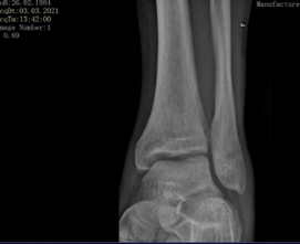
The check-up after 7 days showed the persistence of swelling and redness, so the treatment with the plaster splint was continued and the conversion to a soft cast knee-high was postponed and performed at the 2nd check-up 19 days after the trauma. 40 days after the traumatic episode, an elastic stocking and a bivalve ankle brace were placed and partial and progressive weight bearing was allowed with the aid of two crutches. The radiographic examination showed the correct healing of the two fracture sites but a vague dystrophic "veiling" on the distal epiphysis and metaphysis of the tibia and fibula which was reported by the radiologist colleague as "signs of Sudeck-type atrophy" (Figure 1).
The patchy osteoporosis that can occur in CRPS normally manifests itself after 6-12 weeks of the traumatic event 1 therefore, although it could already represent an alarm bell in itself, it was not taken into consideration as it was considered a finding too close to the traumatic event and therefore not reliable. Clinically, there was no improvement in swelling and redness and a component of impaired sensitivity with hypersensitivity and hyperalgesia had also been added; also, in this case other complications such as compartment syndrome, erysipelas or vasculopathy were excluded. It was therefore decided to prescribe to the patient 1 cycle of 10 sessions of physiokinesi therapy, anti-edema medical therapy and biophysical therapy with pulsed electromagnetic fields (CEMP).
In this phase, therefore, a specific therapy for CRPS was not adopted, probably due to an incautious underestimation of the clinical-instrumental picture which in any case still showed itself with blurred outlines to make the diagnosis of certainty. At the outpatient check-up 10 weeks after the event, the patient appeared satisfied since the pains were of much lower intensity, the attacks were less frequent and the load capacity with the aid of crutches was greater than 80%; furthermore, although she had not started physiotherapy yet, her active and passive motility had also significantly improved and therefore the heparin therapy was stopped.
At the next clinical check, about 4 months after the pathogenic noxa , the subjective and objective picture worsened again: walking without crutches was impossible, both active and passive mobilization of the ankle was very painful and limited, now also the weakness in both the anterior and posterior muscle compartments, the skin was taut, shiny and slightly warm, the bright red skin discoloration had a purplish tone and there was a component of allodynia as well as hyperalgesia in the anterior region. The blood tests remained normal (C-reactive protein 2 mg/dl, furthermore the leukocyte value as well as the differential were normal) and once again infectious, vascular, dermatological and neurological complications were excluded. Following the diagnostic criteria of the International Association for the Study of Pain (IASP) of Orlando (1993) and Budapest (2003) [2] the diagnosis of type I CRPS was established with reasonable certainty.
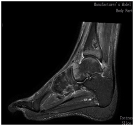
Therefore, intramuscular therapy with neridronic acid bisphosphonate 25 mg 2 ml daily for 16 days was prescribed, the resumption of heparin treatment and therapy with CEMP and the beginning of physiotherapy treatment as a matter of urgency (which is carried out through an internal hospital protocol thanks to the which the patient accesses the rehabilitation service within 7 days of the online request). Nuclear Magnetic Resonance (MRI) without contrast medium confirmed the diagnosis, in fact showing a picture of diffuse edema in T2 on the tibia, talus, calcaneus and tarsal scaphoid on both articular surfaces of each bone segment involved and also the edema of the soft parts adjacent (highly specific element for CRPS). Joint effusion is minimal. Bone edema in the case of CRPS falls within the more general picture of Bone Marrow Edema Lesions, which are classified into 3 categories by Costa-Paz (2001) based on the picture visible in MRI without contrast medium. Our clinical case fell into the 2nd type, namely: patchy with convex margins on contiguous articular surfaces (Figure 2) [3].
A strict clinical and instrumental follow-up began at pre-established deadlines which showed a rapid clinical improvement both subjective and objective; improvement confirmed by a second MRI without contrast medium, performed 3 months after the previous one, which in fact confirms the total regression of the bone edema and adjacent soft tissues. However, both MRIs showed a gap of bone necrosis of 22 mm at the posterolateral margin of the tibia (Figure 2), which is why hyperbaric therapy was recommended 4; therapy which, however, the patient did not tolerate and which he therefore decides to abandon after only three sessions. The bone lesion is in any case investigated with computed tomography without contrast medium which, however, does not add further useful information.
Since the beginning of September 2021, the patient has returned to work but continues to experience episodes of occasional exacerbations (and without new traumas) although with objective and subjective clinical pictures that are less serious than in the first phase; episodes that are regularly treated with relative success through cycles of physiotherapy and pharmacological therapy based on 16 mg intramuscular neridronic acid (daily infiltration for 16 consecutive days) and 25 mg per os prednisolone (daily administration for 2 weeks).
Treatment and outcomes
The patient was contacted by telephone in January 2022 reporting that he had resumed work normally, i.e., at the pace prior to the trauma of January 2021, while however reporting a persistent "discomfort" in the ankle region, i.e.: slight swelling diffused anteriorly and of dyschromia and cutaneous dystrophies. Muscle strength has been recovered, but still not exactly comparable to the contralateral, however he denies dystonias and sensitivity disturbances. When questioned on the global evaluation of his pathology up to that moment, the patient showed himself aware of the fact that the exacerbations, although less serious than in the first phase of the pathology, left results (dyschromia, cutaneous and adnexal dystrophy, reduction of muscle strength) which added to each new attack, without ever a total remission of the subjective and objective picture. The patient made aware from the initial stages of the meaning, nature and complexity of the pathology that afflicted him declared himself satisfied with the clinical result obtained up to now.
Conclusion & final reflections
The rationale and benefits of bisphosphonate therapy in the treatment of CRPS have been known for some time and are well documented in the international literature [5]. The only bisphosphonate for which scientific evidence of efficacy has been demonstrated is the acid neridronico, the excellent results of which have also shown themselves well in this case. In fact, the patient reported a very visible and rapid improvement in swelling and redness, already after the first 3-4 intramuscular injections (skin lesions that had reached the root of the thigh in the first aggressive phase of the pathology); on the contrary, the reduction of the algic component was less rapid. Intramuscular therapy with neridronic acid represents a very manageable and safe therapy, whose complex mechanism of action acts on several levels on bone metabolism. However, the therapeutic success is greater the earlier it is administered; in fact, according to recent studies, the effectiveness of the drug is significantly reduced 6 months after the onset of symptoms.
Cortisone therapy is also used with moderate success, although the enthusiasm of the experts in this regard is lower. The exact dosage and the time frame for a successful therapy are not known, so we often rely on an empirical therapeutic protocol to be adapted to the individual case. A 2015 study reports that a daily administration of 40 mg of prednisolone for 14 days followed by a maintenance dose of 10 mg per week represents the only anti-inflammatory therapy that has shown efficacy in several clinical trials [6]. The Complex Regional Pain Syndrome therefore represents a very complex pathology that can manifest itself in mild or severe, complete or incomplete, transient or persistent forms [7]. In more unique than rare cases (described in the literature) it can lead to disastrous consequences such as the amputation of the limb involved [8].
Despite being a pathology known for at least 100 years, it still arouses the interest of the scientific community today and recent in vivo and in vitro studies have only partially explained the complicated pathogenesis involving both the central and peripheral nervous system and the vegetative and somatic systems. In fact, the disease is supported by a positive feedback mechanism which feeds itself thanks to the action of pro-inflammatory cytokines (for example leukotrienes, interleukins, etc.) which create a circle in which peripheral cutaneous, vascular and muscular receptors are sensitized and excite, in turn, the afferent and efferent nerve fibers [9]. The prevalence and incidence in the world are not known exactly due to the variability of the clinical forms and due to the difficulties of making a diagnosis with certainty.
Diagnosis is largely clinical and is often delayed due to lack of recognition of symptoms; this can lead to serious repercussions and permanent outcomes if early and adequate therapy is not established. The treatment is multidisciplinary and must involve various figures: orthopaedic surgeon, physiatrist, physiotherapist, neurologist, radiologist, rheumatologist, endo-crinologist, anaesthetist, family doctor, etc. [10]. The importance of psychological therapy has been emphasized by recent studies which recommend an early treatment already after 3-4 months of persistence of the pathology. Genetic predisposition undoubtedly plays a decisive role and this can explain the susceptibility of some patients to develop the disease after even trivial traumas (the literature reports that in 10% of cases it is not even possible to recognize any triggering trauma). Physiotherapy treatment is also articulated and varies from mirror therapy to occupational therapies; also, in this case it is more effective the earlier it is started [10].
The literature recognizes dozens of predisposing factors including: fibromyalgia, depression, smoking, atopia, metabolic syndrome, osteoporosis and rheumatic pathologies [11].
Few studies analyse the issue of relapses, their cause and possible prophylactic therapies to avoid them; this is due to the complexity of the subject. In fact, it is often impossible to distinguish between " diffusion ", which can occur in 3 different ways, and " recurrence " of the pathology which, in its natural evolution, can alternate between different phases of remission and recrudescence regardless of the therapies adopted. In fact, in the chronic forms it is not always possible to recognize periods of time completely free from the disease, so as to be able to have a clear definition of recurrence in case the symptoms reappear [12]. Educating the patient on correct behaviours to adopt, making him aware and providing him with adequate therapy and health care minimizes the risk of becoming chronic and certainly of recurrences. Complete and stable recovery is possible but particular "clinical attention" is essential for patients who show the "alarm bells", so as to undertake the diagnostic, instrumental and clinical algorithm that leads to the correct diagnosis as soon as possible and therefore to early establishment of the therapeutic flow which can lead to complete remission of symptoms and therefore to clinical success.
References
- Rios A, Rosenberg Z, Bencardino et al. (2011). Bone Marrow Edema Patterns in the Ankle and Hindfoot: Distinguishing MRI Features. American Roentgen Ray Society.
Publisher | Google Scholor - R. Norman, Stephen B, Roberto S.G et al. (2010). Validation of proposed diagnostic criteria (the “Budapest Criteria”) for Complex Regional Pain Syndrome. Pain. 150(2):268-274.
Publisher | Google Scholor - Costa-Paz M, Musculo DL, Ayerza M et al. (2001). Magnetic resonance imaging follow-up study of bone bruises associated with anterior cruciate ligament ruptures. Arthroscopy. 17:445-449
Publisher | Google Scholor - Katznelson R, Shira C. and Hance C. (2016). Successful Treatment of Lower Limb Complex Regional Pain Syndrome following Three Weeks of Hyperbaric Oxygen Therapy. Pain Res Manag.
Publisher | Google Scholor - Varenna M, Crotti C. (2018). Bisphosphonates in the treatment of complex regional pain syndrome: is bone the main player at early stage of the disease? Rheumatology International Springer-Verlag GmbH Germany, part of Springer Nature.
Publisher | Google Scholor - Resmini L, Ratti C, Canton G, Murena L, Moretti A, Iolascon G. (2015). Treatment of complex regional pain syndrome. Clinical Cases in Mineral and Bone Metabolism. 12(1):26-30.
Publisher | Google Scholor - Iolascon G, de Sire A, Moretti A. (2015). Complex regional pain syndrome (CRPS) type I: historical perspective and critical issues. Clinical Cases in Mineral and Bone Metabolism. 12(1):4-10
Publisher | Google Scholor - Midbari A, Suzan E, Adler T et al. (2016). Amputation in patients with complex regional pain syndrome. Bone Joint J. (98)548–554
Publisher | Google Scholor - Russoa M, Georgiusb P, Santarellia D. (2018). A new hypothesis for the pathophysiology of complex regional pain Syndrome. Med Hypotheses. 119:41-53
Publisher | Google Scholor - The Royal College of Physicians (RCP). Complex regional pain syndrome in adults. 480-484.
Publisher | Google Scholor - Elsharydah A, Nathaniel H. Loo, (2017). Abu Minhajuddin Complex regional pain syndrome type 1 predictors — Epidemiological perspective from a national database analysis. Journal of Clinical Anesthesia. 39:34-37
Publisher | Google Scholor - H. Troeger. (2011). Prophylaxis of CRPS I and recurrent CRPS I Handchir microsurgeon plastic Chir .43:25-31.
Publisher | Google Scholor


