Case Report
Periprosthetic Patellar Fracture Fixation with Locking Star-Plate: A Case Series
- Bryce T. Hrudka BS *
- Jacob T. Hall MD
- John C. Neilson MD
Department of Orthopaedic Surgery, Medical College of Wisconsin, United States.
*Corresponding Author: Bryce T. Hrudka, Department of Orthopaedic Surgery, Medical College of Wisconsin, United States.
Citation: Hrudka B.T, Hall J.T., Neilson J.C. (2024). Periprosthetic Patellar Fracture Fixation with Locking Star-Plate: A Case Series. Clinical Case Reports and Studies, BioRes Scientia Publishers. 6(4):1-9. DOI: 10.59657/2837-2565.brs.24.154
Copyright: © 2024 Bryce T. Hrudka, BS, this is an open-access article distributed under the terms of the Creative Commons Attribution License, which permits unrestricted use, distribution, and reproduction in any medium, provided the original author and source are credited.
Received: July 09, 2024 | Accepted: July 27, 2024 | Published: August 05, 2024
Abstract
Introduction: This study aims to evaluate the effectiveness of a fixed-angle locking star-plate for the fixation of periprosthetic patellar fractures (PPPFs) in individuals with partial or total knee arthroplasty (TKA). The objectives focus on patient outcomes, complication rates, and functional recovery.
Materials and Methods: A retrospective case series design was utilized, encompassing a chart review at a single-center Level 1 Trauma Center. The population included patients with PPPFs who underwent open reduction and internal fixation (ORIF) with a fixed-angle locking star-plate from September 2021 to January 2023. Clinical examinations and radiographs were used to assess outcomes and complications.
Results: Three patients were treated for PPPFs. A 77-year-old male (Patient 1) resumed normal activities and reported complete pain relief at four months post-ORIF. A 51-year-old female (Patient 2) demonstrated enhanced pain control and increased knee flexion, although she presented a non-healing surgical wound. A 70-year-old female (Patient 3) with severe PPPF and osteoporosis experienced an extensor mechanism disruption and infection, leading to hardware removal and permanent knee extension loss. The follow-up period ranged from 2.5 weeks to four months postoperatively.
Conclusions: Fixed-angle locking star-plate fixation for PPPFs generally showed favorable results. However, the treatment of a patient with severe osteoporosis and extensor mechanism impairment resulted in a suboptimal outcome. These findings underscore the importance of tailored approaches to PPPF management and highlight the need for further research to compare treatment methods.
Keywords: periprosthetic patellar fracture; knee arthroplasty; locking star-plate; fixation; open reduction and internal fixation (orif)
Introduction
Primary total knee arthroplasty (TKA) has become one of the most performed elective surgeries in the U.S with annual volumes projected to reach 935,000 to 1.67 million by 2030 [1]. With an increase in life expectancy and the rising volume of annual TKA, there will be a growing number of individuals with artificial knees leading to an expected rise of periprosthetic fractures [2,3]. An increase in periprosthetic patellar fractures (PPPF) is particularly concerning, as they are the second most common periprosthetic fracture post TKA with surgical interventions displaying failure rates as high as approximately 92% [4,5]. Common PPPF management has ranged from nonoperative therapy to surgical interventions such as partial or complete patellectomies with extensor apparatus repair, open reduction and internal fixation (ORIF) with either tension band wiring (TBW), marsupialization with bone graft, and trabecular metal patella augmentation [6,7]. The treatment of PPPF varies and is commonly guided by the Ortiguera and Berry classification (Table 1), yet there is no definitive recommendation regarding the best surgical modality of fixing these periprosthetic fractures [7-10].
Table 1: Ortiguera and Berry classification of periprosthetic patella fracture
| Classification of periprosthetic patella fractures |
|
|
|
|
|
|
|
|
Plate fixation represents a potential new approach for addressing these fractures and has emerged as a promising strategy for managing native patella fractures. Chloros et al., in a comprehensive literature review, have indicated that recent studies support the superiority of plate fixation, predicting it to become the preferred method for native patella fractures [11]. Notably, the fixed-angle locking star-plate specifically has demonstrated enhanced outcomes in complex, comminuted patella fractures when compared to other modalities, such as TBW [2,3,11-13]. Yet, to date, studies specifically examining plate fixation for PPPFs are absent. Thus, considering the suboptimal outcomes historically associated with PPPF surgeries and the encouraging results from plate fixation in native patella fractures, this article presents the results from three cases at a single institution where PPPF was addressed using the fixed-angle locking star-plate. In light of these considerations, we hypothesize that the fixed-angle locking star-plate fixation method will result in improved clinical outcomes for patients with PPPFs.
Methods
The study was designed as a retrospective case series, and a chart review was conducted to evaluate the outcomes of PPPF fixation using a fixed-angle locking star-plate in patients with a partial or total knee replacement. Conducted at a single center Level 1 Trauma Center, this investigation involved patients with PPPFs who had undergone ORIF with a fixed-angle locking star-plate from September 2021 to January 2023. The chart review focused on initial clinic visits and subsequent postoperative follow-ups. The evaluation of outcomes was based on patient-reported assessments, clinical examination, and radiographic findings, aiming to assess the postoperative recovery, complications, and functional outcomes. Given the nature of the case series, the results were descriptive, and no statistical methods were used to analyze the data. The emphasis was on a comprehensive review and synthesis of clinical notes and imaging studies to provide a qualitative understanding of the outcomes following the surgical intervention.
Results
Case 1
A 77-year-old male with a prior surgical history of a left TKA for osteoarthritis, which had been performed in 2013, presented to the clinic on February 4th, 2022, for left knee pain that had begun after a ground-level fall three days prior, but he was unsure if he had landed on his left knee. He reported that the pain was primarily in the front of his knee. Upon examination, swelling was observed without bruising, and the patient was still able to bend his knee. Imaging revealed a left PPPF without disruption of the extensor mechanism, which warranted the placement of a knee immobilizer (KI) until surgical fixation could be performed. The imaging findings are illustrated in fig 1.
Figure 1: Preoperative Radiographs: (a) Anteroposterior view; (b) Lateral view; (c) Sunrise view of the left knee demonstrating a patellar fracture
ORIF was performed on February 15th, 2022, employing a fixed-angle locking star plate secured with 15 screws, as depicted in Figure 2 The surgery, which lasted 2 hours and incurred no perioperative complications, allowed the patient to be discharged the same day with instructions to remain in a KI at all times.
Figure 2: Intraoperative Radiographs: (a) Patella fracture prior to fixation; (b) Postoperative view showing the fixed-angle locking star plate and screws in place
During the initial 2-week follow-up, it was reported that the patient remained in a KI and noted appropriate pain improvement. The incision site was healing well, and the plan was for the patient to remain in the KI, weight-bear as tolerated (WBAT), and to follow up in 2 months. Two months following ORIF, the patient reported complete resolution of pain in the left knee. The radiographs taken at this follow-up visit confirmed a well-fixated patellar fracture with no complications evident, as depicted in Figure 3. The patient was advised to discontinue the use of the KI, with unrestricted knee flexion allowed. However, he was counseled to refrain from weight-bearing activities for an additional month. Physical therapy commenced on the day of the follow-up.
Figure 3: Postoperative Radiograph: Lateral view of the left knee 2 months after ORIF showing the patella with internal fixation
Four months after surgery, the patient reported doing very well and had been back to his normal daily activities, including activities such as climbing ladders and going sailing. On physical examination, the patient exhibited a smooth gait, and his left knee had a full range of motion. Given the patient's recovery and stable post-operative images, routine follow-up appointments were deemed no longer necessary and was encouraged to schedule an appointment if any concerns arose regarding his left knee.
Case 2
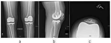
A 51-year-old female with a history of bilateral patellofemoral arthroplasty performed in February 2017 presented to the clinic on September 30th, 2021, complaining of right knee pain. The pain was localized to the anterior aspect of the knee, exacerbated by activities such as climbing stairs and arising from a seated position, and relieved with rest. Significant swelling was also noted. Initial radiographs revealed a right PPPF with evidence of osseous healing, absent any signs of quadriceps tendon detachment, as indicated in Figure 4.
Figure 4: Initial Encounter Radiographs: (a) Anteroposterior view of both knees showing bilateral patellofemoral compartment replacements; (b) Lateral view of the right knee displaying a PPPF; (c) Sunrise view of the right knee confirming the PPPF
After the initial consultation, continuous wear of a KI was advised, along with WBAT. A CT scan conducted on October 8th, 2021, provided further detail on the nature of her PPPF, revealing a comminuted and displaced stellate-type fracture of the patella, depicted in Figure 5. Despite the conservative management approach which included immobilization followed by a gradual increase in range of motion and the initiation of physical therapy, the patient continued to experience significant pain by April 2022. Subsequent imaging six months post-initial management showed no appreciable change. The patient's right knee demonstrated a range of motion from approximately 10 to 135 degrees and was tender to palpation over the medial patellar facet.
Figure 5: CT scan of the Right Knee from October 8th, 2021: (a) Axial cut showing a comminuted and displaced fracture of the patella; (b) Sagittal cut displaying the stellate pattern of the patellar fracture; (c) Coronal cut revealing the extent of displacement and comminution
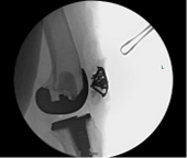
Given the lack of success with conservative management, ORIF was undertaken on August 22nd, 2022. The procedure involved the implantation of a fixed-angle locking star plate along with nine screws to stabilize the right PPPF, as shown in Figure 6. The surgery was completed in 2 hours without any perioperative complications. Postoperatively, the patient was discharged on the same day with instructions to wear a knee brace locked at full extension and to continue with WBAT.
Figure 6: Intraoperative Radiographs: (a) Lateral view of the right knee showing the implanted fixed-angle locking star plate; (b) Axial view of the patella with screws fixating the right PPPF
At the initial follow-up, 2.5 weeks following surgery, the patient's pain was well-managed. By six weeks postoperatively, she demonstrated the ability to flex her knee to approximately 60 degrees and to perform an active straight leg raise. The patient was then advised to ambulate with the brace set to allow flexion from 0 to 50 degrees, with an incremental increase of 10 degrees per week, and was permitted to rest without the brace. At the four-month postoperative mark, her knee flexion reached 100 degrees. Radiographs at this stage indicated stable screw placement and the absence of a persistent fracture line, as seen in Figure 7. However, the patient did encounter a complication: a non-healing eschar at the incision site, which was managed with routine dressing changes. Ongoing physical therapy and subsequent follow-ups were scheduled accordingly.
Figure 7: Four-Month Postoperative Radiographs: (a) Lateral view of the right knee displaying stable positioning of screws; (b) Axial view showing no evidence of a persistent fracture line
Case 3
The final patient in this series was a 70-year-old female with a history of a left TKA conducted in April 2022. She presented to the clinic on September 9th, 2022, for a discussion regarding surgical intervention for her left PPPF with an impaired extensor mechanism. The chief complaint involved left knee pain that began suddenly three months prior on the anterior aspect of her knee, with no clear cause, although she recalled her left knee buckling post-TKA. Pain was exacerbated by activities such as walking, climbing stairs, and rising from a chair, and alleviated by rest and pain medication. At that time, she was utilizing a walker for ambulation. Physical examination revealed tenderness of the left patella upon palpation and compression, and the left leg's range of motion was 0 to 100 degrees, with a 60-degree extensor lag. Preoperative magnetic resonance imaging (MRI) of the patient’s left knee revealed a displaced patellar fracture with the quadriceps tendon largely intact; approximately 75% remained attached to the retracted patellar fragment. The extensor mechanism was maintained, likely due to the attachment of the vastus medialis oblique to the un-displaced portion of the patella, as depicted in Figure 8.
Figure 8: Preoperative Imaging of the Left Knee: (a) Lateral radiograph demonstrating a PPPF with superior retraction of the patellar fragment; (b) Axial MRI view showing muscle attachments to the patella; (c) Sagittal MRI view illustrating the extent of tendon attachment and displacement
On October 28th, 2022, the patient was treated with ORIF, which included comprehensive repair of the extensor mechanism and the placement of a fixed-angle locking star plate secured with 12 screws, as illustrated in Figure 9. The procedure was completed within 3 hours without any perioperative complications. Following surgery, the patient was discharged with instructions to wear a KI consistently and to WBAT. Due to the extensive nature of the extensor mechanism repair, it was recommended that the knee immobilizer be used for an extended duration of two additional months.
Figure 9: Intraoperative Radiograph: Lateral view of the left knee showing ORIF with a fixed-angle locking star plate and screws for a PPPF
Unfortunately, the patient was readmitted on December 8th, 2022, due to disruption of the left knee extensor mechanism and an associated infection, as evidenced in Figure 10a. After the initial treatment, the left quadriceps tendon was repaired, and incision and drainage were performed, along with the removal of the left patellar plate, as detailed in Figure 10b. Alongside the hardware removal, a surgical drain was inserted, antibiotic therapy was commenced, and she was advised to keep the knee immobilizer locked in full extension. On January 6th, 2023, she returned for the explanation of the infected TKA hardware, additional repair of the quadriceps tendon, removal of the polyethylene component from the patella, further irrigation and debridement, and the placement of a rigid antibiotic spacer, which is depicted in Figure 10c. Currently, her pain is well-managed, and the knee remains fixed in extension.
Figure 10: Series of Radiographs Illustrating Postoperative Complications and Interventions: (a) Lateral view of the left knee showing disruption of the extensor mechanism; (b) post-hardware removal showing the knee after patella plate removal; (c) Following TKA hardware removal and insertion of a rigid antibiotic spacer
Discussion
The case series examined the efficacy of fixed-angle locking star-plate fixation in three patients with PPPF, with two demonstrating successful functional recovery and a return to normal activities. These positive outcomes contribute to the emerging evidence that locking plate technology may be superior for managing complex fractures. However, one patient experienced significant postoperative complications, including a failed repair of the extensor mechanism and infection, which ultimately resulted in a permanent knee extension due to the removal of all hardware, including the star-plate. The critical nature of PPPF in the third case was highlighted by an impaired extensor mechanism. The mechanical demands required to overcome the retraction of the fractured patella, alongside osteoporosis as evidenced by specific bone density scores, were likely contributing factors to the adverse outcome. These patient-specific considerations underscore the importance of individual assessments in determining the prognosis of PPPF interventions. A review of the literature, including the study by Deans et al., notes a considerable failure rate associated with conventional ORIF methods for PPPF repair. The findings from the series suggest the potential for improved results with the fixed-angle locking star-plate approach, despite the lack of comprehensive studies examining this method. This gap is particularly notable as management recommendations are often based on the Ortiguera and Berry classifications, which do not currently include considerations for this newer fixation technique (Table 2) [7].
Table 2: The treatment options based on the classification of periprosthetic patella fracture
| Type 1 |
| Extensor mechanism intact |
| Stable patella component |
| Nonoperative treatment |
|
|
| Type 2 |
| Extensor mechanism disrupted |
| Operative repair or reconstruction of the extensor mechanism |
| Stable patella component |
|
|
| Loose patella component with adequate bone |
|
|
| Loose patella component with inadequate bone |
|
|
|
| Type 3 |
| Intact extensor mechanism |
| Loose patella component |
| Asymptomatic with nondisplaced patella component |
|
|
| Asymptomatic with displaced patellar component |
|
| Symptomatic with displaced patella component |
|
|
The limitations of the study, primarily the small cohort size and its retrospective nature, may affect the broader applicability of the results. The absence of a comparative control group and the reliance on patient-reported outcomes, rather than quantifiable measures or statistical analysis, position the conclusions within an exploratory framework. The series indicates a promising direction for PPPF management, offering a potential improvement over traditional method. The findings warrant further investigation with a larger sample size and controlled study designs to validate the benefits of the fixed-angle locking star-plate fixation in PPPF repair.
Conclusion
Given that surgical repairs of PPPFs have historically been associated with failure rates as high as 92%, the investigation into alternative approaches was deemed essential [5]. The study reported on three cases in which patients underwent ORIF for PPPFs with the utilization of a fixed-angle locking star-plate, and it was observed that two of the three cases resulted in excellent outcomes. The failure in the third case could be attributed to the degree of impairment of the extensor mechanism and the patient's underlying osteoporosis. To the best of the authors' knowledge, this study is the first to assess the outcomes of ORIF for PPPFs using a fixed-angle locking star-plate. This technique has shown promise in cases of patella fractures without prosthetic involvement. Future research should aim to expand this body of literature to determine whether the use of a fixed-angle locking star-plate for PPPFs offers improved outcomes over traditional methods.
Take Home Points
- The fixed-angle locking star-plate offers a potential alternative to traditional ORIF in the repair of PPPFs.
- Patient-specific factors, particularly conditions of the extensor mechanism and bone density, are crucial determinants of surgical success.
- There is a significant need for further research to validate the effectiveness of the fixed-angle locking star-plate in PPPFs.
Declarations
Funding
No external funding was received for this manuscript.
Competing interests
The authors declare that they have no competing financial interests or personal relationships that could have appeared to influence the work reported in this paper.
Ethics Statement
This project was reviewed and the need for IRB approval was waived by the MCW Institutional Review Board #5 in accordance with the U.S. Department of Health and Human Services regulations (45 CFR 46) and the Medical College of Wisconsin’s institutional policies and ethical standards. The study was considered minimal risk and involved no direct patient interaction, as it was a retrospective chart review of a three-patient case series.
Informed Consent
Not applicable. This study was a retrospective chart review and did not involve direct patient contact or interaction. The MCW Institutional Review Board approved a waiver of the requirement for informed consent and HIPAA authorization under 45 CFR 164.512 due to the study's minimal risk nature and lack of direct patient engagement. Therefore, informed consents were not required.
Availability of data and materials
The datasets used and/or analyzed during the current study are available from the corresponding author upon reasonable request.
Authors' Contributions
B.T.H: Conducted the literature review, reviewed patient charts, and drafted the manuscript. He compiled and analyzed the data as the primary author.
J.T.H: Refined the manuscript through editing and acquired key references to support the research.
J.C.N: Performed all surgical procedures and provided critical insights and revisions to the manuscript.
References
- Sloan, M., Premkumar, A., & Sheth, N. P. (2018). Projected Volume of Primary Total Joint Arthroplasty in the U.S., 2014 to 2030. JBJS, 100(17):1455-1460.
Publisher | Google Scholor - Singh, C., & Marya, S. K. S. (2022). Periprosthetic Fractures Following TKR. In S. K. S. Marya (Ed.): Knee Arthroplasty. Springer. (275-296).
Publisher | Google Scholor - Singh, S., Surana, R., Rai, A., & Sharma, D. (2020). Outcome Analysis of Fixed Angle Locking Plate in Patella Fractures: A Single Centre Experience from North India. Indian Journal of Orthopaedics, 55(3):655-661.
Publisher | Google Scholor - Berry, D. J. (1999). Epidemiology: hip and knee. Orthopedic Clinics of North America, 30(2):183-190.
Publisher | Google Scholor - Govil, G., Tomar, L., & Dhawan, P. (2020). Peri-prosthetic trans-patellar fractures after Total knee Arthroplasty: a case series and review of literature. Arthroplasty, 2:35.
Publisher | Google Scholor - Canton, G., Ratti, C., Fattori, R., Hoxhaj, B., & Murena, L. (2017). Periprosthetic knee fractures. A review of epidemiology, risk factors, diagnosis, management and outcome. Acta Bio Medica: Atenei Parmensis, 88(S2):118.
Publisher | Google Scholor - Deans, J., & Scuderi, G. R. (2021). Classification and Management of Periprosthetic Patella Fractures. Orthopedic Clinics, 52(4):347-355.
Publisher | Google Scholor - Ortiguera, C. J., & Berry, D. J. (2002). Patellar Fracture After Total Knee Arthroplasty. JBJS, 84(4):532-540.
Publisher | Google Scholor - Parvizi, J., Jain, N., & Schmidt, A. H. (2008). Periprosthetic knee fractures. Journal of Orthopaedic Trauma, 22(9):663-671.
Publisher | Google Scholor - Sayum Filho, J., Lenza, M., Tamaoki, M. J., Matsunaga, F. T., & Belloti, J. C. (2021). Interventions for treating fractures of the patella in adults. Cochrane Database of Systematic Reviews, 2, CD009651.
Publisher | Google Scholor - Chloros, G. D., Kotsarinis, G., Christou, C. D., & Giannoudis, P. V. (2022). What’s new in the management of patella fractures? Injury, 53(6):1730-1736.
Publisher | Google Scholor - Bansal, H., Behera, A., Sharma, V., & Farooque, K. (2022). Patella locking star-plate as salvage to the failed tension band wiring for a patella fracture: a case report. Case Reports in Orthopedic Research, 5:13.
Publisher | Google Scholor - Wurm, S., Bühren, V., & Augat, P. (2018). Treating patella fractures with a locking patella plate - first clinical results. Injury, 49:S51-S55.
Publisher | Google Scholor


