

Case Report
Non-Puerperal Uterine Inversion Secondary to Prolapsed Tumours about Two Cases
- Hanaa Lazhar
- Mariam Mahtate
- Aziz Slaoui *
- Amina Etber
- Najia Zeraidi
- Amina Lakhdar
- Aicha Kharbach
- Aziz Baydada
Gynaecology-Obstetrics and Endoscopy Department, Maternity Souissi, University Hospital Center IBN SINA, University Mohammed V, Rabat, Morocco.
*Corresponding Author: Aziz Slaoui, Gynaecology-Obstetrics and Endoscopy Department, Maternity Souissi, University Hospital Center IBN SINA, University Mohammed V, Rabat, Morocco.
Citation: Lazhar H, Mahtate M, Slaoui A, Etber A, Badreddine N, Sofiane Z. (2023). Non-Puerperal Uterine Inversion Secondary to Prolapsed Tumors: About Two Cases. Journal of Clinical Surgery and Surgical Research, BRS Publishers. 2(1); DOI: 10.59657/2992-9989.brs.23.008
Copyright: © 2023 Aziz Slaoui, this is an open-access article distributed under the terms of the Creative Commons Attribution License, which permits unrestricted use, distribution, and reproduction in any medium, provided the original author and source are credited.
Received: March 21, 2023 | Accepted: April 07, 2023 | Published: April 14, 2023
Abstract
Background: Non-puerperal uterine inversion (NPUI) is a rare gynecological affection whose scarcity makes its diagnosis challenging. Uterine inversion may be -acute- most seen in obstetrics as a complication of the third stage of labor or- chronic- due to uterine benign or malign tumors.
Case report: We proffer two cases of non-puerperal uterine inversion presenting to our emergency department complaining of a protruded mass through the vagina outside the introitus. The first one outlays the second reported case - to our knowledge- in a patient without sexual experience of a uterine inversion due to a leiomyosarcoma, presenting in an acute state, leading to hemorrhagic shock. The second case occurs after recurring uterine inversions due to a known submucosal myoma. Both diagnoses relied on physical examination where not being able to palpate the uterus was the cornerstone of clinical suspicion. Using ultrasound as imaging investigation was pivotal to confirming the diagnosis and an etiology, uterine tumors. For different reasons, both patients underwent transvaginal hysterectomy.
Conclusion: NPUI is a complicated clinical condition where the diagnosis is often exacting. If accurately and timely managed, it has an excellent prognosis. It’s very important to define the tumor’s histopathological profile before choosing one of the underlining known surgical and non-surgical procedures. However, taking into consideration fertility and the patient’s desire should be mandatory before deciding on any course of action.
Keywords: uterine inversion; leiomyosarcoma; fertility prognosis
Introduction
Uterine inversion occurs when the uterine fundus collapses into the endometrial cavity turning the uterus partially or completely inside out [1]. It’s a rare life-threatening condition. Uterine inversion may be classified as puerperal-commonly complicating the third stage of labor- or non-puerperal complicating uterine tumors [2]. Uterine inversion can be seen in acute situations or in chronic forms. Diagnosis may be very challenging. Ultrasound and MRI investigations turn out to be very helpful. Many surgical and non-surgical procedures have been delineated for treatment and management of uterine inversion. Through this article, we hereby present two cases of non-puerperal uterine inversion presenting to our gynecological department, where the first one occurs in an acute form secondary to a leiomyosarcoma presenting as a hemorrhagic shock whereas the second one is chronic and secondary to a submucosal myoma. They were both treated by a vaginal hysterectomy.
Case Report
A 33 years old woman presented to the emergency room in the gynecology and obstetrics department with pelvic pain and an acute protruding mass through the vagina beyond the vulva with active vaginal bleeding. She never had a fever and there was no history of discharge per vaginum. She used to have regular menses that lasted 6 days before an abnormal bleeding carried on for the past year with intermittent pelvic pain. She had already consulted for this complaint to a private clinic where some drugs were prescribed (iron and tranexamic acid pills) and a pelvic ultrasound was performed revealing a submucosal myoma measuring 2cm x4cm x3cm. Her general examination declares signs of hypovolemic pre-shock compromising orthostatic hypotension and syncope following minor head raising, heavy sweating and involuntary tremor in the extremities. Her systemic examination showed a low blood pressure 94/55 mmHg, pulse rate was at 150 BPM with 99% oxygen saturation at room air. Her abdomen was soft and non-tender. The uterus could not be palpated. Gynecologic examination noted a hard irregular pedunculated mass with white patches measuring 10cmX8cm protruding through the vagina (Figure 1).
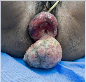
Figure 1: Photography of hard irregular pedunculated mass with white patches protruding through the vagina separated from the vaginal mucosa with thick rim superiorly, presumably the cervix.
It was separate from the vaginal mucosa and had a thick rim superiorly, presumably the cervix. The uterus could not be felt through the vaginal wall. The hymen was torn and its vestiges were actively bleeding). A complete uterine inversion secondary to a submucous uterine fibroid with fundal attachment was suspected. Initial assessment and resuscitation where our priority as the patient was in shock. Her cellular blood count showed a low hemoglobin (7g/dl) with normal platelets and white count. The patient has been transfused with 4 units packed red blood cells. Pelvic ultrasound could not visualize the uterus nor the ovaries in the pelvis and revealed an extruded mass in the vagina confirming the clinical suspicion of acute uterine inversion likely secondary to a suspected submucosal myoma causing hypovolemic shock. The patient consented to a total vaginal hysterectomy with possible oophorectomy. Unfortunately, the patient couldn’t benefit from counseling on the physiologic and psycho-social issues associated with premature menopause and irreversible infertility given the urgent situation. The patient had to undergo surgery comprising excision of the submucous leiomyoma trans vaginally.
A vaginal myomectomy was performed. We tried repositioning the mass. However, since there was a tight ring tissue noted at the cervix, the attempt to reposition failed. We resumed to perform a total inter-adnexal trans-vaginal hysterectomy. After locating the bladder in the cervico-fornico region ultrasonographily, an incision was made to dissect and push up the bladder. The uterosacral ligaments, cardinal ligaments and uterine arteries were ligated. Then we bisected the corpus to access the upper pedicule and completed the hysterectomy. The excised mass was sent for histopathological examination (Figure 2).
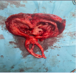
Figure 2: Photography of the excised inverted uterus sent to pathology
The post-operative period was uneventful. After total transfusion before and per-operatively, her haemoglobin level was 8.6g/dl on the second day. The patient was discharged under satisfactory condition on postoperative day 5. After 10 days, she came back for follow-up and histopathology report reveals features of malignant leiomyosarcoma. Secondary to the diagnosis of leiomyosarcoma, the patient underwent further workup examinations concluding to a stage 1 leiomyosarcoma. Therefore, the patients had regular follow ups, every three months for observation. No chemotherapy nor pelvic radiation were performed following surgery.
Second case presentation
We hereby present the uncommon case of a multiparous 45 years old woman that presented to our gynecologic emergency department complaining of a protruded mass through the vagina beyond the vulva. She also grumbled about difficulty in micturition and constipation for the last 4 months. She used to feel pelvic heaviness and pressure accompanied with continuous bleeding per vaginum for 3 months. Her history revealed 5 vaginal deliveries, no previous pap test and an irregular cycle caused by menometrorrhagia for the last 4 months leading to a consultation -at the Outpatient Gynecological department 2 months ago- associated with smelly vaginal discharge. She had had a pelvic ultrasound showing a fundic sub-mucosal fibroid measuring 51mmx47mm (Figure 3). The patient was scheduled for a hysteroscopic myomectomy.
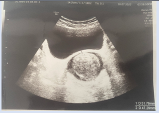
Figure 3: Ultrasound photography of fundic sub-mucosal fibroid measuring 51mmX47mm revealed in an ultrasound
General and systemic examinations were normal. The abdomen was soft. Coming out of the introitus was a necrotic bulky sloughing mass with foul smelling associated with vaginal bleeding (Figure 4). On bi manual examination, the mass could be differentiated from the cervix but the uterus could not be palpated. The cupping of the fundus was palpated. Ergo, a non-puerperal uterine inversion associated with a submucosal fibroid was suspected.
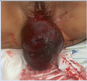
Figure 4: Photography of necrotic bulky sloughing mass coming out of the introitus associated with vaginal bleeding.
Per biologic investigations, her haemoglobin was at 7,8g/dl. Her white count was elevated at 18540 /ul. Her platelets and PT were within normal range. Cytobacteriological examination of the urine (CBEU) showed an acute Escherichia Coli infection. Treatment relied upon antibiotics – Ciprofloxacin 500m-over a 5day course. she also received tranexamic acid and 4 units of PRBC.
Her ultrasound investigation showed a Ushaped uterine cavity with an inverted uterine fundus and bull’s eye configuration on axial image. The patient was a candidate to a vaginal hysterectomy. No repositioning method was tried as the mass was infected and no further fertility was desired. The past underwent a general work-up for surgery and was counseled. Concerned about the location of the ureters, bilateral ureteral stents were placed. Due to complete uterine prolapse, the uterosacral ligament, cardinal ligament, and the uterine artery pedicle were clamped, incised, and suture ligated in a single pass. Good hemostasis was confirmed and then we performed closure of the vaginal cuff. The operation went smoothly. The patient received 1,5 liters of crystalloids and 2 units of PRBC units. Anatomy-pathology of the uterus showed degenerating endometrium with intramural leiomyoma with severe ischemic changes associated with stromal bleeding. Her post-operative care was uneventful. She was discharged on the second day. Her 4 weeks follow up was unremarkable.
Discussion
Uterine inversion is a scarce condition where the fundus of the uterus turns inside out and the latter prolapses through the cervix. Uterine inversion can be puerperal following delivery or miscarriages and non-puerperal where only few cases were reported [1]. Non-puerperal uterine inversion (NPUI) may be acute (case 1) or chronic (case 2). The degree can be classified into 4 stages. Stage 1: The inverted uterus remains in the uterine cavity. Stage 2: complete inversion of the fundus through the cervix. Stage 3: The inverted uterus protrudes through vulva. Stage 4: Inversion of the uterus and vaginal wall through the vulva [2-4]. In our two cases, it was a complete type. The diagnosis is very easy with stage 3 or 4.
NPUI is related to benign or malignant tumors associated with the uterine corpus [2]. A systematic review in 2018 of 170 cases found that benign leiomyomas were the leading cause of chronic uterine inversion (57,2%) followed by leiomyosarcomas at 13,5% [5-6]. The mechanism of non-puerperal inversion remains unclear. The intracavitary tumor distends the myometrium. The latter triggers expulsive contractions leading to cervix dilation and assists tumor’s expulsion by hauling its fundal attachment. Manual traction on the tumor, tumor’s weight, thin uterine wall, tumor attachment to the uterine wall with a thin pedicle, quick expulsion of the tumor and increased intra-abdominal pressure secondary to coughing, straining and sneezing are risk factors to non-puerperal uterine inversion (NPUI) [3-7-8].
Non-puerperal inversions are commonly chronic cases although Das reported 8,6% of non-puerperal inversions as sudden onset [3]. Diagnosis of NPUI is not as easy as it seems. Acute uterine inversion causes severe pelvic pain and hemorrhage as seen in patient n° 1, whereas chronic inversion is insidious and characterized by pelvic pressure and discomfort, irregular bleeding leading to anemia and vaginal discharge, symptoms shown in patient n°2 [9]. There have been a few cases where a suprapubic catheterization was needed due to acute urinary retention [10]. There was a reported case of NPUI due to fibroid presenting in a hypovolemic shock like our first patient [11].
On physical examination, the mass is protruded or the mass can be felt in the vaginal canal. Either way, the uterus cannot be palpated. During our initial assessment, not palpating the uterus was key to suspecting uterine inversion. The inverted uterus forms an inverted pyriform swelling occupying the upper part of the vagina. As seen in patient n°2, It’s smooth, dark red and usually bleeds on palpation [12]. Since 1968, Lascardies has noted three important clinical signs of NPUI. Firstly, it’s sometimes hard to recognize along the mass the presence of a cervical ring. Secondly, the uterine cervix could not be found and the endometrial cavity cannot be probed especially where the NPUI is not complete. At last, the cupping of the fundus can be sometimes palpable which was the case in patient n°2 [13].
Pelvic ultrasound is the first imaging investigation line considering its availability and simplicity. However, transvaginal ultrasound’s diagnostic value is limited mainly in cases where a large mass is protruded into the vagina. The probe could not be inserted into a choked vaginal canal. Besides, ultrasonic beams could not get through the mass [14]. In these cases, MRI is a better diagnostic tool [14]. An ultrasound was performed for our two patients. Alongside physical examination, it was sufficient to make a proper diagnosis.
The suggestive features include a Y shaped uterine cavity in the longitudinal plane in incomplete inversions whereas in complete inversion, the uterus is U shaped associated with thickened and inverted uterine fundus on sagittal section and bull’s eye configuration on axial image [15-16]. Our ultrasound performed in patient n°2 revealed enough suggestive features to diagnose a non-puerperal uterine inversion due to a submucosal fibroid.
In order to overcome the challenging diagnosis, an examination under anesthesia associated with histological sampling of the protruded mass can be very helpful [17]. In a 2020 systemic review, 32,02% of NPUI are associated with malignancies [18]. Unless the submucosal fibroid etiology is obvious, histological evaluation is justifiable. The flower-vase appearance results from the appearance of ovaries and tubes projecting out of the indented uterine fundus [19].
NPUI is constrained by whether the condition is acute or chronic, reproductive wish of the patient and the cause of inversion. In acute inversion, the uterus can generally be reverted by intravaginal manipulation whereas chronic NPUI, surgery is imperative. Nevertheless, initial assessment and resuscitation would be the priority in cases of septic or hemorrhagic shock. Surgical or non-surgical repositioning is only tempted if uterine preservation is considered preventing pain, bleeding, infections and gangrene. Before repositioning, excision of the fibroid is essential. In one hand, Spinelli and Kustner are trans-vaginal surgical reposition techniques. On the other hand, Huntington and Haultain procedures are repositioning performed trans-abdominally via laparotomy [20].
Heath RP et al. [19] suggest a guide shown in their algorithm to investigate and plan treatment. In the first case, non-surgical repositioning was tempted to preserve patient’s fertility but in vain. Serendipitously, it was the best procedure since the anatomopathological results was malignant leiomyosarcoma. In the second case, given that the patient was perimenopausal, and had her family completed we went ahead and performed a vaginal hysterectomy. Following Heath RP systematic review,19.6% underwent vaginal hysterectomy and 39.8% underwent abdominal hysterectomy [19].
Conclusion
Non-puerperal uterine inversion secondary to uterine tumors is a scarce condition leading to a sparse experience among health professionals making its diagnosis and management exacting. Acute forms entail focusing first on resuscitation before attempting treatment. Chronic forms are on the other hand subject to surgical treatment including both vaginal and abdominal approaches. We hereby highlight the importance of tumor biopsy before definitive surgery given the incidence of tumor malignancy. Fertility desire should be considered before leaning towards a treatment path. This work has been reported in line with the SCARE 2020 criteria [16].
Abbreviations
NPUI: Non-Puerperal Uterine Inversion
MRI: Magnetic Resonance Imaging
Declarations
Guarantor of Submission
The corresponding author is the guarantor of submission.
Acknowledgements: None.
Funding: There are no funding sources to be declared.
Availability of data and materials
Supporting material is available if further analysis is needed.
Competing interests: The authors declare that they have no competing interests.
Consent for Publication
Written informed consent was obtained from the patient for publication of this case report and any accompanying images. A copy of the written consent is available for review by the Editor-in-Chief of this journal.
Ethics Approval and Consent to Participate
Ethics approval has been obtained to proceed with the current study. Written informed consent was obtained from the patient for participation in this publication.
References
- H. W. Jones, (1951). “Non-puerperal inversion of the uterus,” The American Journal of Surgery. 5:492-495.
Publisher | Google Scholor - V. Gowri, (2000). “Uterine inversion and corpus malignancies: a historical review” Obstetrical & Gynecological Survey. 55(11).703-707.
Publisher | Google Scholor - P. Das, (1940). “Inversion of the uterus,” BJOG: An International Journal of Obstetrics and Gynaecology. (47)5.525-548.
Publisher | Google Scholor - Spinelli PG. (1897). “Inversion of the uterus.” Riv Ginec Comtemp. 11:567-570.
Publisher | Google Scholor - M. de Vries and D. A. M. Perquin, (2010). “Non-puerperal uterineinversion due to submucous myoma in a young woman: a casereport,” Journal of Medical Case Reports. 4(1).1-3.
Publisher | Google Scholor - Lupovitch, E. R. England, and R. Chen, (2005). “Non-puerperaluterine inversion in association with uterine sarcoma: casereport in a 26-year-old and review of the literature,” Gyneco-logic Oncology. (97) 3:938-941.
Publisher | Google Scholor - Rosa Silva B, de Oliveira Meller F, Uggioni ML, Grande AJ, Chiaramonte Silva N, Colonetti T, et al. (2018). “Non-puerperal uterine inversion: a systematic review.” Gynecol Obstet Invest. 83(5):428-436.
Publisher | Google Scholor - Kesrouani A, Cortbaoui E, Khaddage A, Ghossein M, Nemr E. (2021). “Characteristics and outcome in non-puerperal uterine inversion”. Cureus. 13(2):13345.
Publisher | Google Scholor - Y. J. Song, J. Yang, H. S. Yun et al. (2016). “Non-puerperal uterine inversion presented with hypovolemic shock,” Journal of Menopausal Medicine. (22) 3:184-187.
Publisher | Google Scholor - Y.-L. Chen, C.-A. Chen, W.-F. Cheng et al. (2006). “Submucous myoma induces uterine inversion,” Taiwanese Journal of Obstetrics and Gynecology. (45)2.159-161.
Publisher | Google Scholor - T. Ashraf-Ganjooie, (2005). “Nonpuerperal uterine inversion: a case report,” Archives of Iranian medicine. (8) 1:63-66.
Publisher | Google Scholor - R. P. Herath, M. M. Hosni, M. Rashid, and M. (2011). Hassanaien, Chronic nonpuerperal uterine inversion: laparotomy assisted vaginal hysterectomy case report chronic nonpuerperal uterine inversion: laparotomy assisted vaginal hysterectomy,” American Journal of Obstetrics & Gynecology. 7-10.
Publisher | Google Scholor - E. Lascarides and M. Cohen, (1968). “Surgical management of nonpuerperal inversion of the uterus,” Obstetrics & Gynecology.32(3)376-381
Publisher | Google Scholor - M. A. Atalay, B. Ç. Demir, N. Solak, F. O. Atalay, andŞ. Küçükkömürcü, (2013). “An unusual presentation of a submucousleiomyoma accounting to a non-puerperal uterine inversion:a case report,” Journal of the Turkish German Gynecological Association. 14(2).116-118.
Publisher | Google Scholor - C.-F. Hu and H. Lin, (2012). “Ultrasound diagnosis of complete uterine inversion in a nulliparous woman,” Acta Obstetricia et Gynecologica Scandinavica. 91(3).379-381.
Publisher | Google Scholor - Bertrand S, Randriamarolahy R. (2011). “Uterine inversion caused by submucous leiomyoma” Clin Imaging. 35:478-479
Publisher | Google Scholor - O. S. Umeononihu, J. I. Adinma, N. J. Obiechina, G. U. Eleje, O. I. Udegbunam, et.al. (2013). “Uterine leiomyoma associated non-puerperal uterine inversion misdiagnosed as advanced cervical cancer: a case report,” International Journal of Surgery Case Reports. 4(11).1000-1003.
Publisher | Google Scholor - R. P. Herath, M. Patabendige, M. Rashid, P. S. Wijesinghe, (2020).
Publisher | Google Scholor - R. P. Herath, M. Patabendige, M. Rashid, P. S. Wijesinghe,
Publisher | Google Scholor - Umeounonihu OS. (2013). “uterine leiomyoma associated non-puerperal uterine inversion misdiagnosed as advancedcervical cancer.” Int J Surg Case Rep. 4:1000-1003
Publisher | Google Scholor - Agha R.A, Franchi T, Sohrabi C, Mathew G. (2020). for the SCARE Group the SCARE 2020 guideline: updating consensus Surgical CAse REport (SCARE) guidelines. Int. J. Surg. 84:226-230.
Publisher | Google Scholor


