Case report
Multiple Cutaneous Features Associated with Primary Sjogren’s Syndrome
- Rosales Sotomayor A
- Castro-Molina SA
- Mendez Flores Silvia
- Barrera Godinez A
- Carrillo Cordova Dulce Maria
- López-Loya
- Dominguez Cherit Judith *
Department of Dermatology, Instituto Nacional de Ciencias Médicas y Nutrición Salvador Zubirán, Mexico.
*Corresponding Author: Dominguez Cherit Judith, Department of Dermatology, Instituto Nacional de Ciencias Médicas y Nutrición Salvador Zubirán, Mexico.
Citation: Rosales Sotomayor A, Castro Molina SA, Mendez F Silvia, CCD Maria, Dominguez C Judith, et al. (2022). Multiple Cutaneous Features Associated with Primary Sjogren’s Syndrome. Dermatology Research and Reports, BRS publishers 1(1); DOI: 10.59657/2993-1118.brs.22.001
Copyright: © 2022 Dominguez Cherit Judith, this is an open-access article distributed under the terms of the Creative Commons Attribution License, which permits unrestricted use, distribution, and reproduction in any medium, provided the original author and source are credited.
Received: October 07, 2022 | Accepted: October 21, 2022 | Published: November 01, 2022
Abstract
Sjogren’s syndrome is a systemic autoimmune disorder that mainly affects exocrine glands, although the clinical manifestations include both exocrine gland involvement and extraglandular disease features. In addition to dryness, musculoskeletal pain and fatigue are the hallmarks of this disease and constitute the classic symptom triad presented by the vast majority of patients. The authors present the case of a 59-year-old woman with achalasia, sicca syndrome, subcutaneous nodules affecting the knees, purpuric plaques on the palms and vitiligo. Serological tests were consistent with Sjögren′s syndrome, therefore the diagnosis of primary Sjögren’s syndrome with extensive cutaneous manifestations as extraglandular organ involvement was made.
Keywords: sjogren’s syndrome; erythema nodosum; neutrophilic vasculitis; vitiligo
Introduction
We present a new case of Sjögren′s syndrome who developed clinically and histologically compatible cutaneous lesions, these findings have a low incidence worldwide. It had seen in Departament of Dermatology in Instituto Nacional de Ciencias Médicas y Nutrición “Salvador Zubirán”, Mexico City.
Case
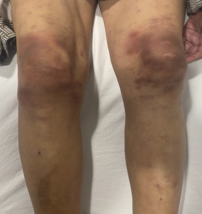
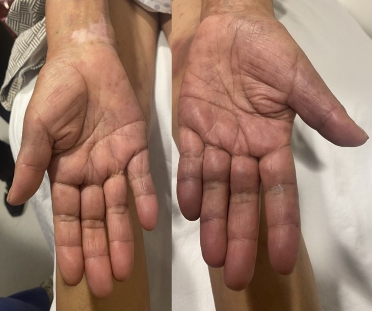
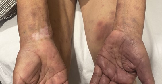
We describe the case of a 59-year-old woman with history of achalasia, she was referred to our emergency department for the presence of bilateral, tender, erythematous to purpuric subcutaneous confluent nodules affecting the knees (Figure 1), measured up to 2 cm in diameter and had been worsening in the past month, on physical examination we also found purpuric plaques on the palms (Figure 2) and irregular, ill-defined, amelanotic macules on both wrists. She also referred fatigue, dryness of the eyes and mouth, arthralgia, dysphagia and dyspnea. (Figure 3).
Figure 1: Bilateral, tender, erythematous to purpuric subcutaneous confluent nodules affecting the knees consistent with erythema nodosum
Figure 2: purpuric plaques on the palms consistent with neutrophilic vasculitis of a small and medium vessels
Figure 3: irregular, ill-defined, amelanotic macules on both wrists consistent with vitiligo
Laboratory studies were remarkable for leukocytosis, lymphopenia, anemia, high C-reactive protein, positive antinuclear antibodies (ANAs) with a fine mottled pattern 1:2560 and cytoplasmatic pattern 1:80, anti-Scl-70 antibodies of 5.8 U/mL (n.: <= 3.7), anti-SSb of 328.6 U/mL (n.: <= 7.0) and anti-SSa of 236.9 U/mL (n.: <= 9.1), Schirmer test was positive (right eye lessthen 0.2mm, left eye 0.1mm. Anti-centromere antibodies (ACA) and anti-U1-RNP antibodies were negative. Two biopsies were performed, the first taken from the knee showed neutrophilic and granulomatous septal paniculitis, nodular cystic fat necrosis, without vasculitis compatible with erythema nodosum, and the second from the palm demonstrated neutrophilic vasculitis of a small and medium vessels. A diagnosis of Sjögren syndrome with erythema nodosum, neutrophilic vasculitis and vitiligo as extraglandular manifestations was made and she started treatment with prednisone 30 mg daily and hydroxychloroquine 200 mg daily. She underwent Heller myotomy for achalasia without complications.
Discussion
Regarding Sjögren syndrome, cutaneous manifestations are rare, incidence has been reported as allow as 4.1% in some cohorts but are associated with activity in the other ESSDAI domains, these include mainly purpuras, other forms of vasculitis [1], livedo reticularis [2], xerosis, Reynaud phenomenon and annular erythema. Less common findings include vitiligo, Sweet syndrome, granulomatous paniculitis and erythema elevatum diutinum [3]. Panniculitis associated with Sjögren syndrome is a rare cutaneous manifestation, as reported by Yu-Chen Huang et al. only 9 cases have been published, clinical manifestation usually shows classic tender erythematous plaques over the extremities with symmetrical distribution [4]. Biopsy specimens of erythema nodosum classically demostrate edematous septa and mild lymphocytic infiltrates although neutrophils may predominate in early lesions, vasculitis is not generally seen. Although well-developed erythema nodosum is classified as a lymphocytic or granulomatous septal panniculitis, early cases often consist of a neutrophilic component with predominantly septal pattern like subcutaneous Sweet syndrome, it may be associated with a wide variety of systemic disorders [5]. Addressing the rare distribution, erythema nodosum affecting extensor surfaces has been reported caused by progesterone injection [6], during hydroxyurea therapy [7] and associated with Lofgren’s syndrome [8]. Nodular cystic fat necrosis is an unusual localized form of fat necrosis characterized by very distinct histology demonstrating lipomembranous changes of the subcutaneous fat, with cystic fat necrosis and calcification in later stages of the process, previously associated with erythema nodosum [9] and one case of dermatomyositis [10]. In pSS cutaneous vasculitis has been reported in up to 9.7% of cases [11], 90% of them presenting with leucocytoclastic vasculitis. Although esophageal involvement is common in scleroderma, there are few reports of the association between achalasia and Sjögren syndrome [12]. Sjögren syndrome is an autoimmune disease with diverse clinical features, interestingly cutaneous findings can support and precede diagnosis, and more importantly it is known that they often correlate with severity of systemic involvement and prognosis.
Conclusion
Sjögren’s syndrome is a systemic autoimmune disorder with diverse clinical features, cutaneous findings can support and precede diagnosis, and often correlate with severity of systemic involvement and prognosis.
Conflict of Interest
None
References
- Orgeolet L, Foulquier N, Misery L, et al. (2020). Can artificial intelligence replace manual search for systematic literature? Review on cutaneous manifestations in primary Sjögren’s syndrome. Rheumatol (United Kingdom). 59(4):811-819.
Publisher | Google Scholor - Villon C, Orgeolet L, Roguedas AM, et al. (2021). Epidemiology of cutaneous involvement in Sjögren syndrome: Data from three French pSS populations (TEARS, ASSESS, diapSS). Jt Bone Spine. 88(4).
Publisher | Google Scholor - Jhorar P, Torre K, Lu J. (2018). Cutaneous features and diagnosis of primary Sjögren syndrome: An update and review. J Am Acad Dermatol. 79(4):736-745.
Publisher | Google Scholor - Lin, Ming-Hsiu; Huang Y-C. (2012). Panniculitis associated with Sjögren′s syndrome. Indian Journal of Dermatology. Indian J Dermatol Venereol Leprol.
Publisher | Google Scholor - Chan MP. (2014). Neutrophilic panniculitis: Algorithmic approach to a heterogeneous group of disorders. Arch Pathol Lab Med. 138(10):1337-1343.
Publisher | Google Scholor - Jeon HC, Choi M, Paik SH, Na SJ, Lee JH, Cho S. (2011). A case of assisted reproductive therapy-induced erythema nodosum. Ann Dermatol. 23(3):362-364.
Publisher | Google Scholor - Ogawa Y, Akiyama M. (2016). Non-infectious panniculitis during hydroxyurea therapy in a patient with myeloproliferative disease. Acta Derm Venereol. 96(4):566-567.
Publisher | Google Scholor - Chauhan A, Jandial A, Mishra K, Sandal R. (2021). Acute arthritis, skin rash and Lofgren’s syndrome. BMJ Case Rep. 14(6):2020-2022.
Publisher | Google Scholor - S K Ahn , B J Lee, S H Lee WSL. (1995). Nodular cystic fat necrosis in a patient with erythema nodosum. Clin Exp Dermatol.
Publisher | Google Scholor - Ferenczi K, Summers P, Aubert P, et al. (2008). E XTRAORDINARY C ASE R EPORT A Case of CD30 + Nasal Natural Killer / T-Cell Lymphoma. 30(6):567-571.
Publisher | Google Scholor - Ramos-Casals M, Brito-Zerón P, Seror R, et al. (2015). Characterization of systemic disease in primary Sjögren’s syndrome: EULAR-SS Task Force recommendations for articular, cutaneous, pulmonary and renal involvements. Rheumatol (United Kingdom). 54(12):2230-2238.
Publisher | Google Scholor - Poglio F, Mongini T, Cocito D. (2007). Sensory ataxic neuropathy and esophageal achalasia in a patient with Sjogren’s syndrome. Muscle and Nerve. 35(4):532-535.
Publisher | Google Scholor


