Case Report
Massive Liver Abscess with Septic Peritonitis Drainage in A Cardiac Compromised Patient Under Regional Anaesthesia - A Case Report
- Venu Varun Sri Krishna P
- Dilip Kumar G
- Hemaa S
- Krishna Prasad T *
Shri Sathya Sai Medical College and RI, Sri Balaji Vidyapeeth, Ammapettai, Kanchipuram, India.
*Corresponding Author: Krishna Prasad T, Shri Sathya Sai Medical College and RI, Sri Balaji Vidyapeeth, Ammapettai, Kanchipuram, India.
Citation: Krishna P V V S, Dilip G K, Hemaa S, Prasad T K. (2024). Massive Liver Abscess with Septic Peritonitis Drainage in A Cardiac Compromised Patient Under Regional Anaesthesia - A Case Report. Clinical Case Reports and Studies, BioRes Scientia Publishers. 7(3):1-5. DOI: 10.59657/2837-2565.brs.24.192
Copyright: © 2024 Krishna Prasad T, this is an open-access article distributed under the terms of the Creative Commons Attribution License, which permits unrestricted use, distribution, and reproduction in any medium, provided the original author and source are credited.
Received: October 12, 2024 | Accepted: October 25, 2024 | Published: November 02, 2024
Abstract
A significant risk of complications and death exists for patients with end-stage liver disease during the perioperative phase. The pharmacokinetics and pharmacodynamics of anesthetic medications are greatly impacted by liver dysfunction in the geriatric population. Controlling coagulopathy, intravascular volume, and the extra-hepatic consequences of liver disease were essential before surgery. We are discussing here about a massive liver abscess with a poor cardiac reserve, and we need an alternative anesthetic approach.
Keywords: massive liver abscess; septic peritonitis drainage; cardiac compromised patient; anaesthesia
Introduction
The approach to managing pyogenic hepatic abscesses has significantly evolved over the past thirty years. In the past, liver abscesses were considered highly dangerous, typically requiring open drainage, with mortality rates ranging from 9% to 80%. However, advancements in interventional radiology have led to a shift in treatment methods. Ultrasound and computed tomography (CT) imaging now enable early diagnosis and precise guidance for percutaneous aspiration or drainage. Alongside modern antibiotics, these percutaneous techniques have become the primary treatment for liver abscesses [1,2]. Patients with end-stage liver disease face a high risk of complications and death during the perioperative period. The liver disease significantly impacts the pharmacokinetics and pharmacodynamics of the anaesthetic drugs. Before surgery, it's crucial to manage coagulopathy, intravascular volume, and the extra-hepatic effects of liver disease. For such major surgeries in a patient with coronary artery disease with a poor ejection fraction of 22%, invasive monitoring was advised. Careful monitoring of liver blood flow, renal function, encephalopathy, and the prevention of sepsis is essential.
We have written our experience managing liver abscesses over the past seven years, with severe cardiac compromise with resting dyspnoea to document the current causes of this condition and the outcomes of routine percutaneous aspiration and drainage. Regional techniques have been introduced to minimize the use of opioid analgesics [3,4]. New nerve block techniques have emerged in the search for safer and less invasive methods of postoperative pain management. The erector spinae regional block has gained attention as a promising alternative to traditional neuraxial analgesia for various thoracic, intra-abdominal, and joint surgeries. This was partly due to its lower risk of spinal hematoma, infection, and negative hemodynamic effects. Experience indicates that the erector spinae block was easier to perform than other thoracic nerve blocks, like the paravertebral block, and it can be safely administered to patients on therapeutic anticoagulation without absolute contraindications [3,4,5].
Case Report
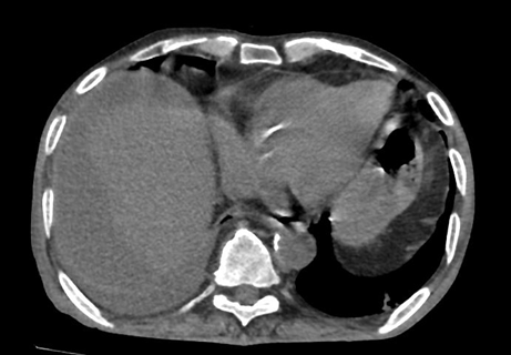
Written informed consent was obtained from a 75-year-old male patient (weighing 76.5 kg, height 172.9 cm) who has coronary artery disease, EF of 22% with obstructive sleep apnea, arthritis, chronic kidney disease, type II diabetes mellitus, controlled hypertension, and hyperlipidemia. He presented for an open right extended hepatectomy. A CT scan of the abdomen revealed a subhepatic anechoic collection measuring approximately 324 cc with internal separations and no signs of internal vascularity, located next to the right lobe of the liver. (Fig 2). On the day of surgery, the patient's vital signs were as follows: temperature 36.6°C, blood pressure 130/72 mmHg, respiratory rate 28 breaths per minute, pulse oximetry 86 % on room air, with intermittent CPAP support. The airway examination showed limited neck extension a small oral opening, poor buccal muscles and edentulous, so supported ventilation with a nasal airway. An electrocardiogram revealed rate-controlled atrial fibrillation, premature ventricular complexes, and signs of a previous septal infarct.
Figure 1: CT Abdomen showed a subhepatic anechoic collection measuring around 324 cc with internal separations and no evidence of internal vascularity noted adjacent to the right lobe of the liver.
After obtaining informed consent, the operating provider marked the patient, a timeout was conducted, and the patient was positioned for a unilateral erector spinae block in the left lateral position with standard monitors in place and oxygen administered via a nasal cannula. The skin was sterilized using a chlorhexidine cleansing solution. The T7 spinous process was delineated after the relevant anatomy was palpated. To find the transverse process, a curvilinear ultrasound probe (Mindray) was then positioned posteriorly on the left side. At the level of the T4 transverse process, a 23-gauge spinal needle was placed in-plane beneath the erector spinae muscle from the cranial to the caudal direction. Five ml of saline was used to hydro-dissect the plane until the needle reached the transverse process.
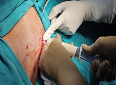
One millilitre of local anaesthetic was administered once it was confirmed that no blood had been aspirated, and no harmful effects were seen. Further normal saline was added to get visual confirmation of the catheter's position in the muscle plane. After that, the ultrasonography was moved to the right side. The process was repeated on the right side once the pertinent anatomy and landmarks were located with the use of ultrasonography assistance. After that, 5 mL increments of 15 mL of 0.25% ropivacaine were given, and the patient was repeatedly checked for negative blood aspiration The patient underwent the surgery with good tolerance, and their vital signs stayed consistent. For the intercostal block, we used a 22 G, 3.5 or 5 cm needle that was advanced while the patient was in a left lateral position to puncture both the internal and external intercostal muscles. To ensure that the needle tip stays superficial to the parietal pleura, the ideal target needle endpoint is situated just inside the internal intercostal muscle. Once more, the deepest intercostal muscle is not always visible, making it an unreliable landmark for injection guidance. 2.5 ml of 0.5 ropivacaine in two levels given, level decided after discussion with the surgeon. The patients were arranged laterally in a way that promoted good health. ropivocaine was used to provide anaesthesia intraoperatively. Pain score assessed and given to surgeon. The liver abscess was drained successfully without any complications, as shown in fig 1.
Figure 2: Pus being drained after anaesthetizing with combined blocks.
Discussion
A useful indicator of hepatic function, prothrombin time (PT) is used to predict outcomes in patients with acute liver failure and chronic liver disease following surgery. On the other hand, conditions like vitamin K insufficiency disseminated intravascular coagulation, or warfarin medication can cause increased PT levels that are unrelated to liver function. Vitamin K should be given for a few days before surgery if at all possible. An ECG should be part of any cardiac evaluation, and if there are any risk factors for cardiomyopathy, valvular lesions, left ventricular dysfunction, or pulmonary vascular disorders, echocardiography should be taken into consideration. A dynamic evaluation of left ventricular function, an exercise ECG, or both may be helpful if severe coronary artery disease is suspected. An ultrasound or chest X-ray can help locate pleural effusions that might need to be drained before surgery. Urgent optimization is crucial for patients in need of emergency surgery, and this should prioritize intravascular volume status, coagulation function, neurological evaluation, and infection screening. Premedication with an H2 receptor antagonist, such as ranitidine, is advised instead of sedatives, as the latter may cause encephalopathy. Ensuring adequate hepatic blood flow and oxygen delivery should be the key priorities during surgery. Hypoxemia or relative hypoperfusion might result in decompensation and further hepatocellular damage. Because prolonged prothrombin time following liver surgery can lead to an epidural hematoma, the use of epidurals has been controversial. The way epidurals function is by producing sympathectomy, which lowers blood pressure and increases the need for fluids and vasopressors.
In comparison to patients who underwent conventional anaesthesia, our patients who received ESP block in conjunction with acetaminophen, magnesium sulfate, xylocaine, and dexmedetomidine as part of a multimodal anaesthesia regimen without opioids showed intraoperative hemodynamic stability in terms of blood pressure, pulse, and cardiac output. Similarly, Fiorelli et al [6] discovered that following an open thoracotomy, ESP blocks increased patient satisfaction and decreased the use of intraoperative opioids. They also resulted in lower 48-hour postoperative pain scores and fewer pulmonary problems. After hepatic surgery, Maddineni et al [7] reported a good result when employing ESP instead of the gold standard epidural analgesia for efficient pain management. Comparably, research by Gürkan [8] showed that PVB and ultrasound-guided ESP block gave patients having breast surgery adequate analgesia and had an opioid-sparing impact by lowering morphine intake. As part of an improved recovery regimen for major abdominal surgery, the ESP block has demonstrated considerable potential in reducing the uncommon but serious risk of pneumothorax associated with paravertebral block [9]. The rhomboid intercostal block is another useful alternative block that can relieve both somatic and visceral pain, but its maximum distribution is limited to the area between T2 and T9 [10] and quadratus lumborum block is a more complex, intrusive technique that may even cause motor weakness.
Given the established advantages of regional trunk blocks, it would be unethical to compare ESPB to a placebo in the absence of a regional anaesthetic. The effectiveness of the ICNB has been evaluated in a sizable meta-analysis comprising 66 relevant trials. In the first 24 hours following surgery, the ICNB is said to be slightly less effective than paravertebral block, non-inferior to thoracic-epidural anaesthesia, and superior to systemic analgesia. According to the research, the ICNB's analgesic effects gradually disappear 24 to 48 hours following surgery. It was discovered that ESPB and TPVB, which have comparable analgesic effects, can both effectively relieve pain in elderly individuals who have undergone thoracoscopic lobectomy. According to some research, thoracic epidural analgesia in thoracic surgery can be effectively substituted with ultrasonic-guided thoracic erector spinae plane block, which has analgesic effects comparable to the former. After an epidural block fails during a thoracotomy, the continuous erector spinae plane block approach can also be used for analgesic remedial treatment. We discovered that older individuals with poor cardiac and lung reserve could benefit from regional blocks like ESPB and ICBs safely and efficiently. In such complications, where giving General anaesthesia was also harmful, we suggest combined regional blocks be a useful technique for postoperative analgesia.
References
- Ziser A, Plevak DJ, Wiesner RH, Rakela J, Offord KP, Brown DL. (1999). Morbidity and mortality in cirrhotic patients undergoing anesthesia and surgery. Anesthesiology, 90(1):42-53.
Publisher | Google Scholor - Maze M Bass, NM Miller RD. Anaesthesia and the hepatobiliary system Anesthesia 2000 5th Edn. Philadelphia Churchill Livingstone, 1960-1972.
Publisher | Google Scholor - Tsui BCH, Fonseca A, Munshey F, et al. (2019). The erector spinae plane (ESP) block: A pooled review of 242 cases. J Clin Anesth, 53:29-34.
Publisher | Google Scholor - Luis-Navarro JC, Seda-Guzmán M, Luis-Moreno C, Chin K. (2018). Erector spinae plane block in abdominal surgery: Case series. Indian J Anaesth, 62(7):549-554.
Publisher | Google Scholor - Kot P, Rodriguez P, Granell M, et al. (2019). The erector spinae plane block: A narrative review. Korean J Anesthesiol, 72(3):209-220.
Publisher | Google Scholor - Fiorelli S, Leopizzi G, Menna C, et al. (2020). Ultrasound-guided erector spinae plane block versus intercostal nerve block for post-minithoracotomy acute pain management: a randomized controlled trial. J Cardiothorac Vasc Anesth, 34(9):2421-2429.
Publisher | Google Scholor - Maddineni U, Maarouf R, Johnson C, Fernandez L, Kazior MR. (2020). Safe and effective use of bilateral erector spinae block in patient suffering from post-operative coagulopathy following hepatectomy. Am J Case Rep, 21:e921123.
Publisher | Google Scholor - Gürkan Y, Aksu C, Kuş A, Yörükoğlu UH. (2020). Erector spinae plane block and thoracic paravertebral block for breast surgery compared to IV-morphine: a randomized controlled trial. J Clin Anesth, 59:84-88.
Publisher | Google Scholor - Hamilton DL, Manickam B. (2017). Erector spinae plane block for pain relief in rib fractures. Br J Anaesth, 118(3):474-475.
Publisher | Google Scholor - Elsharkawy H, Saifullah T, Kolli S, Drake R. (2016). Rhomboid intercostal block. Anaesthesia, 71(7):856-857.
Publisher | Google Scholor


