

Short report
Improvement Of Graphene Induced Abdominal Distension with Melena, Nephropathy by Graphene Exfoliator Nacl + Kcl Solution
Department of Emergency Medicine, New life Hospital Bokhyun-dong, Bukgu, Daegu, Korea.
*Corresponding Author: Chur Chin,Department of Emergency Medicine, New life Hospital Bokhyun-dong, Bukgu, Daegu, Korea.
Citation: C. Chin. (2024). Improvement of graphene induced abdominal distension with melena, nephropathy by graphene exfoliator NaCl + KCl solution, International Journal of Medical Case Reports and Reviews, BioRes Scientia Publishers. 3(1):1-2. DOI: 10.59657/2837-8172.brs.24.042
Copyright: © 2024 Chur Chin, this is an open-access article distributed under the terms of the Creative Commons Attribution License, which permits unrestricted use, distribution, and reproduction in any medium, provided the original author and source are credited.
Received: February 13, 2024 | Accepted: February 29, 2024 | Published: March 04, 2024
Abstract
A 63-year-old man with right corona radiata infarct presented to our hospital with a history of abdominal distension, 500 cc melena, vital signs: Blood pressure 90 (systolic)/60 (diastolic), heart rate 90, total leukocyte count of 11500/cmm, hemoglobin 11.3 g/dl, platelet 414000/ µl, stool occult blood: Positive, a prothrombin time 15.5 sec (normal range: 10-15), Blood Urea Nitrogen (BUN) 41.7 mg/dL, Creatinine (Cr) 2.28 mg/dL, serum sodium/potassium/chloride 137/4.9/108 nmol/L, aspartate transaminase and alanine transaminase 45 IU/L and Erythrocyte Sedimentation Rate (ESR) 40 mm in the first hour using Westergren method.
Keywords: corona; bowel; blood
Description
The intravenous infusion of a solution consisting of 250 mL normal saline over 6h for 3 days with supportive care resulted in recovery of the symptoms with BUN/Cr 22.8/1.69 mg/dL, platelet 264000/ µl with negative stool blood [1-7]. Ischemic bowel disease is due to inadequate oxygenated blood supply to the bowel walls. Ischemic bowel disease includes acute and chronic mesenteric ischemia affecting the small bowel and colon ischemia with embolism, arterial or venous thrombosis. Embolism to the visceral vessels is the most common cause of mesenteric ischemia. Atrial Fibrillation (AF) is a risk factor for ischemic bowel disease in patients with atrial fibrillation. AF-related irregular heart rhythm may predispose to hypoperfusion and ischemia of the bowel walls. CT findings of the ischemic bowel disaese emcompass the presence of pericolic fluid was found in the acute phase and bowel wall thickening in the subacute phase [8].
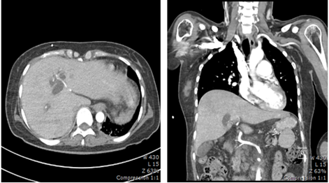
Figure 1: Computed Tomogram: Gastric varix
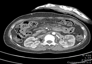
Figure 2: Computed Tomogram: Small bowel wall edema
References
- Chin C. (2023). Comparison of 50 Cases of the Anti-Cancer Effects of NaCl with KCl as a Potent Graphene Exfoliator, Prehydrated Patients to NaCl-Only Prehydrated Patients on the Terminal Stage Cancer Patients. Case Reports in Clinical Medicine, 12:425-431.
Publisher | Google Scholor - Chin C. (2023). Changes in electrocardiogram after intramuscular injection of graphene using salt- intercalation exfoliation. J Clin Exp Cardiol. 14:1-15.
Publisher | Google Scholor - Chin C. (2021). Cell entry inhibitor with sulfonated colloid gold as new potent broad spectrum virucides. J Infect Dis Ther, 9:1-4.
Publisher | Google Scholor - Chin C. (2023). The Anti-Inflammatory Effects of NaCl with KCl as a Potent Graphene Exfoliator in a Patient with Guillaine-Barré Syndrome and Facial Nerve Palsy. Case Reports in Clinical Medicine, 12:447-451.
Publisher | Google Scholor - Chin C. (2023). Improvement of renal functions, graphene-induced rapid progression of prediabetes in an elderly woman with arthritis by graphene-exfoliator NaCl with KCl solution, 3 cases. Journal of Clinical Images and Medical Case Reports, 4:1-3.
Publisher | Google Scholor - Chin C. (2023). Improvement of graphene induced pulmonary edema by graphene exfoliator NaCl with KCl solution, Journal of Clinical Images and Medical Case Reports, 4:4-5.
Publisher | Google Scholor - Chin C. (2023). The anti-inflammatory effects of NaCl with KCl as a potent graphene exfoliator in a patient with interstitial pneumonia by epithelial-mesenchymal transition. Journal of Clinical Images and Medical Case Reports. 4:36-37.
Publisher | Google Scholor - Francesca Iacobellis, Daniela Berritto, Dominik Fleischmann, Giuliano Gagliardi, Antonio Brillantino, et al. (2014). CT Findings in Acute, Subacute, and Chronic Ischemic Colitis: Suggestions for Diagnosis, Biomedical Research International, 895248:1-7.
Publisher | Google Scholor


