Research Article
Determining the Condition of the Abdominal Cavity Tissues using Coherent Radiation
1Department of Surgery no 1, Bukovinian State Medical University, Chernivtsi, Ukraine.
2Department of optics and publishing and printing, Educational and Scientific Institute of Physical, Technical and Computer Sciences, Yuriy Fedkovych Chernivtsi National University, Chernivtsi, Ukraine.
3Department of Surgery no1, Bukovinian State Medical University, Chernivtsi, Ukraine.
4Department of Surgery, Chernivtsi Regional Clinical Hospital, Chernivtsi, Ukraine.
*Corresponding Author: Fedir Grynchuk, Department of Surgery no 1, Bukovinian State Medical University, Chernivtsi, Ukraine.
Citation: Grynchuk F., Besaha R., Grynchuk F., Horditsa V. (2024). Determining the Condition of the Abdominal Cavity Tissues using Coherent Radiation. Journal of BioMed Research and Reports, BioRes Scientia Publishers. 5(5):1-8. DOI: 10.59657/2837-4681.brs.24.076
Copyright: © 2024 Fedir Grynchuk, this is an open-access article distributed under the terms of the Creative Commons Attribution License, which permits unrestricted use, distribution, and reproduction in any medium, provided the original author and source are credited.
Received: February 21, 2024 | Accepted: August 14, 2024 | Published: October 21, 2024
Abstract
Background: Determining the condition of abdominal tissues is an important task, because tissues condition often affects the choice of volume and ways of surgical intervention. Incorrect tissues condition determination causes serious problems. But there is no sufficiently reliable way of determining the condition.
Aim: To investigate changes in the parameters of coherent radiation in the tissues of the abdominal cavity.
Materials and methods: To achieve the research objectives were used white rats with models of intestinal obstruction (40) and the acute peritoneum inflammation (40), 30 intact rats. The intestine walls photoluminescence intensity (PLI) and the laser beams scattering zone width (LBSZW) in small and large intestine and parietal peritoneum were investigated. An LGN-503 argon laser (wavelength of 458 nm) and laser LEDs (wavelength of 0.63 and 0.4 nm) were used. Tissues of the intestines and peritoneum were taken for histological examination.
Results: PLI and LBSZW increase with the intestines and peritoneum lesions. But the absolute parameters are variable. PLI ratio at wavelengths λ=474/λ=489 nm and LBSZW ratio at wavelengths λ=0.63/λ=0.4 μm in the intact intestines are not significantly different. The both ratio in the necrotized intestines are not significantly different. The both ratio in the dystrophically affected intestines are not significantly different. The both ratio in the dystrophically affected intestines are significantly less than ratio in the intact intestines (p<0.05). The both ratio in the necrotized intestines are significantly less than ratio in the dystrophically altered intestines (p<0.05) and in the intact intestines (p<0.01). LBSZW ratio in the intact peritoneum are significantly less than in the inflammatory peritoneum (p<0.01).
Conclusions: 1. PLI ratio parameters at the wavelengths λ=474/λ=489 nm and LBSZW ratio parameters at wavelengths λ=0.63/λ=0.4 μm in the intestines accurately determine the condition of their tissues in the experiment.
2. LBSZW ratio parameters at wavelengths λ=0.63/λ=0.4 μm in the parietal peritoneum accurately determine the condition of its tissues in the experiment.
Keywords: coherent radiation; luminescence; laser beams scattering; small intestine; large intestine; parietal peritoneum; necrosis; inflammation
Introduction
The tissue condition is determinate during every surgical intervention, but the biggest problem arises during emergency surgery, when it is almost impossible to determine the tissue condition before the operation [1,2]. The condition of the abdominal tissues often affects the choice of volume and ways of surgical intervention. This is most relevant during surgery for acute peritonitis, acute intestinal obstruction, acute mesenteric ischemia [1-3].
Currently, various ways of determination the abdominal tissues condition are used. Most often, radiography, computed tomography (CT), ultrasound sonography, magnetic resonance imaging (MRI) is used for this [4-7]. Injections of dyes or X-ray contrasts into the blood vessels are used to determination the intestines tissues condition [3,8]. Laboratory markers are also used (leukocytes, C-reactive protein, procalcitonin, etc.) [2].
But these ways have a number of shortcomings. Laboratory criteria allow only indirect determination. X-ray examination can reveal only the consequences of organ and tissue disorders, or indirect signs of disorders. CT and MRI are much more informative. But these ways can, for the most part, determinate the main lesions of organs and tissues. Determination of small local lesions is problematic. Sonography determines the swelling, some anatomical disorders, but is not able to clearly determine the tissues condition, especially the intestines and peritoneum. Dye or contrast injection determines only blood circulation, not the summary tissues condition.
Some authors suggest ways of comprehensive tissues condition determination. This, for example, measurement of tissue saturation [9,10] or electrical impedance [11]. A disadvantage of these ways is comparison with an intact tissue, but the intact tissues are determinate subjectively. Therefore, these ways are not reliable enough.
So, the main way of the condition tissues determination is still visual. But this way is very subjective [3]. In the same time, incorrect tissues condition determination causes serious problems. The damaged tissues may be left in the abdominal cavity. This causes complications such as peritonitis, abscess, etc. Severe digestive disorders, pathological syndromes, etc. occur due to excessive resection.
To determine the state of abdominal tissues, it is also suggested to use lasers [12-14]. However, such ways, for various reasons, have no practical application. It is known that lasers have long been used for diagnosis in various fields of medicine [15-18]. The measurements using lasers is high accuracy [15,17]. The measurements parameters are able to assess the tissues condition at the molecular level [16].
Therefore, the study of the coherent radiation possibilities for determination the condition of abdominal tissues is relevant and promising.
Abbreviations
computed tomography – CT; large intestine – LI; laser beams scattering zone width – LBSZW; ligated intestinal loops – LIL; magnetic resonance imaging – MRI; photoluminescence intensity – PLI; small intestine – SI.
Materials and methods
Study area
The study was conducted at Department of Surgery № 1, Bukovinian State Medical University is located at Chernivtsi, Ukraine. The presented research is part of the scientific work of the Department of Surgery № 1, and is one of the scientific work series on ways to determine the abdominal tissues condition. Bukovinian State Medical University was established in 1944 as one of modern educational, scientific and medical center in the country. Nowadays, it runs graduate, postgraduate, scientific and medical programs. University is furnished with various scientific and medical facilities.
Study period
This study was conducted over a period of 6 months from May to December, 2023 G.C.
Study design
110 intact white non pedigree female sexually mature (age 6 months) rats. The rat’s weight was 180-200 g. Before the start of the experiment, the rats were in a vivarium. Housing and feeding conditions were the same for all rats.
3 series of experiments were conducted. The informativeness of the luminescence intensity parameters for assessing the intestinal tissue condition was studied in the 1st series. The informativeness of the laser beams scattering parameters for assessing the intestinal tissue condition was studied in the 2nd series. The informativeness of the laser beams scattering parameters for assessing the parietal peritoneum condition was studied in the 3rd series.
Samples size
10 rats were the control group and 20 rats were the research group in the 1st series and in the 2nd series of experiments. 10 intact rats were the control group, 40 rats were the research group in the 3rd series of experiments.
Inclusion criteria
Rats for each control and research group were randomly selected from the rats taken for the experiment. The control groups and the research groups were homogeneous in terms of age and weight. After the start of the experiment, the rats were in the same conditions and had the same drink. Therefore, the control groups and the research groups were comparable. This made it possible to obtain comparable data and make reliable conclusions.
Data collection
Laparotomy was performed in the 1st and 2nd series. A loop of the small intestine (SI) with the mesentery in middle part was ligated in 10 rats and f loop of the large intestine (LI) with the mesentery in middle part was ligated in 10 rats each research group.
The luminescence spectra in the middle part of SI and LI walls were measured in the control group in the 1st series. The luminescence spectra in ligated intestinal loops (LIL) were measured in 6 h since intestine were ligated in the research group.
The laser beams scattering zone width (LBSZW) in the middle part of SI and LI walls were measured in the control group in the 2nd series. LBSZW in LIL of the intestines were measured in 6 h since intestine were ligated in the research group.
In 3rd series, acute inflammation of the peritoneum was simulated by intraabdominal injection of 20% autofaeces suspension in the dose of 10 ml per 100 g of mass in research group. Laparotomy was performed in control group and in 6, 12, 24, 48 h since the peritoneum inflammation was simulated in research group. LBSZW in parietal peritoneum were measured. Measurements were made in 4 quadrants of the abdomen: upper left, upper right, lower left, lower right.
The examined tissues were taken for histological examination after the measurements and were fixed in the 10% formalin solution, dehydrated in an ascending alcohol battery, and embedded in paraffin for histological examination. Sections were made on a microtome with a thickness of 5 μm. Deparaffinized sections were stained with hematoxylin-eosin. Stained preparations were studied in a Delta Optical Evolution Pro 100 light microscope.
The walls of intestines were irradiated with a monochromatic laser beam in 1st series. Its source was an LGN-503 argon laser with a wavelength λ=458 nm. The laser was powered by an AC 220 W 50 Hz electrical network. The laser radiation scattered by the intestine wall was focused on the input slit of the MDR-12 monochromator, behind which the ZhS-16 light filter was mounted. At the out from the monochromator, the laser beam fell on a photodetector connected to a universal voltmeter V-7-21A, which was used to determine the output radiation parameters. The photodetector was powered by an AC 220 W 50 Hz electrical network. The temperature lamp TRSh 2850-3000 was used as a reference radiation source for decoding the luminescence spectrum.
The walls of intestines and the parietal peritoneum were irradiated with the laser beams in the 2nd and 3rd series. Laser LEDs with radiation wavelengths λ=0.63 μm and λ=0.4 μm were used for irradiation. Laser LEDs was powered by a standard DC source with a voltage of 1.5 V. LBSZW (in millimeters) in the intestines walls and peritoneum was measured during irradiation with a standard ruler. Measurements were made from distance 20 cm. Measurements were made with an accuracy of 1 mm.
Ethical clearance
Inhalational sevoflurane anesthesia was used for analgesia. Animals were removed from the experiment by an overdose of anesthetic.
While performing the work, the norms of conducting research in the field of biology and medicine were observed: the Vancouver Conventions on Biomedical Research (1979, 1994), the Council of Europe Convention on the Protection of Vertebrate Animals Used in Experiments and for Other Scientific Purposes (1986).
Data analysis
The hypothesis of normal data distribution (Gaussian distribution) was tested in samples by Shapiro-Wilk test. Verification of the hypothesis of average data equality was carried out by Wilcoxon, Mann-Whitney-Wilcoxon, Student tests, depending on the data distribution in the samples. The significance level (alpha) 0.05 was set in the study. The results of the study were statistically processed by the Microsoft® Office Excel (build 11.5612.5703) tables.
Results
Histological examinations in 1st and 2nd series in the control groups found intact intestine structure. Histological examinations in each intestines LIL found necrosis.
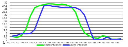
The photoluminescence intensity (PLI) parameters in the intact intestines are shown in Figure 1.
Figure 1: PLI parameters in the intact intestines
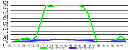
PLI parameters in the necrotized intestines parts are shown in Figure 2.
Figure 2: PLI parameters in the necrotized intestines
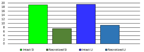
PLI parameters ratio at different wavelengths in the walls of the intact and necrotized intestines parts were calculated. It was established that the ratio of PLI parameters at the wavelengths λ = 474 nm and λ = 489 nm informatively showed the condition of the intestinal tissues. The parameters of this ratio are shown in Figure 3.
Figure 3: PLI ratio parameters at wavelengths λ = 474 / λ = 489 nm
PLI ratio in necrotized SI was significantly (p less than 0.01) less than PLI ratio in intact SI. PLI ratio in the necrotized LI was significantly (p less than 0.01) less than PLI ratio in intact LI. PLI ratio in the intact SI and LI did not differ significantly. PLI ratio in the necrotized SI and LI did not differ significantly.
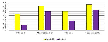
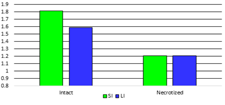
LBSZW parameters in the intestines are shown in the Figure 4.
Figure 4: LBSZW parameters (mm) in the intestines
LBSZW parameters in intact SI at the wavelength λ=0.63 μm were significantly less than LBSZW in LIL (p less than 0.01). LBSZW parameters in intact SI at the wavelength λ=0.4 μm were significantly less than LBSZW in LIL (p less than 0.01). LBSZW parameters in intact LI at the wavelength λ=0.63 were significantly less than LBSZW in LIL (p less than 0.01). LBSZW parameters in intact LI at the wavelength λ=0.4 μm were significantly less than LBSZW in LIL (p less than 0.01). But LBSZW parameters at both wavelengths were different in SI and LI, both in intact and necrotic ones.
We also calculated the ratio of LBSZW at wavelength λ=0.63 μm to LBSZW at wavelength λ=0.4 μm. This ratio parameters are shown in Figure 5.
Figure 5: LBSZW ratio parameters at wavelengths λ = 0.63 / λ = 0.4 μm in the intestines
The LBSZW ratio in the necrotized SI were significantly less than ratio in intact SI (p less than 0.01). The LBSZW ratio in the necrotized LI were significantly less than ratio in intact SI (p less than 0.01). The LBSZW ratio in the intact SI and LI did not differ significantly. The LBSZW ratio in the necrotized SI and LI did not differ significantly.
Histological examinations in 3rd series in the control group showed intact peritoneum structure. Histological examinations in 6 h since the peritoneum inflammation was simulated showed serous peritoneum inflammation. Histological examinations in 6,12,24 end 48 h since the peritoneum inflammation was simulated showed purulent peritoneum inflammation.
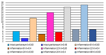
LBSZW parameters in the parietal peritoneum are shown in the Figure 6.
Figure 6: LBSZW parameters (mm) in the parietal peritoneum
LBSZW parameters in the intact peritoneum at the wavelength λ=0.63 were significantly (p less than 0.01) less than LBSZW in 6, 12, 24, 48 h since the peritoneum inflammation was simulated. LBSZW in 6 h were significantly less than LBSZW in 12 h (p less than 0.01). LBSZW in 12 h were significantly less than LBSZW in 24 h (p less than 0.01). LBSZW in 24 h and 48 h did not differ significantly. LBSZW parameters in the intact peritoneum at the wavelength λ=0.4 were significantly (p less than 0.01) less than LBSZW in 6, 12, 24, 48 h since the peritoneum inflammation was simulated. LBSZW in 6 h were significantly less than LBSZW in 12 h (p less than 0.05). LBSZW in 12 h were significantly less than LBSZW in 24 h (p less than 0.01). LBSZW in 24 h and 48 h did not differ significantly.
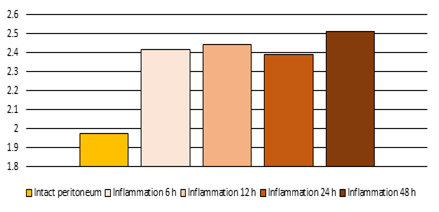
We also calculated the ratio of LBSZW at wavelength λ=0.63 μm to LBSZW at wavelength λ=0.4 μm. This ratio parameters are shown in Figure 7.
Figure 7: LBSZW ratio parameters at wavelengths λ = 0.63 / λ = 0.4 μm in the parietal peritoneum
Control LBSZW ratio parameters (1.97±0.08 un) were significantly (p less than 0.01) less than LBSZW ratio in 6 h (2.41±0.06 un), in 12 h (2.44±0.05 un), in 24 h (2.39±0.05 un), in 48 h (2.51±0.04 un) since the peritoneum inflammation was simulated. The LBSZW ratio in 6 h, in 12 h, in 24 h, in 48 h did not differ significantly.
Discussion
Comparing PLI parameters with the histological examinations data in 1st series of experiment shows that due to the development of the intestinal wall’s necrosis, parameters of its PLI in the wavelength range λ = 470-490 nm decrease. This is a result of different processes: blood circulation disruption, oedema, tissue destruction etc. The intact tissues are well filled with blood and these tissues luminesce more strongly [12,15]. Due to the intestine wall’s tissues destruction, their blood supply decreases, blood circulation slows down in and blood stagnation occurs viable tissues. The intestinal tissues PLI decreases. So, the PLI parameters show the total changes in the tissues and accurately describes the intestinal walls condition.
But absolute PLI parameters are highly variable (Figure 1). Numerous factors affect the PLI parameters. Among such factors are individual, local, the degree of intestine filling, stressful situation, time of day, season etc. [12]. Of course, there are species differences – the parameters will differ in animals and people, because the histological structure of the tissues is some different [17].
But mentioned factors do not affect the relative parameters. PLI ratio at wavelengths λ=474/λ=489 nm in the intact SI was 19.09±1.89 un and the ratio in the intact LI was 19.35±1.27 run (р>0.05). The ratio in the necrotized SI was 7.40±0.66 un and in the necrotized LI was 9.04±1.27 run (р>0.05).
Comparing LBSZW parameters with the histological examinations data in 2nd series of experiment shows that due to the intestinal condition disruption, LBSZW increases. This is a consequence of the distancing of the dimensions intestine’s walls structural elements h from the laser beams’ wavelength λ. The intact intestines structures are closer to the solid condition than necrotizing intestines, their dimensions are smaller. Therefore, intact structures scatter laser beams less [18]. As a result of the intestines structures destruction, the cellular contents go beyond them, the tissues become saturated with plasma, swell. The intestinal structures become more homogeneous. The scattering of beams is carried out by the volume, so LBSZW increases [14,18]. So, an increase in LBSZW parameters indicates the intestine walls condition disruption. But absolute LBSZW parameters cannot be used to determine condition. This is due to the fact that the LBSZW are affected by numerous factors, which we mentioned above.
But these factors do not affect the relative parameters. LBSZW ratio at wavelengths λ=0.63/λ=0.4 μm in the intact SI (1.58±0.08 un) and LI (1.61±0.07 un) was not significantly different (р>0.05). The ratio in LIL of SI (1.20±0.03 un) and LI (1.19±0.02 un) was also not significantly different (p>0.05), but it was significantly different from the both intact intestines (p<0>
Comparing LBSZW parameters with the histological examinations data in 3rd series of experiment shows that due to the peritoneum inflammation, LBSZW in the peritoneum increases. This is a consequence of the distancing of the dimensions peritoneum’s structural elements h from the laser beams’ wavelength λ. The intact peritoneum’s structures are closer to the solid condition, their dimensions are smaller. So, intact structures scatter laser beams less [13,18]. As a result of the inflammation, the tissues are more filled with blood, become saturated with plasma, swell. The peritoneum’s structures become more homogeneous. The scattering of beams is carried out by the volume, so LBSZW increases. So, an increase in LBSZW indicates the inflammatory changes of the peritoneum condition.
But absolute LBSZW parameters cannot be used to determine the peritoneum condition. This is due to the fact that LBSZW are affected by numerous factors, which we mentioned above. But the use of relative parameters neutralizes these factors effect. As research has shown, LBSZW ratio at wavelengths λ=0.63/λ=0.4 μm in intact peritoneum was significantly (p<0>
Therefore, PLI ratio at the wavelengths λ=474/λ=489 nm and LBSZW ratio at wavelengths λ=0.63/λ=0.4 μm in the intestinal walls allows to evaluate the intestine’s tissues condition. LBSZW ratio in the parietal peritoneum allows to evaluate its condition.
Conclusions
1. PLI ratio parameters at the wavelengths λ=474/λ=489 nm and LBSZW ratio parameters at wavelengths λ=0.63/λ=0.4 μm in the intestines accurately determine the condition of their tissues in the experiment.
2. LBSZW ratio parameters at wavelengths λ=0.63/λ=0.4 μm in the parietal peritoneum accurately determine the condition of its tissues in the experiment.
Recommendation
Based on the findings the following recommend-dations are forwarded. This study shows the principal possibility of using coherent radiation for determining the condition of the abdominal cavity tissues. In order to further use and implement the research results, it is necessary to create unified industrial devices. Such devices can be created in cooperation with engineers. The study was conducted on site therefore there is need of further research that could be conducted in other to generalize outside the participating institution.
Strength
This paper has relatively large sample size. This is the previous study and it can be used as base line for future studies.
Limitation
The small sample sizes were used in study. It is necessary to conduct additional experiments on a larger number of animals to confirm the data. Female rats were used in the previous study. Therefore, male rats should be used to confirm the data in future studies. The study used rats. To confirm the data, it is advisable to conduct experiments with other species of animals. Research data must be tested in humans to definitively determine the validity of experimental data after the creation of unified industrial devices. Taking into account the known physiological and pathological regularities common to all warm-blooded mammals, these limitations will not have a significant affect the generalizability of the findings. It can be expected that the basic regularities will be confirmed, but some indicators may change slightly.
References
- Jackson, P., Vigiola Cruz, M. (2018). Intestinal Obstruction: Evaluation and Management. American Family Physician., 98(6):362-367.
Publisher | Google Scholor - Sartelli, M. et al. (2021). WSES/GAIS/SIS-E/WSIS/AAST global clinical pathways for patients with intra-abdominal infections. World Journal of Emergency Surgery., 25;16(1):49.
Publisher | Google Scholor - Bryski, M. G., et al. Techniques for intraoperative evaluation of intestine viability in mesenteric ischemia: A review. Am J Surg. 2020;220(2):309-315.
Publisher | Google Scholor - Tam, W. (2023) Current abdominal X-rays practice in accident and emergency. Journal of Medical Imaging and Radiation Sciences., 17:47.
Publisher | Google Scholor - Delaplain, P. T., et al. (2020). The use of computed tomography imaging for abdominal seatbelt sign: A single-center, prospective evaluation. Injury., 51(1):26-31.
Publisher | Google Scholor - Al Ali, M., Jabbour, S., Alrajaby, S. (2019). Acute abdomen systemic sonographic approach to acute abdomen in emergency department: a case series. Ultrasound Journal., 11(1):22.
Publisher | Google Scholor - Yu, H. S., et al. (2016). Emergency abdominal MRI: current uses and trends. British Journal of Radiology., 89(1061):20150804.
Publisher | Google Scholor - Ryu S, Hara K, Goto K, Okamoto A, Kitagawa T, Marukuchi R, et al. Fluorescence angiography vs. direct palpation for intestine viability evaluation with strangulated intestine obstruction. Langenbecks Arch Surg. 2022;407(2):797-803.
Publisher | Google Scholor - Sugiura, T., et al. (2021). Predicting intestinal viability by consecutive photoacoustic monitoring of oxygenation recovery after reperfusion in acute mesenteric ischemia in rats. Scientific Reports., 11(1):19474.
Publisher | Google Scholor - Suzuki, Y., et al. (2021). Usefulness of a finger-mounted tissue oximeter with near-infrared spectroscopy for evaluating the intestinal oxygenation and viability in rats. Surgery Today., 51(6):931-940.
Publisher | Google Scholor - Hou, J., et al. (2021). Assessing Ischemic Injury in Human Intestine Ex Vivo with Electrical Impedance Spectroscopy. Journal of Electrical Bioimpedance., 12(1):82-88.
Publisher | Google Scholor - Besaha, R. M., Hrynchuk, F. V., Polyansky, I. Yu. (2006). Diagnostic meaning of intestinal wall photoluminescence changes. Proceedings of SPIE. 6254:62541U-1.
Publisher | Google Scholor - Grynchuk, A. F., Grynchuk, F. V., Polianskyi, I. Iu. (2017). Objective diagnostics of the acute peritonitis spread. Kharkiv Surgical School., 1:31-34.
Publisher | Google Scholor - Shurma, A. I., Grynchuk, F. V. (2023). Experimental study of the use of coherent radiation to determine the bowels viability. Clinical and Experimental Pathology., 22(1(83):57-62.
Publisher | Google Scholor - Barnes, F. S. (1975). Applications of lasers to biology and medicine. Proceedings of the IEEE., 63(9):1269-1278.
Publisher | Google Scholor - Simone, N. L., et al. (1998). Laser-capture microdissection: opening the microscopic frontier to molecular analysis. Trend in Genetics., 14(7):272-276.
Publisher | Google Scholor - Legres, L. G., et al. (2014). The Laser Technology: New Trends in Biology and Medicine. Journal of Modern Physics., 5(5):267-279.
Publisher | Google Scholor - Grynchuk, F. V., Dutka, I. I., Besaga, R. M. (2021). Diagnostics of haemostasis efficiency with laser illumination scattering pattern analysis. Proceedings of SPIE., 12126, 121262N:117.
Publisher | Google Scholor

