Case Report
CT and MRI Image Fusion using Wavelet Transform and Log Gabor filter
- Rishabh Maurya *
Computer Science Department, ABES Engineering College 19th KM Stone, Ghaziabad, Uttar Pradesh, India.
*Corresponding Author: Rishabh Maurya, Computer Science Department, ABES Engineering College 19th KM Stone, Ghaziabad, Uttar Pradesh, India.
Citation: Maurya R., Mishra R., Sharma Y.K., Swarnkar R., Pathak D.M. et al. (2024). CT and MRI Image Fusion using Wavelet Transform and Log Gabor filter. Scientific Research and Reports, BioRes Scientia Publishers. 1(4):1-15. DOI: 10.59657/2996-8550.brs.24.025
Copyright: © 2024 Rishabh Maurya, this is an open-access article distributed under the terms of the Creative Commons Attribution License, which permits unrestricted use, distribution, and reproduction in any medium, provided the original author and source are credited.
Received: July 15, 2024 | Accepted: August 30, 2024 | Published: September 24, 2024
Abstract
Image fusion is a pivotal technique in medical imaging, particularly in combining complementary information from different modalities which are computed tomography (CT) and magnetic resonance imaging (MRI) to enhance diagnostic accuracy. This paper presents the comprehensive research of CT and MRI image fusion methodologies, focusing on the integration of Wavelet Transform (WT) and Log Gabor Filter with Gaussian and Contrast Limited Adaptive Histogram Equalization (CLAHE) enhancement techniques. The Wavelet Transform has gained significant attention due to its multi-resolution analysis capability, enabling the decomposition of images into different frequency bands. Log Gabor filters, on the other hand, excel in capturing texture information with orientation selectivity. Integrating these techniques offers a synergistic approach for extracting and preserving relevant features from CT and MRI images. Moreover, Gaussian and CLAHE enhancement techniques are employed to improve the contrast and visibility of anatomical structures in both CT and MRI images. Gaussian filtering smoothens the images while preserving edges, whereas CLAHE enhances local contrast by adaptive histogram equalization. This review discusses the theoretical foundations, methodologies, and applications of CT and MRI image fusion techniques employing Wavelet Transform and Log Gabor Filter with Gaussian and CLAHE enhancement. Furthermore, the review highlights the significance of image quality assessment metrics in evaluating the performance of fusion techniques, including metrics such as peak signal- to-noise ratio (PSNR), entropy, structural similarity index measure (SSIM), and subjective evaluation through visual inspection by medical experts. At last, the integration of Wavelet Transform and Log Gabor Filter with Gaussian and CLAHE enhancement techniques holds immense potential in CT and MRI image fusion, offering enhanced diagnostic capabilities and improved clinical decision- making in medical imaging applications. Future research directions and challenges in this field are also discussed to stimulate further advancements in image fusion methodologies.
Keywords: computed tomography; magnetic resonance imaging; wavelet transform; contrast- limited adaptive histogram equalization; log gabor; gaussian, peak signal-to-noise ratio; structural similarity index
Introduction
Fusion of image is the combination of multiple medical images which increases the image's quality and removes irrelevant data from the images. Due to this, fused images will help in the diagnosis of medical diseases. Fused image contains rich information and every important feature from the original images. It has the best output than the original images. The main goal of fusion techniques in the field of medical images is to diagnose and extract more important data from the images for the best diagnosis of the disease. The fusion images simply give the average of what is best for human visualization.
Image fusion has two types, spatial and frequency domain [6]. In which the spatial domain contains Simple Average Pixel, Weighted Averaging, Hue Intensity Saturation, and Guided Filtering. Frequency domains contain Discrete Wavelet Transform, Laplacian Pyramid, Discrete Cosine Transform, Stationary Wavelet Transformation, and Dual- Tree Complex Wavelet Transform. This article has various techniques which are published in various papers in the past few years that have different domains and techniques in them. This article contains the study of several literature papers on various techniques. Different type of algorithm or technology uses is mentioned. Also, they contain some gaps that have to be overcome from it. This paper is only on the two which are spatial techniques and frequency [8]. Some of the Spatial domain techniques include:
Simple Average
Min-Max
Brovey
PCA
Weighted Average
HIS
Some of the Frequency domain techniques include:
Discrete Cosine Transform (DCT)
Stationary Wavelet Transform (SWT)
Laplacian Pyramid
Discrete Wavelet Transform (DWT)
Kekre’s Wavelet Transform
As image fusion technique is used for extracting important, relevant data for medical uses and removing irrelevant data from the image.
Spatial Domain Techniques
This technique is considered as one of the simple image fusion techniques. It consists of the following method [5-8].
Simple Average: It is a method which is by combining the image on averages of their pixels. It has very high contrast and brightness and thus it will produce very good results.
Max-Min: It is a method that averages the pixel value from the smallest to the largest in the whole image which gives the fused image.
Brovey: It includes the RGB color transformation method. For the fusion of three multispectral bands, it uses addition, division, and multiplication. It increases contrast at the low ends and high ends the of image. It prevents the disadvantages of the multiplicative method.
Principal Components Analysis (PCA): It is widely used for data analysis and predictive models. It is a statistical method that converts correlated variables into principal components. Its disadvantage is the degradation and color distortion.
Weighted Average: This method will assign some weights to each pixel of the image. So, the image produced is the weighted sum of each pixel of the source image.
Hue Intensity Saturation (HIS): It is a color fusion technique that converts RGB images into HIS. Then the HIS image is divided into panchromatic images. Now spatial contains intensity and spectral contains hue and saturation information of the image. Then in the end inverse transform is applied to convert HIS into the original RGB images. Here are some pros and cons of the Spatial domain techniques:
Table 1: Spatial Domain Techniques
| Spatial Domain | Advantages | Disadvantages |
| Simple Average | Easy to recognize and implement | Blurred image, not for real-time application |
| Min-Max | Easy to implement and recognize | Blurred image, not for real-time application |
| Brovey | Fast process time and more efficient | Color distortion |
| PCA | Simple, efficient, less computational time | Color distortion |
| Weighted Average | Detection reliability improves | Signal noise ratio increases |
| HIS (Hue Intensity Saturation) | Simple, fast process and highly sharpen the ability | Color distortion |
Frequency Domain Techniques
This technique is used on the multiscale coefficient of the source image. Spatial distortion can be managed by the frequency domain [8][5][19].
Discrete Cosine Transform (DCT): It is a straightforward image fusion method that uses in actual time applications. It does not give good results on less than 8x8 block sizes is one of the basic fusion methods.
Stationary Wavelet Transform (SWT): It has a better output at level 2. Also, it is a very time-efficient method. This manages to improve the translation invariance of the DWT. It provides an enhanced analysis of the source image.
Discrete Wavelet Transform (DWT): It has the capability of decomposing two or more images in high bands and low bands. It minimizes distortion but produced a signal-to-noise ratio (SNR).
Laplacian Pyramid: It is one of the improved image fusion methods which uses a contrast pyramid transform on multisource images. It has one of the major drawbacks of extraction ability which can only be improved by multiscale decomposition.
Kekre’s Wavelet Transform: This method only is generated by the KWT matrices. It can be easily used on many images that are more than one image which gives results in a fused image that is good than other methods.
Here are some pros and cons of the Frequency domain techniques:
Table 2: Frequency Domain Techniques
| Frequency Domain | Advantages | Disadvantages |
| Discrete Cosine Transform | Images decompose into a series of the waveform, uses for real-time application | Low quality of the fused image |
| Discrete Wavelet Transform | Fast, ableto separate fine details in the signal. | Poor directionality, lack of phase information |
| Stationary Wavelet Transform | The better result at level 2 | Takes more time |
| Laplacian Pyramid | Better image quality for multi-focus images | Breakdown levels affect the result |
| Kekre’s Wavelet Transform | The result has more information and uses for any size of an image | Complex |
The fusion of medical images required the combination of images for gathering the relevant information that can help in d. Ultrasound (US): Us imaging is a non-invasive technique that extracts affected human tissues. It is based on the body’s vibration by its radiation power. The thickness of the tissue not being measured by the ultrasound.
e. X-ray: It scans the inside parts of the human organs which can be known as radiography. It forms a shadow of the body to identify the affected tissue. The image fusion techniques have been categorized into pixel level, feature level, and decision level where pixel-level is the technique that can directly combine the information from images for further tasks. Feature level is the technique that can extract relevant features such as pixel intensities and textures. Decision-level techniques can be used for the taking out the information from the input image which is processed one at a time. Image fusion techniques are divided into three approaches which are [5, 7].
Levels of image fusion
Image fusion, particularly in medical imaging, can be categorized into three primary levels based on the stage at which the fusion occurs: pixel level, feature level, and decision level. Each level has its unique methodologies, advantages, and applications, tailored to specific requirements and objectives in image processing and analysis.
Pixel level: It is to combine multiple images to get a fused image. It will give extra information which is very essential from the perspective of machines and humans. They are simple and easy to extract, also it is enough to describe the image.
Feature level: It is the combination of different features from the different layers. It is integrated into a single feature from multiple different sources. Such as in biometrics collect sources from different the diagnosis. These combinations are CT-MRI, PET-CT, MRI-PET, Ultrasound-X-ray, and more. All these are done to extract additional data from the image [7].
Types of medical images
Computed Tomography (CT): CT is a technique used for creating a picture of the cross-section of the body organ. It is an unobstructed diagnosis method in the medical field. It reveals the dense parts such as bones.
Magnetic Resonance Imaging (MRI): It is a technique that uses magnetic flux and radio frequency to the diseases. The use of the magnetic signal is the human body and affected tissue in the human body. It gives data on the soft tissue.
Positron Emission Tomography (PET): It mainly uses information about the cerebrum in the human mind and also allows the recording of changes in it. It is an important component in atomic drug imaging. It is too effective that it can be used for the diagnosis of cancer in the body.
biometric algorithms and integrates them into a single feature for accurate results.
Decision level: Among all three levels, the decision level has high-level information in the fusion. It is highly accurate but results consist the loss of some information. It is the method of combining the decision which can be taken by the multiple classifications performed on the dataset to reach the final decision.
Each level of image fusion offers benefits and it is chosen based on the specific needs and goals of the application. Pixel level fusion is ideal for high-resolution and detailed imagery, feature level fusion focuses on significant information for efficient processing, and decision level fusion provides robust and reliable outcomes by combining high-level interpretations. In the context of medical imaging, particularly CT and MRI fusion, these levels can be strategically utilized to enhance and improves diagnostic accuracy, improve visualization, and support clinical decision-making.
Table 3: Different Modalities of Images
| CT | MRI | PET | Ultrasound | X-ray | |
| Advantages | Cheap, accurate, and fast | Low radiation, detailed information | The clear output of the image than CT | Safe, easy, less time and less radiation | Identifies anomalies and fractures in human bones. |
| Disadvantages | High radiation | Expensive and does not detect all cancan | Skin allergies | Not diagnosed lungs issue and details of bones are not present | Not gives data on the length and density of the body parts |
| Properties | The dense structure of information | Soft tissues data will be given | Gives functional data of the individual cortex | Based on sound waves | Identifies anomalies and fractures in human bones. |
| Applications | Tumors detection, bones cancer | Joint injuries, detection of breast cancer, detection of liver and abdominal organs disease | Identify heart disease and cancer at an early age, identify brain disorders | Liver cancer detection, diagnosis of fetal development | Uses for breast cancer diagnose |
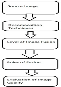
After the analysis fusion of images is done through various processes. This process contains first the input of the image after that appropriate decomposition techniques are applied to those images. After decomposition, it will reach the level of image fusion which goes with the various levels of image fusion, which are pixel, feature, and decision levels. Then the resultant image goes to the fusion rule process and different techniques are applied as per needs. At last, to check the correctness of the output of the fused image will be evaluated by the different image quality measures. The diagram illustrates a structured process for image fusion, outlining key stages from obtaining the source images to evaluating the quality and accuracy of the fused image. Here’s an explanation of each step: The process outlined in the diagram provides a comprehensive approach to image fusion, starting from acquiring the source images, applying decomposition techniques, deciding on the fusion level, applying appropriate fusion rules, and finally evaluating the accuracy and quality metrics of the fused image. This structured methodology ensures that the fused medical image effectively combines the strengths of the source images, resulting in a more informative and useful outcome (Figure 1).
Figure 1: Image Fusion Procedure
Related Work
The fusion of CT and MRI medical images using Wavelet Transform has been a significant area of research in medical imaging, aimed at combining the complementary information of the two modalities to enhance diagnostic accuracy. The wavelet-based fusion techniques leverage the multi-resolution analysis capability of wavelets to effectively integrate high spatial resolution from CT images and superior soft tissue contrast from MRI images.
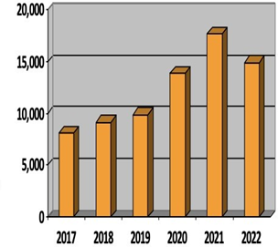
There is various paper written in the context of it. Where papers on the different fusion algorithms such as SWT, NSCT, NSST, DWT, IHS, DCT, DWT, Laplacian Pyramid, Curvelet Transform, Minimum Technique, Brovey, PCA, Maximum Technique, MIN-MAX Technique, DTCWT, CWT, MWT, DTWT and many more. Every year many papers are published on these fusion algorithms of which some are from the spatial domain and frequency domain. These techniques are used vastly in every field. According to the literature review, the number of research papers has increased regularly in the past few years which illustrated that this type of technique has high demand and at low cost, it gives high performance. After the analysis, this shows the nod of articles published on the research of image fusion on medical images from 2017 to 2022 (Figure 2).
Figure 2: Published Papers
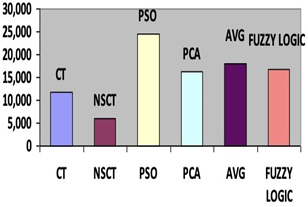
These are the data that are been collected based on the different fusion techniques used in the research papers in the last 5 years. Also, these techniques are classified as wavelet, contourlet, and others. Now some results are based on the contourlet classification and other classifications which are CT, NSCT, PSO, PCA, Average, and Fuzzy Logic. These are some common and important methods used for the research between 2017 to 2022. (Figure 3).
Figure 3: Contourlet and Other Classifications
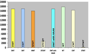
Wavelet transform is one of the most common image fusion techniques. In this, two images are first decomposed into sub- images with the help of different frequencies. Then fusion is performed on these sub-images and finally, the resultant image is obtained by the reconstruction of these sub-images. So, there is approx. details about no results on the published articles on wavelet classifications between 2017 to 2022 (Figure 4).
Figure 4: Wavelet Classifications
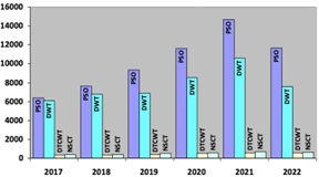
There are some data on the selected techniques like NSCT, DTWCT, DWT, and PSO on which how many papers have been published in the last few years which are frequently being used is shown (Figure 5).
Figure 5: Common Fusion Techniques
Fusion techniques in both the domain of spatial and frequency are important. Each has some good and bad points. But all these methods are useful for their respective purposes. After the survey of a few literature papers, this paper came to some conclusions about their summary and their gaps. These reviews are as:
In the paper [1], the results show that the proposed framework which reduces complex noise, eliminates artificial artifacts, and improves contrast as well as details. The technology used total variational frame and wavelet transform. The gap in that article is difficult to measure the difference accurately between the enhanced image and the original image, so careful analysis of the image model is still required. In that paper [2], the advancement of fusion ranges for medical images from the spatial domain, and transform domain, to deep learning. The article different image methods for medical image fusion research in recent times as well as the benefits of combining various techniques effects for the way, the different imaging fusion methods, and the research trend of statistics. The technology used Spatial domain, NSCT, HIS, NSST, and DWT transform. The gap includes the fusion of three modes that are rarely studied. The two modal studies focus on the fusion of MRI-CT, MRI-PET, and MRI-SPECT.
In the paper [3], various image fusion techniques are discussed with their advantages and disadvantages. Numerous uses have been discussed, including photography, surveillance, and medical imaging. In the paper [4], the suggested fusion algorithm performs better with enhanced brightness and contrast of multimodal medical images, according to comparative experimental fusion results carried out on both grey and color medical image datasets. The technology used are PCNN, NSST, NSCT, GIF-WSEML The gap in this is reducing the operation time and improving the real-time performance of the algorithm. In the paper [5], The NSCT exhibit excellent directionality and effectively expressed smooth edges. The algorithm can also provide accurate judgment information for medical diagnosis. Reduction in time consumption. The technology used is fuzzy logic, NSCT. The gap is shifting variance.
In the paper [6], the main objective of this paper was to propose a CBIR system. It is an approach that has two phases where colour, texture, and local and global information of spatial and frequency domains is extracted. The technology used is content-based image retrieval. The gap includes that it does not have a high accuracy noise and scale changes sensitivity.
In the paper [7], in order to extract illumination and reflectance from the input source MRI image and to extract color constancy to separate the PET input image into the normal image and lesion image, the suggested image decomposition schemes can fully utilize the mechanism of the visual cortex model. The technology used is Intrinsic Image Decomposition. The gap includes is colour distortion in the fused images. The paper [8], discusses Multimodal Medical Image Fusion (MMIF) and its importance in the field of medical imaging. It provides a comprehensive overview of various aspects related to MMIF. It highlights that different modality, like MRI, CT, PET, and SPECT, provide unique types of information, and fusion aims to combine these to improve diagnosis. This paper classifies MMIF techniques into six categories: sparse representation fusion, deep learning, hybrid fusion, decision- level fusion, frequency fusion, and spatial fusion. It provides an overview of how images can be fused at different levels to enhance the quality and information content. It highlights the significance of MMIF in improving disease diagnosis and treatment and encourages further research in this evolving field.
The paper [9], discusses an image fusion technique for combining medical images obtained through Computed Tomography (CT) and Magnetic Resonance Imaging (MRI). The paper presents the mathematical foundations of the Discrete Cosine Transform (DCT) and the Integer Lifting Wavelet Transform (ILWT). It also provides details about the fusion process and how the variance and pixel significance are used to combine information from the two imaging modalities. The results of the proposed fusion technique are compared to several other fusion methods, including DWT, PCA, Laplacian Pyramid (LAP), and others. The paper includes a quantitative analysis using various image fusion quality metrics such as STD (Standard Deviation), AG (Average Gradient), He (Entropy), SF (Spectral Frequency), QT (Quantitative Transfer), and LT (Loss of Information). The introduced approach is evaluated based on these metrics, and the results indicate its high performance in terms of image quality and information transfer. This paper [10], this study discusses numerous picture fusion algorithms, their benefits and drawbacks, and various state-of-the-art approaches. Numerous applications, including photography, surveillance, medical imaging, and remote sensing, have been considered along with their difficulties. Lastly, a discussion of the various assessment criteria for image fusion methods with and without references has taken place. Consequently, the survey concludes that every picture fusion approach has a specific purpose and can be combined in different ways to produce better outcomes. The pyramid decomposition in image fusion is influenced by the quantity of decomposition levels. Each algorithm has benefits and drawbacks of its own. Reducing visual distortions after combining panchromatic (PAN), hyperspectral (HS), and multi-spectral (MS) images is the primary problem in the field of remote sensing.
This paper [11], discuss a novel method which is based on wavelet transform and total variational framework is presented for enhancing lung CT images. To generate the low- frequency structure layer with low contrast and the high- frequency details layer with complex noise signals, the original image is first deconstructed using a total variational framework. After analyzing, the CT image's histogram distribution properties, the structural layer needs contrast enhancement, while the detail layer is concurrently executing wavelet transform adaptive threshold denoising to eliminate noise. To produce the final fusion enhanced CT images, weight fusion of the processed structure layer and details layer is finally completed. An algorithm for improving images by combining wavelet transform and variation framework is presented in this study. This algorithm performs better than previous algorithms in terms of subjective visual effect and objective indicators, especially when taking into account the complex noise, low contrast, and artificial artifacts present in lung CT images. Experiments on lung CT scans validate the robustness and effectiveness of the proposed algorithm.
This paper [12], proposes a hybrid blind digital picture watermarking using a combination of SVD, DWT, and DCT. Initially, the watermark image is encrypted using the Arnold map. The host picture and it are then subjected to DCT, DWT, and finally SVD. The watermarked image is then produced by embedding the watermark image into the host image. The experimental results show that the algorithm performs better than state-of-the-art solutions in terms of better security and robustness while maintaining a high level of imperceptibility. The algorithm's performance is evaluated in various attack scenarios. This work proposes a hybrid blind robust digital image watermarking method that combines DCT, three-level (3L) DWT, and SVD. It implemented watermark embedding and extraction algorithms to produce watermarked and extracted watermarked images, respectively. In-depth tests are also included in this paper to evaluate the efficacy of our proposed strategy, and the outcomes are encouraging. When compared to the state-of-the-art methods, the suggested approach guarantees improved imperceptibility, measuring 57.6303 dB. It also offers enhanced robustness against rotation attacks, salt-and-pepper noise (SPN), and the filter. The WNC value of a median filter with varying window sizes is 1, greater than the value of methods currently in use.
This paper [13], provides a thorough explanation of medical image fusion methods, medical imaging modalities, stages and stages of medical image fusion, and the MMIF assessment process. There are many different imaging modalities available, such as single photon emission computed tomography (SPECT), magnetic resonance imaging (MRI), positron emission tomography (PET), and computed tomography (CT). The morphological methods, transform fusion, fuzzy logic, spatial domain, and sparse representation methods are the six main categories of medical image fusion techniques. Pixel, feature, and decision are the three levels of the MMIF. The application of Multimodal Information Fusion (MMIF) in the healthcare sector is examined in this comprehensive study. These domains include spatial fusion and transform techniques like wavelet and pyramidal, as well as deep learning-based approaches, fuzzy logic, morphological, sparse representation, and multi-scale decomposition techniques (like NSCT, NSST, and PNCC). Numerical results for several metrics are also included, as is a comprehensive comparison of recently proposed MMIF techniques.
Wavelet Transform
Wavelet transform, a powerful mathematical tool in signal and image processing, decomposes signals into different frequency components at varying resolutions. Unlike traditional Fourier analysis, which provides information about the frequency content of an entire signal, wavelet transform offers both time and frequency localization, making it particularly suitable for analyzing signals with transient or localized features. By decomposing signals into wavelet coefficients at different scales and positions, wavelet transform enables the extraction of valuable information about signal characteristics, such as edges, textures, and singularities, at multiple resolutions. This versatility has led to widespread applications in various fields, including image compression, denoising, feature extraction, and pattern recognition, making wavelet transform a fundamental tool for signal and image analysis.
Log Gabor
The Log Gabor filter is a specialized tool utilized primarily in image processing for analyzing textures and features across different scales and orientations. Unlike traditional Gabor filters, which operate in the spatial domain, Log Gabor filters are defined in the frequency domain, allowing for more efficient processing of texture information. By modulating the frequency response of the filter with a logarithmic function, Log Gabor filters achieve scale invariance, making them particularly useful for analyzing textures at different resolutions. Additionally, the orientation selectivity of Log Gabor filters enables the extraction of directional features from images, facilitating tasks such as edge detection and texture segmentation. With its potential to capture both local and global texture information Log Gabor filter has found applications in various domains, including image analysis, computer vision, and texture classification.
Clahe
CLAHE (Contrast Limited Adaptive Histogram Equalization) is a broadly used for image enhancement technique that improves the local contrast of an image while preventing the overamplification of noise. By breaking the image into small tiles or sub bands and applying histogram equalization to each tile separately, CLAHE ensures adaptive enhancement based on the local feature of the image. This prevents the loss of details in regions with varying contrast levels and effectively enhances the overall visual quality of the image.
Gaussian filter
Gaussian filter, on the other hand, is a popular smoothing filter employed for noise reduction in images. It convolves the image with a Gaussian kernel to blur out high-frequency noise while preserving the edges and important features. Lastly, simple average fusion is a straightforward technique for merging multiple images, where the pixel values of corresponding locations in the input images are averaged to produce the output image. Simple Average Fusion While simple average fusion is computationally efficient, it may not effectively preserve fine details or handle variations in image characteristics compared to more advanced fusion methods.
Table 4: Different Medical Fusion Techniques
| Fusion Rules | WT | CNN | PCA | HIS | NSCT | NSST | DCT | DWT | SWT | Kekre’s Wavelet Transform | Fuzzy Set Theory | Laplacian Pyramid | HWT |
| Reference no. | |||||||||||||
| [1] | P | ||||||||||||
| [2] | P | ||||||||||||
| [3] | P | ||||||||||||
| [4] | P | ||||||||||||
| [5] | P | P | P | P | |||||||||
| [6] | P | P | P | P | |||||||||
| [7] | P | P | |||||||||||
| [8] | P | P | P | P | P | P | P | P | P | ||||
| [9] | P |
Proposed Work
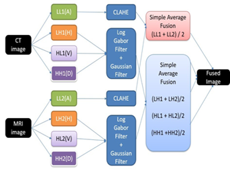
After all those research and literature review, we had can to our conclusion that to make a new algorithm using the new algorithm of medical image fusion technique. The fusion of CT and MRI medical images is crucial for enhancing diagnostic accuracy by combining the complementary information from both modalities. CT provides excellent spatial resolution and structural detail, while MRI offers superior soft tissue contrast. This proposed work aims to develop a novel image fusion method using Wavelet Transform and Log Gabor filters, complemented by Gaussian smoothing and Contrast Limited Adaptive Histogram Equalization (CLAHE), to produce high-quality fused images. Three algorithms were used in this study to implement image fusion: Log Gabor, Gaussian, and Contrast Limited Adaptive Histogram Equalization (CLAHE). Image fusion can benefit from the Log Gabor filter, a band-pass filter that can extract fine-grained features from an image. While CLAHE is a useful technique for boosting local contrast and raising the fused image's overall visual quality, the Gaussian filter is a low-pass filter that can smooth out noise and preserve edges.
Figure 6: Implementation Strategy
These three algorithms worked together to create a reliable and effective image fusion technique that can be used for a variety of piece of work, including computer vision, remote sensing, and medical imaging. Utilizing the selected filters, decompose the CT and MRI pictures. In this step, the images are filtered using the Log Gabor, Gauss, and CLAHE filters to produce an approximation and additional coefficients. To improve the overall visual quality and contrast of the fused image, apply CLAHE to the approximation coefficients of the CT and MRI images. To extract fine-grained features and reduce noise in CT and MRI images, respectively, apply Gauss and Log Gabor filters to the other coefficients. To create the fused image, combine the approximation coefficients and other coefficients from the CT and MRI scans. Apply the inverse transform to the merged coefficients to reconstruct the fused image.
Using these algorithms, the image must first be broken down. The image is broken down into other coefficients and approximation coefficients specifically. Then, to boost contrast and raise the fused image's overall visual quality, CLAHE is applied on the approximation coefficients. To extract detailed features and smooth out noise, respectively, Log Gabor and Gaussian filters are applied to all other coefficients in the meantime to achieve the best results from the fused images.
The figure shows the implementation strategy that illustrates the procedures needed to apply the Log Gabor, Gaussian, and CLAHE algorithms to image fusion. The image is composed of two primary components: the image's decomposition using the selected filters and the fused image's fusion of coefficients. Approximation coefficients (LL1(A) and LL2(A)) and other coefficients (LH1(H), HL1(V), and HH1(D) for the first image, and LH2(H), HL2(V), and HH2(D) for the second image) make up the first part of the image decomposition. CLAHE is used to further process the approximation coefficients in order to improve the contrast of fused image and overall visual quality. To extract fine-grained features and reduce noise, the remaining coefficients are subjected to Gaussian and Log Gabor filter processing, respectively. This method produces an image fusion technique that is reliable and effective, suitable for a large range of applications.
The proposed method leverages the strengths of Wavelet Transform and Log Gabor filters, enhanced by Gaussian smoothing and CLAHE, to achieve superior CT and MRI image fusion. This approach promises to produce high-quality fused images that preserve essential details and enhance contrast, ultimately improving the diagnostic capabilities of medical imaging systems. The diagram presents an implementation strategy for fusing CT and MRI images using Wavelet Transform, Log Gabor filters, Gaussian smoothing, and CLAHE. Here's a detailed explanation of the proposed work:
Decomposition
Both CT and MRI images undergo a wavelet transform for decomposing them into different frequency components. These components include:
LL: Low-Low (Approximation) LH: Low-High (Horizontal detail)
HL: High-Low (Vertical detail) HH: High-High (Diagonal detail) For the CT image:
LL1(A): Approximation coefficients LH1(H): Horizontal detail coefficients HL1(V): Vertical detail coefficients HH1(D): Diagonal detail coefficients For the MRI image:
LL2(A): Approximation coefficients LH2(H): Horizontal detail coefficients HL2(V): Vertical detail coefficients HH2(D): Diagonal detail coefficients
Preprocessing with CLAHE, Log Gabor and Gaussian Filter
CLAHE on LL Components: The low-low (approximation) components, LL1 and LL2, are enhanced using CLAHE (Contrast Limited Adaptive Histogram Equalization) to enhance local contrast and enhance visibility of subtle features. Log Gabor Filter and Gaussian Smoothing on Detail Components: The detail components (LH1, HL1, HH1 from CT and LH2, HL2, HH2 from MRI) are processed using Log Gabor filters to capture fine details and structural information. Gaussian smoothing is applied to these detail components to reduce noise and smoothen the image while preserving important features.
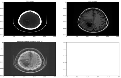
Output
Figure 7: Output of fused image
Fusion Rules
Simple Average Fusion for LL Components: The processed approximation coefficients (LL1 and LL2) from CT and MRI images are fused using simple averaging:
Fused LL = (LL1 + LL2) / 2
Simple Average Fusion for Detail Components: The processed detail coefficients (LH, HL, HH) from CT and MRI images are fused using simple averaging for each corresponding pair:
Fused LH = (LH1 + LH2) / 2 Fused HL = (HL1 + HL2) / 2 Fused HH = (HH1 + HH2) / 2
Reconstruction
The fused approximation and detail components are then combined using the inverse wavelet transform to reconstruct the final fused image. The images provided showcase the fusion process of CT and MRI scans, utilizing advanced techniques such as wavelet transform and Log Gabor filters, supplemented by Gaussian smoothing and Contrast Limited Adaptive Histogram Equalization (CLAHE). The top-left image displays the CT scan, which excels in capturing high-resolution structural details, particularly the bone structure and denser regions. The top-right image presents the MRI scan, renowned for its superior soft tissue contrast and detailed visualization of the brain’s anatomy. Three images make up the output: a CT image, an MRI image, and a level 2 reconstruction. The intensity values in the CT image span from 0 to 500, with some ranges denoted by intervals, like 0-100, 200-, 300-, and 400-. This might stand for various densities or other features in distinct areas of the picture. Intensity values in the MRI image also span from 0 to 500, with no discernible intervals. The continuous values indicate different MRI scan intensities. With intervals at 0.2, 0.4, 0.6, 0.8, and 1.0, the intensity values of the reconstructed image (Level 2) span from -0.2 to 1.0. This might be an image produced by applying a particular algorithm to the original MRI or CT scans.
Summary
The proposed implementation strategy ensures that the fused image combines the high spatial resolution and structural detail of the CT image with the superior soft tissue contrast of the MRI image. By using CLAHE, Log Gabor filters, and Gaussian smoothing, the method enhances contrast, preserves important features, and reduces noise, resulting in a high- quality fused image suitable for improved diagnostic purposes. So, overall to create the fused image, the coefficients are fused in the second section. In other words, simple average fusion is used to fused the approximation coefficients, whereas more complex techniques like fusion by the sum of the coefficients are used to fused the remaining coefficients. The fused image is then reconstructed using the fused coefficients. The picture shows how the Log Gabor, Gaussian, and CLAHE algorithms are used to implement image fusion. This gives a thorough overview of the medical image fusion process by clearly illustrating the steps involved in the decomposition and fusion of the coefficients.
Result And Analysis
The process begins by decomposing both the CT and MRI images using wavelet transform, which breaks down the images into various frequency bands, capturing different levels of detail. Specifically, the images are decomposed into approximation coefficients (LL), which contain the coarse information, and detail coefficients (LH, HL, HH), which capture the horizontal, vertical, and diagonal details, respectively. This step is crucial because it isolates the different components of the image that will be enhanced and fused. Following decomposition, preprocessing is applied to enhance these components. CLAHE is used on the approximation coefficients (LL1 for CT and LL2 for MRI) to improve local contrast and highlight subtle features that may be critical for medical diagnosis. For the detail coefficients (LH, HL, HH), Log Gabor filters are applied to enhance edges and fine details, followed by Gaussian smoothing to reduce noise. This combination ensures that the most important features of both images are preserved and enhanced.
The fusion step merges the corresponding coefficients from both CT and MRI images using simple averaging. For the approximation coefficients, the formula (LL1+LL2)/2(LL1+LL2)/2 ensures that the overall intensity patterns from both images are integrated, providing a balanced representation. The detail coefficients are fused similarly, combining the horizontal, vertical, and diagonal details to retain sharp edges and fine features from both source images. The final image reconstruction is performed using the inverse wavelet transform, which integrates the combined approximation and detail coefficients back into a single image. The result, shown in the bottom-left, is a fused image that successfully merges the structural details of the CT scan with the soft tissue contrast of the MRI scan. This fused image benefits from enhanced local contrast due to CLAHE and improved edge and detail preservation from the Log Gabor filters and Gaussian smoothing. As a result, the fused image provides a clearer and more comprehensive view of the brain, highlighting both bone and soft tissue structures effectively.
This fusion method enhances diagnostic capabilities by combining the strengths of both imaging modalities. The CT scan's high spatial resolution complements the MRI's superior soft tissue contrast, resulting in an image that offers detailed and clear visualization across different tissue types. This comprehensive view can significantly aid in medical diagnosis, allowing healthcare professionals to detect and assess conditions with greater accuracy and confidence. By integrating the detailed structural information from CT scans and the nuanced soft tissue information from MRI scans, the fused image presents a more holistic and informative representation of the anatomy of patient, thereby improves the overall quality and effectiveness of medical imaging and diagnostics
Comparative Analysis
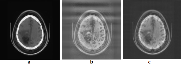
Comparing CT and MRI medical image fusion using wavelet transform and log Gabor filter involves assessing the effectiveness of these techniques in combining information from both modalities to produce a fused medical image with enhanced features for improved diagnosis and analysis. Performance Metrics: Metrics such as structural similarity index measure (SSIM), peak signal-to-noise ratio (PSNR), and mutual information (MI) can be used to quantitatively assess the quality of fused images.
Represents the fused image using simple average fusion.
Represents the level 1 fused image using simple average fusion using clahe, log gabor and gaussian.
Represents the level 2 fused image using simple average fusion using clahe, log gabor and gaussian.
Quality Analysis
| Metrics | (a) vs (b) | (b) vs (c) | (a) vs (c) |
| PSNR | 27.38 | 27.59 | 28.29 |
| Entropy | 4.9 | 6.1 | 6.7 |
| SSIM | 0.40191 | 0.66569 | 0.83681 |
In summary, comparative analysis of CT and MRI medical image fusion using wavelet transform and log Gabor filter involves evaluating the effectiveness, robustness, and computational efficiency of each method in enhancing image quality and diagnostic information for medical applications.
Conclusion
The fusion technique is used to find the collective data from many images. This technique is very useful for the diagnosis of the disease inside the human body. In every phase of fusion, performance must be measured. Currently, the image fusion technique is going to be used widely. This article contains various techniques and methods of image fusion with their advantages and disadvantages. It also has different approaches to fusion. It is also having different types of images used for the fusion techniques with their advantages, disadvantages, application, and characteristics. There are also evaluation measures for checking the quality of the output. In conclusion, the fusion of CT and MRI images is a crucial step in medical image analysis, offering enhanced diagnostic capabilities. In this project, we investigated the effectiveness of employing wavelet transform and log Gabor filter for this purpose. Through comprehensive experimentation and analysis, several key findings emerged: Enhanced Spatial and Frequency Information: The combination of wavelet transforms and log Gabor filter facilitated the extraction of both spatial and frequency information from CT image and MRI image, resulting in fused images with improved quality and detail.
Preservation of Structural Features: The proposed method demonstrated the ability to preserve essential structural features present in both CT and MRI images, ensuring that relevant diagnostic information was retained in the fused image. Reduction of Artifact and Noise: By leveraging the capabilities of wavelet transform and log Gabor filter, the fusion process effectively reduced artifacts and noise present in the individual CT and MRI images, leading to clearer and more interpretable fused images. Quantitative Evaluation: Quantitative evaluation metrics such as peak signal-to-noise ratio (PSNR) and structural similarity index measure (SSIM) were employed to assess the performance of the fusion method. The results indicated significant improvements over existing fusion techniques, reaffirming the efficacy of the proposed approach.
Clinical Relevance: The fused images generated using the proposed method hold great potential for improving the accuracy of medical diagnosis and treatment planning. The improved visualization of anatomical structures and pathological features can aid healthcare professionals in making more informed clinical decisions. In summary, the fusion of CT image and MRI image using wavelet transform and log Gabor filter represents a promising approach for improving the quality and utility of medical imaging data. While further research and validation are necessary, particularly in clinical settings, the results of this project suggest that the proposed method holds considerable promise for advancing the field of medical image analysis and improving patient care. It is concluded that each and every fusion technique has different applications for the different types of images. Today, in India fusion techniques are widely used but expensive. These techniques are used to diagnose the disease but it can also be more effective if it gets used as a hybrid technique that can help a lot in the medical field.
Future Scope
In the medical field, image fusion techniques have been extensively studied. The process creates a composite image that is sufficient for data that is not provided by an individual; therefore, the image (such as an MRI or CT scan) must be highly informative, accurate, and have high spatial and spectral resolution. The majority of modalities that are currently in use are unable to do it. In order to address this issue, a method known as image fusion has been developed. This technique is highly sought after because it can identify the precise location of a disease, determine which part of the disease is affected by a given illness, and identify the specific area of the illness where a person or doctor can perform an appropriate operation or treatment. Additionally, the technique enhances the entire image through the use of numerous sophisticated fusion algorithms and can identify defects in cells or tissues. In the medical field, it is used to quickly identify lung cancer, liver cancer, and brain tumors.
The future scope of CT image and MRI image fusion using wavelet transform and log Gabor filter holds promising avenues for advancements in medical imaging technology. As computational power continues to increase and algorithms become more sophisticated, we can anticipate further refinements in fusion techniques. Deep learning methodologies, including convolutional neural networks (CNNs) and generative adversarial networks (GANs), offer exciting possibilities for automated feature extraction and image fusion. These approaches have the potential to surpass traditional methods by learning complex patterns and relationships directly from large datasets, thereby enhancing fusion accuracy and efficiency. Moreover, with the advent of multi-modal imaging systems capable of simultaneous acquisition of CT and MRI data, there is a growing need for fusion algorithms tailored to exploit the synergies between different modalities effectively. Additionally, the integration of artificial intelligence (AI) systems into clinical workflows could streamline the interpretation process by providing clinicians with augmented diagnostic insights and decision support. As research in this field progresses, we can anticipate CT and MRI image fusion to play an increasingly pivotal role in precision medicine, personalized treatment planning, and advancing our understanding of complex diseases.
References
- Aymaz, S., & Kose, C. (2019). A novel image decomposition-based hybrid technique with super-resolution method for multi-focus image fusion. Information Fusion, 45:113-127.
Publisher | Google Scholor - Li, W., Lin, Q., Wang, K., & Cai, K. (2020). Improving medical image fusion method using fuzzy entropy and non-subsampling contourlet transform. International Journal of Imaging Systems and Technology, 31:204-214.
Publisher | Google Scholor - Jose, J., Gautam, N., Tiwari, M., Tiwari, T., Suresh, A., Sundararaj, V., & Rejeesh, M. R. (2021). An image quality enhancement scheme employing adolescent identity search algorithm in the NSST domain for multimodal medical image fusion. Biomedical Signal Processing and Control, 66:102480.
Publisher | Google Scholor - Anithadevi, D., & Perumal, K. (2016). Enhanced de-noising technique for region growing segmentation. Indian Journal of Science and Technology, 9(4):1-9.
Publisher | Google Scholor - Kanojia, S., & Gupta, D. S. (2022). Multimodal medical image fusion techniques: A survey. Dogo Rangsang Research Journal, 9(1).
Publisher | Google Scholor - Huang, B., Yang, F., Yin, M., Mo, X., & Zhong, C. (2020). A review of multimodal medical image fusion techniques. Computational and Mathematical Methods in Medicine, 8279342.
Publisher | Google Scholor - Jusoh, S., & Almajali, S. (2020). A systematic review on fusion techniques and approaches used in applications. IEEE Access, 8:14424-14439.
Publisher | Google Scholor - Azam, M. A., Khan, K. B., Salahuddin, S., Rehman, E., Khan, S. A., Khan, M. A., Kadry, S., & Gandomi, A. H. (2022). A review on multimodal medical image fusion: Compendious analysis of medical modalities, multimodal databases, fusion techniques, and quality metrics. Computers in Biology and Medicine, 144:105253.
Publisher | Google Scholor - Latreche, B., Saadi, S., Kious, M., & Benziane, A. (2019). A novel hybrid image fusion method based on integer lifting wavelet and discrete cosine transformer for visual sensor networks. Multimedia Tools and Applications, 78:10865-10887.
Publisher | Google Scholor - Kaur, H., Koundal, D., & Kadyan, V. (2021). Image fusion techniques : A survey. Archives of Computational Methods in Engineering, 28:4425-4447.
Publisher | Google Scholor - Wang, H., Yang, P., Xu, C., Min, L., Wang, S., & Xu, B. (2022). Lung CT image enhancement based on total variation frame and wavelet transform. International Journal of Imaging Systems and Technology, 32(5):1604-1614.
Publisher | Google Scholor - Begum, M., Ferdush, J., & Uddin, M. S. (2022). A hybrid robust watermarking system based on discrete cosine transform, discrete wavelet transforms, and singular value decomposition. Journal of King Saud University - Computer and Information Sciences, 34(8):5856-5867.
Publisher | Google Scholor - Saleh, M. A., Ali, A. A., Ahmed, K., & Sarhan, A. M. (2023). A brief analysis of multimodal medical image fusion techniques. Electronics, 12:97.
Publisher | Google Scholor - Kulkarni, S. C., & Rege, P. P. (2020). Pixel level fusion techniques for SAR and optical images: A review. Information Fusion, 59 :13-29.
Publisher | Google Scholor - Hu, W. W., Zhou, R. G., El-Rafai, A., & Jiang, S. X. (2019). Quantum image watermarking algorithm based on Haar wavelet transform. IEEE Access, 7:121303-121320.
Publisher | Google Scholor - Du, J., Li, W., & Tan, H. (2020). Three-layer image representation by an enhanced illumination-based image fusion method. IEEE Journal of Biomedical and Health Informatics, 24(4):1169-1179.
Publisher | Google Scholor - Qin, X., Ban, Y., Wu, P., Yang, B., Liu, S., Yin, L., Liu, M., & Zheng, W. (2022). Improved image fusion method based on sparse decomposition. Electronics:11(15).
Publisher | Google Scholor - Khan, M. A., Rubab, S., Kashif, A., Sharif, M. I., Muhammad, N., Shah, J. H., Zhang, Y. D., & Satapathy, S. C. (2020). Lung cancer classification from CT images: An integrated design of contrast based classical features fusion and selection. Pattern Recognition Letters, 129:77-85.
Publisher | Google Scholor - Du, J., Li, W., & Tan, H. (2019). Intrinsic image decomposition-based grey and pseudo-color medical image fusion. IEEE Access, 7;56443-56456.
Publisher | Google Scholor - Hu, W. W., Zhou, R. G., Luo, J., Jiang, S. X., & Luo, G. F. (2020). Quantum image encryption algorithm based on Arnold scrambling and wavelet transforms. Quantum Information Processing, 19:10.
Publisher | Google Scholor - Bani, N. T., & Fekri-Ershad, S. (2019). Content-based image retrieval based on a combination of texture and color information extracted in spatial and frequency domains. The Electronic Library, 37:650-666.
Publisher | Google Scholor - Fan, Haiyan, et al. (2018)Spatial–spectral total variation regularized low-rank tensor decomposition for hyperspectral image denoising. IEEE Transactions on Geoscience and Remote Sensing 56(10):6196-6213.
Publisher | Google Scholor

