

Case Report
Cattle Hydatidosis Occurrence, Cyst Characterization and Associated Risk Factors at Wolaita Soddo Municipality Abattoir, Southern Ethiopia
1Department of Veterinary Medicine, College of Agriculture, Woldia University, Ethiopia.
2Department of Veterinary Medicine, College of Veterinary Medicine, Samara University, Ethiopia.
*Corresponding Author: Tsedalu Yirsa, Department of Veterinary Medicine, College of Agriculture, Woldia University, Ethiopia.
Citation: Y. Tsedalu, M. Terefe, B. Abebe. (2024). Cattle Hydatidosis occurrence, cyst characterization and associated risk factors at Wolaita Soddo Municipality Abattoir, Southern Ethiopia. Journal of BioMed Research and Reports, BioRes Scientia Publishers. 5(1):1-9. DOI: 10.59657/2837-4681.brs.24.092
Copyright: © 2024 Tsedalu Yirsa, this is an open-access article distributed under the terms of the Creative Commons Attribution License, which permits unrestricted use, distribution, and reproduction in any medium, provided the original author and source are credited.
Received: June 03, 2024 | Accepted: June 21, 2024 | Published: June 28, 2024
Abstract
Background: Hydatidosis is a zoonotic infection of many mammalian species produced by Echinococcus granulosus larvae which are distributed globally specifically in Ethiopia.
Methods: A cross-sectional study was conducted from February 2023 to August 2023 to determine the occurrence of cattle hydatidosis, cyst characterization and associated risk factors at Wolaita Soddo Municipality Abattoir, Southern Ethiopia. The antemortem investigation was intended to identify some of the associated risk factors for hydatid cysts in the tested animals. A post-mortem examination and laboratory methods were also used to determine the presence and characteristics of hydatid cysts in various organs.
Results: A total of 15% (60/400) had harbored hydatid cysts. No related risk variables (P ≥ 0.05) were found in hydatic cysts. In terms of organ distribution, the lung (20.5%) had the highest frequency of these cysts, followed by the liver (15.25%), kidney (4.75%), and spleen (3.5%). Out of the 176 hydatic cysts that were counted, the small-sized cysts (47.7%) were more observed than medium-sized (29.5%) and large-sized (22.7%) cysts. Furthermore, of these, more fertile cysts (40.9%) than infertility ones (59.1%) were seen. However, out of 72 fertile cysts that were tested for viability, more non-viable cysts (41.7%) were observed than viable cysts (58.3%).
Conclusions: Overall, even with the small size of the infection detected, hydatidosis seems to be supported by both a socioeconomic setting and a public health concern. Because of this, it continues to rank among the most significant illnesses at the study site, requiring significant preventative and control measures.
Keywords: abattoir; cattle; hydatidosis; prevalence; wolaita sodo
Introduction
Hydatidosis is a zoonotic infection of various mammalian species caused by Echinococcus granulose (E. granulose) metacestodes, which is widespread worldwide, especially in developing countries such as Ethiopia [1,2]. The disease causes public health risks and major economic losses in Ethiopia as animal parts are condemned and milk production is reduced [3]. It also diminishes carcass weight, offal, and fertility in cattle [4]. There are four species of Echinococus including Echinococcus granuloses (E. granuloses), Echinococcus multilocularis (E. multilocularis), Echinococcus vogeli (E. vogeli), and Echinococcus oligathers (E. oligathers) [5]. Furthermore, several distinct strains of E. granuloses and E. multilocularis are recognized [6]. They can infect various intermediate hosts including livestock such as sheep, goats, cattle, camels, buffaloes, pigs and also humans [3]. Echinococcus is the smallest parasite (7 mm long) which usually does not have an intestine, and metabolism occurs in the syncytial outer shell of the body, the epidermis, two hook rows and muscle suction [7]. Its life cycle consists of a common definitive host, domestic dogs, and an intermediate host such as domestic ungulates and humans [8]. A dog has adult tapeworms in its small intestine and passes parasitic eggs in its feces, which are consumed by livestock and humans. After the egg has been consumed by the intermediate hosts, the oncosphere penetrates the wall of the small intestine and travels to various internal organs [7]. Finally, infected animals and humans develop hydatid cysts in their lungs, livers or other organs [9]. Man is an accidental ingestion of onchospheres from contaminated food, water and environments, whereas the dog is the commonest final host E.granulosus, which becomes infected by ingestion of infected offal [10].The Echinococcus cysts in the intermediate host are typically asymptomatic, except for a small number of cases with chronic and heavy infections [11]. The location and size of the cyst also affect how this infection manifests clinically. Early on in the infection, especially if the cyst is small, it might not cause any symptoms. As the illness worsens, symptoms like nausea, vomiting, hepatomegaly, and epigastric or upper quadrant stomach pain may occur [12]. The control method of the disease in dogs using praziquantel which is in the effective adult stage of E. granulosus while in the case of humans’ warrants surgical excision of cysts from the affected organ [13]. Hydatidosis is the most important zoonotic parasite illness, causing both direct and indirect economic losses in the livestock sector, particularly in cattle and sheep [14]. The diseases are located more in developing countries like Ethiopia, where the unhygienic conditions are coupled with poor cattle management practices and lack of meat inspection [15]. Furthermore, it is believed that the unsanitary environment, a high number of stray dogs, inadequate legislation regarding control, and inadequate meat inspection protocols all play a major role in the disease's prevalence in the nation, particularly in the study area [16]. Abattoir surveys have been used in several studies carried out throughout Ethiopia to ascertain the prevalence of bovine hydatidosis and the characteristics of its cysts [17,18]. The incidence of this disease ranged from 13.7 to 72.4% in cattle slaughtered at Dire Dawa, Gonder, Adama, and Asella [19,2-22]. Hydatidosis is the major cause of organ condemnation resulting in a huge economic loss in various parts of Ethiopia [1,5,9,15,22,23]. This demonstrates its critical importance, and the presence of this disease necessitates additional investigation in the country specifically in the study site. Despite its great economic and public health significance, there is a lack of new updated information about Bovine hydatidosis and its characteristics and associated risk factors at the study abattoir. As a result, it is critical to acquire information on the current status of the occurrence of cattle hydatidosis, cyst characteristics and associated risk factors with their appropriate preventative and control approaches. Thus, the purpose of this study was to assess the occurrence of hydatidosis, cyst characteristics and potential risk factors in slaughtered cattle at the Wolaita Soddo municipality abattoir in Southern Ethiopia.
Materials and Methods
Study setting, design and period
The study was carried out at Wolayta Soddo Municipal Abattoirs (WSMA), situated in the Southern Nations Nationality and People's Regional State. Geographically, Wolayta Zone, Wolayta Soddo town is positioned 383 km southwest of Addis Ababa. It is recognized as one of the certified towns in the region with Municipality status. The town is positioned 390 km south of Addis Ababa. It is located at a latitude of 8°50°N and a longitude of 37°45°E, with an altitude of 2025 meters above sea level. The study area experiences an average annual temperature of 20°C and receives rainfall ranging from 450 to 1446 mm. The total population of the area is approximately 1,581,650. The primary occupation of the rural population is mixed farming, involving the management of both crops and livestock. The livestock population in the area is estimated to include 68,900 cattle, 1992 sheep, 382 goats, 121 horses, 131 mules, 488 donkeys, and 55,191 chickens (24). It has also a population of 2,044,079, consisting of 1,040,710 females and 1,003,369 males, and covers an area of 4,471.3 square kilometres. The population density is 356.67, with 366,567 or 11.49 percentage being urban inhabitants, and 1,196 or 0.08 percentage being pluralists. There are a total of 310,454 households in the zone, averaging 4.84 persons per household, and 297,981 housing units (25).A cross-sectional abattoir study was conducted from February 2023 to August 2023 to assess the occurrence of Cattle Hydatidosis, cyst characterization and associated risk factors at WSMA, Southern Ethiopia.
Population and sample
The study involved cattle that were slaughtered at the Wolaita Sodo municipal abattoir. Most of the cattle slaughtered at this abattoir belonged to local or indigenous breeds. Apart from a small number of female animals, namely cows and heifers with reproductive challenges, the majority of the cattle slaughtered at the abattoir for the study were males. It was commonly understood that the majority of the slaughtered animals were sourced from locations like Arbaminch, Dawiro, Jinka, Goffa, and Borana, with many of them being brought in from the neighbouring woreda.
Sample size and sampling methods
The determination of the sample size was conducted by applying the formula provided in Thrusfield's (26) study, considering a previously expected prevalence of 40% (16). Additionally, a 5 percentage absolute precision and 95% confidence intervals were utilized in the calculation.
n= 
Where: n= required sample size; Pexp = expected prevalence d2= absolute precision.
Therefore, a total of 368 animals were initially required for the survey. However, to enhance the precision of the study, the sample size was increased to 400 based on case accessibility. Simple random sampling was employed to select animals slaughtered at the abattoir to determine the presence of cattle hydatidosis, cyst characteristics and associated risk factors. Cattle were chosen using a lottery method every three slaughtering days per week during the regular visitation period. The selection of the work area was purposefully done considering the distribution of animal’s slaughtered, animal availability, and agroecological zones. The interviewers managing the abattoirs were tasked with collecting
Data collection and laboratory methods
Ante mortem inspection
Regular visits were conducted to perform ante-mortem examinations on the animals designated for slaughter. A total of 400 cattle underwent examination at the abattoir throughout the study. The ante-mortem inspection involved assessing and documenting the origin and body condition score (BCS) of each animal (Annex 1). Animals were categorized as fat, medium, or lean based on their body condition, following the classification by Butterworth and Nicholson (1986). Additionally, the age of the animals was estimated by examining their dentition, as outlined by De Lahunta and Habel (27), and conventionally grouped into three categories: young (4 to 6 years), adult (7 to 9 years), and old (>10 years). Each animal was identified by specific marks on their body surface using ink. These procedures were carried out upon the animals' arrival in large groups at the abattoir, as described by Chambers and Herenda [28].
Post mortem examination
During the postmortem inspection, animals were chosen based on their identification numbers and underwent thorough examination through visualization, palpation, and systematic incision of each visceral organ, focusing on the liver, lung, spleen, and kidney. The diameter of each randomly selected cyst was measured in centimeters, and the number of cysts per organ was tallied and documented along with the respective organs. The selected cysts from various organs were gathered and brought to the laboratory for fertility and viability tests, with the results being recorded in the data specifically prepared for this purpose (Annex 3). The hydatid cysts were categorized based on size as small (less than 4cm), medium (4-8cm), and large (above 8cm in diameter) (28) (Annex 3).
Characterization of hydatid cysts
After collecting each hydatid cyst randomly from various organs, it was transported to the laboratory to conduct fertility and viability tests. The fluid within the hydatid cyst was extracted and examined under a microscope to identify protocolizes, while its viability was determined through an eosin exclusion test. The contents of the cyst were then transferred to a petri dish and assessed for cyst status under 40X magnification. If protocolizes were observed as white points on the germinal epithelium or brood capsules within the suspension, the cysts were classified as fertile. Conversely, if no protocolizes were present, the cysts were sterile [6]. The cysts that were gathered underwent viability staining using 0.1% eosin. This process involved placing a drop of sediment containing protocolizes on a microscopic glass slide. Subsequently, a drop of 0.1% eosin solution was introduced to the hydatid fluid on the slide, which was then covered with a cover slide and examined at 10x and 40x magnification. The principle behind this method is that viable protocolizes will repel the dye, while non-viable or deceased protocolizes will absorb it. As a result, the technique allows for the differentiation between dead (red-stained) and living (unstained) protocolizes [29].
Data analysis methods
The information gathered from the abattoir survey and physical assessment of hydatid cysts was inputted into Microsoft Excel spreadsheets and analyzed utilizing SPSS version 25. Following data input, the accuracy of the database was verified against the original documents. Additionally, descriptive statistics (frequencies and percentages) were computed. Furthermore, chi-square (X2) test analysis was employed to identify potential risk factors linked to hydatidiosis. The strength of these associations was determined by calculating odds ratios (OR) at a 95% confidence interval (CI), with a P-value ≤ 0.05 considered statistically significant.
Findings
Overall prevalence of hydatidosis and its potential associated risk factors
The overall prevalence of hydatidosis was estimated at 60 (15%) out of the 400 slaughtered cattle examined at the Wolaita Sodo municipal slaughterhouse. There was no associated significant (P ≥ 0.05) risk factor within the presence of hydatid cyst in the case of breeds, sex, age and body condition scores of slaughtered cattle in the study region (Table 1).
Table 1: Overall prevalence of hydatid cyst and its potential risk factors
| Risk factors | Attributes | N | N (%) | Chi-square (X2) | P-value |
| Age | Young | 31 | 4 (12.9) | 0.244 | 0.885 |
| Adult | 183 | 29 (15.8) | |||
| Old | 186 | 27 (14.5) | |||
| Sex | Male | 393 | 59 (15.0) | 0.003 | 0.957 |
| Female | 7 | 1 (14.3) | |||
| Breed | Local | 391 | 57 (14.6) | 3.996 | 0.136 |
| Exotic | 5 | 1 (0.2) | |||
| Cross | 4 | 2 (0.5) | |||
| BCs | Medium | 173 | 30 (17.3) | 1.600 | 0.449 |
| Fat | 154 | 19 (12.3) | |||
| Lean | 73 | 11 (15.1) | |||
| Total | 400 | 60 (15) |
NB: No: Number; BCS: Body condition scores; n: Total number of examined cattle; N: numbers of positive cattle
Distribution of hydatid cysts in different organs
The distribution of cysts in various organs among 60 positive cattle that were slaughtered was reported as follows: 18 (30%) had cysts exclusively in the liver, 15 (25%) in the lungs, 5 (8.33%) in the spleen, and 3 (5%) in the kidney. The remaining 41 (31.66%) infections were found to affect multiple organs (Figure 1).
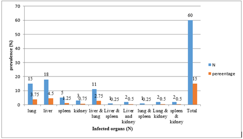
Figure 1: Distribution of hydatid cysts in different organs of infected cattle at WSMA.
Characterization of hydatid cysts
Among the 176 cysts analyzed for fertility, it was found that 32 (39.00%) cysts were from the lungs, 29 (47.5%) cysts from the liver, 7 (36.8%) cysts from the kidney, and 4 (28.6%) cysts from the spleen contained protoscolices, indicating their fertility. The rest of the cysts were determined to be infertile (Table 2).
Table 2: Distribution of hydatid cysts size and counts in different organs of infected cattle
| Organ affected | Occurrence | Small cyst (%) | Medium cyst (%) | Large cyst (%) | ||||
| N | % | N | % | N | % | N | % | |
| Lungs | 82 | 20.5 | 43 | 52.4% | 26 | 31.7% | 13 | 15.9% |
| Liver | 61 | 15.25 | 32 | 52.5% | 7 | 11.5% | 22 | 36.1% |
| Kidney | 19 | 4.75 | 6 | 31.6% | 11 | 57.9% | 2 | 10.5% |
| Kidney | 14 | 3.5 | 3 | 21.4% | 8 | 57.1% | 3 | 21.4% |
| Total | 176 | 44 | 84 | 47.7% | 52 | 29.5% | 40 | 22.7% |
NB: N: Number of slaughtered cattle infected; %: percentage
A total of 72 cysts from various organs such as the lung, liver, kidney, and spleen were assessed for viability. Out of these, 14 cysts from the lung, 10 from the liver, 5 from the kidney, and 1 from the spleen showed viable protoscolices with amoeboid-like peristaltic movement. When stained with 0.1% eosin solution, the viable protoscolices partially excluded the dye, while the dead ones absorbed it (Table 3).
Table 3: Characterization of cysts collected from different organs of infected cattle
| Organ involved | Fertile cyst N (%) | Sterile cyst N (%) | Viable cyst N (%) | Non-viable cyst N (%) | Total |
| Lung | 32 (39.0) | 50 (61.0) | 14 (43.8) | 18 (56.2) | 114 |
| Liver | 29 (47.5) | 32 (52.5) | 10 (34.5) | 19 (65.5) | 90 |
| Kidney | 7 (36.8) | 12 (63.2) | 5 (71.4) | 2 (28.6) | 26 |
| Spleen | 4 (28.6) | 10 (71.4) | 1 (25) | 3 (75) | 18 |
| Total | 72 (40.9) | 104 (59.1) | 30(41.7) | 42 (58.7) | 248 |
Discussion
The overall prevalence of hydatidosis in cattle slaughtered at the municipal abattoir of Wolaita Soddo is estimated to be 60 (15%). This result aligns with previous studies reporting rates of 13.9%- 24% from different regions of Ethiopia [1-3,7,16,30]. Conversely, it is lower than earlier findings, such as 33.3% in Yabello, 40.2% in Hawasa (16), 44.6% in Shashemene, 55.7% in Jimma, 96.6% in Hawsa (32) from various parts of Ethiopia. This study is also higher than the earlier reports of 5.6 % in Asella and 6.5% in Debre Berhane, 10.7 in Haramaya and 10.9 in Wolaita Sodo from Ethiopia [9,15,16,22,23,31,33,34]. The variations in the prevalence mentioned above in different regions of the country can be attributed to the differences in the types of Echinococcus found in different geographical locations, as well as differences in culture, social behaviour, and attitudes towards dogs in those areas. The occurrence of hydatidosis was lower compared to other regions due to the origin of the slaughtered animals, with the majority of cases coming from lowland areas near the woreda. The environmental conditions, such as high temperature and low humidity, were unfavourable for the survival of Echinococcus eggs, contributing to the lower prevalence. Thompson and Mc Manus suggested that environmental factors like humidity and temperature influence the natural selection of infective eggs from adult dog tapeworms [35]. On the other hand, the higher prevalence can be attributed to information obtained from the Soddo veterinary clinic and Soddo town municipality, which have implemented a program to control rabies and associated diseases by destroying stray dogs without legal owners in the study area every 2-3 years. This program carried out in previous years, has helped reduce the dog population and interrupt the life cycle of hydatidosis. Urquhart et al [36]. also argued that in areas without specific measures for hydatidosis control, the incidental benefit of destroying stray dogs for rabies control has a significant impact on reducing hydatidosis in both humans and animals. There is no statistically significant risk factors were identified for hydatidosis (P ≥ 0.05) in this study. [1,2,16,37]. This finding is consistent with previous studies and contradicts the earlier reports [2,7,8,15,34]. Nevertheless, animals were exposed to hydatidosis infection regardless of their body shape and condition, as people in the area preferred meat from fat animals, particularly for cutting meat consumption (‘’Kurt’’). The prevalence of hydatidosis tends to increase with the age of the animal (cattle) due to the practice of culling infected young cattle early by selling or slaughtering them before they reach old age, as older cattle have lower acquired immunity against hydatid infection. This is supported by the fact that Taeniid eggs cannot develop into metacestode in older animals [1,16,38]. Additionally, the number of ingested eggs by the intermediate host depends on the level of contamination and the infectivity of the eggs [39]. Regarding the size of hydatid cysts, out of the 176 cysts recovered from 60 cattle with hydatidosis in this study, it was more observed in small sizes (47.7%) than medium (29.5%), and large size cysts (22.7%). This finding is supported by the findings of Belachew et al [7]. and Kumsa [2]. However, Gautama and Pal and Kebede and Zekarias contradict these findings who stated that large and medium cyst cysts were more observed than the small size cyst of Bovine Hydatidosis harboring. This distribution may be attributed to the fact that a higher number of cysts are small in size due to the infected cattle being slaughtered before the cysts grow larger, and the limited immune response of the hosts to contain these cysts [8,23,40]. The intermediate hosts and the characteristic lesions of hydatidosis are fluid-filled cysts that develop slowly over several months, with large cysts often found in older animals [41]. The predilection sites of hydatid cysts in the internal organs of examining cattle were more observed in the lungs (20.5%) followed by the liver (15.25%), spleen (3.5%) and kidney (4.75%) in the study area. This suggests that the presence of cysts was more prominent in the lungs compared to the liver and other organs. This observation is consistent with previous studies [1-3,7-9,16,23]. The higher prevalence of calcified cysts in the liver may be due to the relatively higher reticuloendothelial cells and abundant connective tissue reaction of the organ [42]. Additionally, the lungs and liver serve as the primary capillary sites for migrating oncospheric echinococcus (hexane embryo) [17,43]. A total of 176 cysts were recovered from 60 cattle harboured by hydatidosis. Among these cysts, the percentage of sterile cysts (59.1%) was higher than fertile ones (40.9%). This sterile cyst was also more encountered in the spleen than in other internal organs in the study area. The findings of this study align with those of previous research [1,2,8,16,17]. Nevertheless, this finding is contradicted by earlier studies in Ethiopia which stated that fertile cysts are more observed than sterile cysts [7,15,23]. The reduced fertility of hydatid cysts observed in this research could be attributed to the younger age of the cattle under examination, as older animals tend to have a higher number of fertile cysts. Previous studies have indicated that fertile cysts take longer to develop in livestock [44]. This notion was further supported by Soulsby, who suggested that fertile cysts are more commonly found in older animals, while sterile cysts are prevalent in younger animals [41]. It is also known that cysts can be either fertile or sterile, with the ratio of sterile cysts varying among different host species: over 90% in cattle, 20% in pigs, and 8% in sheep [20]. However, the fertility rates of cysts may differ based on factors such as geographical location, host species, cyst site, size, and type [45]. Hydatidosis is a significant zoonotic disease with implications for public health and the economy in many countries worldwide. The prevalence of hydatidosis in a particular area may be influenced by various social, cultural, environmental, and epidemiological factors [43]. The limitations of this study included the security problem as well as constraints related to funding, equipment, and reagents.
Conclusion
A significant moderate rate of hydatidosis was observed in the study area. This finding suggests that the presence of these diseases has not been eliminated, which could potentially lead to public health problems and substantial economic loss in the study area. The economic loss in livestock can be attributed to the condemnation of organs and the denied weight gain of affected cattle. No significant potential risk factors were found to be associated with the harbouring of these cysts in this study. However, it was observed that small cysts frequently occurred in the liver, while large cysts were also found in the lungs. Upon characterization, the majority of the cysts were found to be sterile and non-viable. Most of these cysts were simultaneously localized in the liver and lungs of infected cattle. Based on these conclusions, the following recommendations are suggested: the construction of a routine abattoir in the township should be planned and executed, immediate attention should be given to the safe disposal of all condemned slaughterhouse materials and contaminated offal, and the ban on rear-yard killing should be enforced. Regular deworming of dwellings and stray dogs should be carried out, and public awareness regarding the zoonotic and economic significance of the disease should be increased. Additionally, awareness creation programs should be established for butchers, abattoir workers, and meat inspectors to ensure proper enforcement of these measures.
Abbreviations
AMI : Ante mortem inspection
BCS : Body condition score
M.a.s.l : Meter above sea level
PMI : Post mortem inspection
WSMA : Wolaita soddo municipality abattoir
WZFEDD : Wolaita zone Finance and Economic Development department
Declarations
Ethics approval and consent to study participate
All necessary measures were taken to minimize any suffering experienced by the animals involved in this study. It is important to note that there were no known risks or discomforts associated with the collection of specimens from the study animals.
Consent for publication
Not applicable
Conflict of interest
The authors declare no conflict of interest exist
Funding
No funding was received for this study.
Authors’ contribution
TY: Conceptualizations of the study, Methodology, validation, Statistical analysis coordinates data collection.
TM: Data collection and Laboratory activities; and AB performed the statistical analysis, software, and supervision. The author(s) read and approved the manuscript.
Data availability statement
The data used for analysis is fully available in the manuscript file without restriction.
Acknowledgements
We acknowledge Samara University and Wolaita Sodo University, Wolaita Soddo municipality abattoirs for their support of reagents and materials to accomplish this work.
References
- Guduro GGaD, A.H. (2019). Cyst Viability and Economic Significance of Hydatidosis in Southern Ethiopia. J Parasitol Res, 7.
Publisher | Google Scholor - Kumsa B. (2019). Cystic echinococcosis in slaughtered cattle at Addis Ababa Abattoir enterprise, Ethiopia. Veterinary and Animal Science, 7:100050.
Publisher | Google Scholor - Mesfin Mathewos DD, Metages Yirgalem, Tesfaye Denano. (2022). Haben Fesseha Cystic echinococcosis in cattle slaughtered at a slaughterhouse in Gessa, southern Ethiopia. Parasite Epidemiology and Control, 1818(e00262):8.
Publisher | Google Scholor - Sariozkan SaY, C. (2009). Estimating the production losses due to cystic echinococcosis in ruminants in Turkey. Vet Parasitol, 163:330-334.
Publisher | Google Scholor - Mekuriya MEM. (2023). Prevalence, Organ Distribution, and Economic Importance of Bovine Hydatidosis in Gimbichu Municipal Abattoir, Hadiya Zone. Ethiopia Journal of Innovations in Medical Research. 2(6):55-60.
Publisher | Google Scholor - FAO. (2023). Diagnostic manual on meat inspection for developing counters. FAOUN. Rome, 160-164.
Publisher | Google Scholor - Belachew T AM, Gunse T. (2019). Bovine Hydatid Cyst: Prevalence, Characterization, Public Health and Economic Importance at Adama Abattoir, Central Ethiopia. Int J Vet Sci Res, 5(1):14-18.
Publisher | Google Scholor - Pal KPGaM. (2021). Prevalence, Fertility, and Viability of Cystic Echinococcosis in Cattle Slaughtered at Adaba Abattoir, West Arsi Zone, Ethiopia. Int J Med Parasitol Epidemiol Sci, 2(1):3-9.
Publisher | Google Scholor - Asefa HFaI. (2022). Co-infection of fasciolosis and hydatidosis and their financial loss in cattle slaughtered at Wolaita Sodo municipal abattoir. southern Ethiopia Animal Diseases. 2-27.
Publisher | Google Scholor - Biniamin T.AH. (2018).The Prevalence of Cystic Echinococcosis in Cattle Slaughtered in Sebeta Municipal Abattoir, Central Ethiopia. Biomed J Sci&Tech Res, 6(1):4955-4959.
Publisher | Google Scholor - Fufa Abunna SF, Bekele Megersa, Alemayehu Regassa. (2012). Prevalence of bovine hydatidosis in Kombolcha ELFORA abattoir, North Eastern Ethiopia. Open Journal of Animal Sciences, 2:281-286.
Publisher | Google Scholor - Moro PaS, P.M. (2009). Echinococcosis: a review. International Journal of Infectious Diseases, 13 (2):125-133.
Publisher | Google Scholor - Mahendra Pal NZ. (2020). Tefera Woldemariam and Gemechu Berhanu. Prevalence of Cystic Echinococcosis in Various Food Animals Slaughtered at Selected Abattoirs in Ethiopia Veterinary Research International. 8(3):118-123.
Publisher | Google Scholor - Jobrey LF, Trune, Abebe, G. and Dorchies, P. (1996). Hydatidosis in three selected regions in Ethiopia: An assessment trial on its prevalence, economic and public health importance. Rev Med Vet, 147.
Publisher | Google Scholor - Tariku Beyene AH. (2019). Zoonotic metacestodes and associated financial loss from cattle slaughtered at Yabello municipal abattoir, Borana-Oromia, Ethiopia. Parasite Epidemiology and Control, 3:e00096.
Publisher | Google Scholor - Yohannes GaM, S. (2019). Study on the Prevalence and Associated Risk Factors of Bovine Hydatidosis in Hawassa Municipal Abattoir, Hawassa, Ethiopia. Gen Surg, 1(3):1012.
Publisher | Google Scholor - Kebede N, Mekonnen, H., Wossene, A., Tilahun, G. (2009). Hydatidosis of slaughtered animals in wollaita soddo Abattoir, Southern Ethiopia. Trop Anim Health and prod, 41(4):629-33.
Publisher | Google Scholor - Regassa A, Abunna, F., Mulugeta, A. and Megersa, B. (2009). Major metacestodes in cattle slaughtered at Wolaita Soddo Municipal abattoir, Southern Ethiopia: Prevalence, cyst viability, organ distribution and socioeconomic implications. Trop Anim Health Prod.
Publisher | Google Scholor - Abebe FaY, J. (2011). Infection prevalence of hydatidosis (Echinococcusgranulosus, Batsch, 1786) in domestic animals in Ethiopia, A synthesis report of previous surveys, JUCAVM, Jimma, Ethiopia. Vet J, 15:11-33.
Publisher | Google Scholor - Jobre Y, Labago, F., Tirone, G., Abebe, G. and Dorchies, P. Hydatidosis in three selected sites in Ethiopia.An assessment trial on its prevalence, economic and public health significance. H Rev Med Vet,147:797-804.
Publisher | Google Scholor - Kebede N, Mitiku, A. and Tilahun, G. (2010). Retrospective survey of human hydatidosis in Bahir Dar, North-Western Ethiopia. East Mediterr Health J, 16:937-941.
Publisher | Google Scholor - Mohammed Abrahim Ahmed CAaAM. (2024). A Study on Prevalence and Economic Significance of Bovine Hydatidosis in Haramaya Muncpial Abattoir. vet Med Animal Sci, 7(1):1135.
Publisher | Google Scholor - Zekarias TKaT. (2020). Prevalence and Financial Loss Estimation of Hydatidosis of Cattle Slaughtered at Shashemene Municipal Abattoir, South Central Oromia, Ethiopia European Journal of Biological Sciences, 12(2):54-63.
Publisher | Google Scholor - WZFEDD. (2012). Wolaita Zone Socio-Economic information.Wolaita Zone Finance and Economic Development Department, Unpublished.
Publisher | Google Scholor - CSA. (2020). Federal democratic republic of Ethiopia Ethiopian Statistics Service Agricultural Sample Survey 2020 [2013 E.C.] Volume II report on livestock and livestock characteristics (private peasant holdings), Addis Ababa, Ethiopia.
Publisher | Google Scholor - Thrusfield BM. (2005). Veterinary epidemiology. Black Well Science Ltd, U.K.
Publisher | Google Scholor - DeLahunta AaH, R. E. (1986). Teeth.Applied Veterinary Anatomy.WBSaunerrs Xompany, USA.
Publisher | Google Scholor - Herenda DaC, P.G. (1994). Manual in meat inspection for developing countries. 1994.
Publisher | Google Scholor - Dalimi A, Motamedi, G.H and Hosseini, M. (2002). chinococcosis/hydatidosis in western Iran. E Vet Parasitol, 105:161-171.
Publisher | Google Scholor - Aschalew Tadesse BA, Ashenafi Asefa and Belete Haile. (2016).Prevalence and Economic Significance of Bovine Cystic Echinococcosis in Debra Tabor Municipal Abattoir, North West Ethiopia. Acta Parasitologica Globalis, 7(3):114-120.
Publisher | Google Scholor - Ayub Temam BDMA. (2016). Study on Prevalence and Monetary Loss Attributed to Hydatidosis in Cattle Slaughtered at Jimma Municipal Abattoir. Southwestern Ethiopia 16, 2:9.
Publisher | Google Scholor - Alemu TZaB. (2017). Major Gross Lesions of Lung in Cattle Slaughtered at Hawassa Municipal Abattoir, Southern Ethiopia. Journal of Veterinary Medicine, 2-7.
Publisher | Google Scholor - Jemalo A HG, Furgasa W. (2018). Major Causes of Organ Condemnation and Their Economic Loss in Beef Cattle Slaughtered at Assella Municipal Abattoir. J Vet Sci Ani Husb, 6(2):208.
Publisher | Google Scholor - Akeberegn D AT, Kassa T. (2017). The Prevalence of Bovine Hydatidosis among Slaughtered Cattle at Debre Berhan Municipal Abattoir, North Shewa Zone, Ethiopia. J Veter Sci Med, 5(1):5.
Publisher | Google Scholor - Thompson RCaM, D.P. (2002). Towards a taxonomic revision of the genus Echinococcus..18: 452-5. Trends Parasitol. 18:452-425.
Publisher | Google Scholor - Urquhart GM, Armour, J., Duncan, J.L., Dunn, A.M. and Jennings, F.W. (1996). Veterinary parasitology, 2nd ed. Blackwell Science, United Kingdom, 07.
Publisher | Google Scholor - Asfaw AaA, B. (2014). Prevalence of Hydatid Cyst in Cattle at Municipal Abbatoir of Shire. J Vet Sci Technol, 5:186.
Publisher | Google Scholor - Gemmell MA, Roberts, M. G., Beard, T. C., Campano, D. S., Lawson, J. R. and Nonnemarker, J. M. (2001). Control of Echinococcosis in WHO/OIE Manual on Echinococcosis in Humans and Animals: a Public Health Problem of Global Concern. World Organization for Animal Health (Office International des Epizooties), Paris and World Health Organization, Geneva.
Publisher | Google Scholor - Thompson RCaA, C.E. (1988). Hydatidosis, veterinary perspective and annotated. Bibliography C.A.B international, 1-16.
Publisher | Google Scholor - Dawit G, Adem, A., Simenew, K. and Tilahun, Z. (2013). Prevalence, cyst characterization and economic importance of bovine hydatidosis in Mekelle municipality abattoir, northern Ethiopia. J Vet Med and Animal Health, 5:81-86.
Publisher | Google Scholor - Soulsby EJL. (1982). Helminths, arthropods and protozoa of domesticated animals 7th ed. English Language Book Society/ BilliereTindall, 123-127.
Publisher | Google Scholor - Torgerson P. (2002). Transmission dynamics of taeniid parasites in animal hosts, 221-235.
Publisher | Google Scholor - Macpherson CN. (1985). Epidemiology of hydatid disease in Kenya: a study of domesticated intermediate hosts in Masai land”. Trans R soc Trop Med Hyg, 79:193-195.
Publisher | Google Scholor - OIE. (2008). Hydatidosis 5thedition. Officeinterna des- Ephizotices, Paris. Pp. 175-185.
Publisher | Google Scholor - Ibrahim MM. (2010). Study of cystic echinococcosis in slaughtered animals in Al Baha region, Saudi Arabia: Interaction between some biotic and abiotic factors. Acta Tropical. 113:26-33.
Publisher | Google Scholor


