Case Report
Ascariasis: A Common Disease with Uncommon Presentation in A Resource Limited Setting: A Case Report
1Department of Radiology, Menelik II comprehensive specialized Hospital, Addis Ababa, Ethiopia.
2Department of Radiology, College of Health Science, Mizan Tepi University, Mizan, Ethiopia.
3Department of Radiology, School of Medicine, Addis Ababa University, Ethiopia.
4University of Texas Medical Branch, Galveston, TX, USA.
*Corresponding Author: Messay Gebrekidan, Department of Radiology, Menelik II comprehensive specialized Hospital, Addis Ababa, Ethiopia.
Citation: M. Gebrekidan, Shimalis T. Fayisa, Samuel S. Hailu, Abel T. Abebe. (2024). Ascariasis: A Common Disease with Uncommon Presentation in A Resource Limited Setting: A Case Report. Clinical Case Reports and Studies, BioRes Scientia Publishers. 5(1):1-2. DOI: 10.59657/2837-2565.brs.24.096
Copyright: © 2024 Messay Gebrekidan, this is an open-access article distributed under the terms of the Creative Commons Attribution License, which permits unrestricted use, distribution, and reproduction in any medium, provided the original author and source are credited.
Received: December 19, 2023 | Accepted: January 09, 2024 | Published: January 19, 2024
Abstract
Ascaris-induced intestinal obstruction is a rare complication primarily seen in children in areas with a high prevalence of worm infestations. It can occur through two mechanisms: immune-mediated reactions releasing neurotoxins that cause contractions and inflammation in the small intestine (aperistalsis), or mechanical obstruction by adult worms, commonly at the ileocecal valve. Partial obstructions are managed conservatively, while complete obstructions often require surgical intervention. In a recent case, a 19-year-old male presented with persistent abdominal pain, vomiting, and inability to pass stools and gas. Imaging revealed partial obstruction, and conservative management with fluids, a nasogastric tube, and antibiotics led to the spontaneous passage of worms, relieving symptoms. The patient was discharged with anthelmintics and advised on follow-up and sanitary measures. This case is notable for the uncommon occurrence of Ascaris-induced intestinal obstruction in adults and the successful conservative management resulting in early worm expulsion.
Keywords: ascaris lumbricoides; bowel obstruction; x-ray; ultrasound
Introduction
Ascariasis, which is caused by the nematode Ascaris lumbricoides, is the most prevalent and largest helminth infection in humans worldwide [1]. In regions endemic to Ascariasis, clinical manifestations predominantly arise in individuals with high worm burdens and those who experience frequent infections, despite the fact that the majority of infections are asymptomatic. It presented often with non-specific abdominal symptoms, such as abdominal pain, distention, nausea, and occasional diarrhea [2]. Intestinal obstruction caused by Ascaris is a rare complication, primarily observed in children with a high worm burden. It is exceptionally rare in adults [3]. The obstruction occurs when a mass of aggregated worms obstructs the bowel, particularly in the terminal ileum near the ileocecal valve. This obstruction is usually partial, but in some cases, it can progress to complete obstruction, requiring surgical intervention to treat acute intestinal obstruction [4].
Case presentation
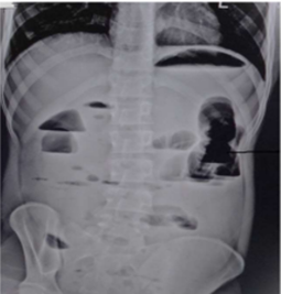
A 19-year-old male patient arrived to emergency department with sudden exacerbation of crampy abdominal pain, inability to have a bowel movement for two days, and vomiting. The patient mentioned experiencing long-standing abdominal pain before the acute episode. And have seen motile worms in his feces 3 days prior. He has no history of previous surgery. During the physical examination, the patient appeared acutely ill, but vital signs were within normal ranges. The chest examination revealed clear and resonant sounds, while the heart examination showed no abnormalities. Upon examining the abdomen, it was found to be distended with active bowel sounds, but there were no signs of a fluid thrill or shifting dullness. Hernial sites are free. Laboratory tests, specifically a complete blood count (CBC), showed leukocytosis with a predominance of neutrophils. Diagnostic imaging was performed, including an erect abdominal film, which revealed centrally distended bowel loops with multiple air-fluid levels and focal distension of the large bowel. Small rectal gas shadow also seen. The pattern of obstruction was indistinct. No abnormalities in the soft tissues were observed (Figure 1).
Figure 1: Plain abdominal X ray showing central abdomen multiple air fluid levels, with small rectal gas shadow.
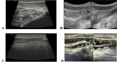
An ultrasound examination showed distended small bowel loops, primarily involving the ileal loops, along with a stranded mesentery. Multiple linear curly hypoechoic lesions with peripheral echogenic walls were also seen. These findings were consistent with worm impaction, most likely due to Ascaris lumbricoides. Some worms were observed to be moving, while others appeared sluggish near the ileocecal valve, which explained the patient's clinical and radiological presentation. No significant free peritoneal fluid collection was noted, and the appendix had normal wall thickness and compressible. All solid organs appeared normal (Figures 2).
Figure 2: (A & B), partially distended bowel loops with stranded mesentery and omentum are visible. Two worms can be observed within the lumen at the level of the Ileocecal valve. There is no significant presence of peritoneal fluid (A, D). The image also depicts a gaseous abdomen in most of the abdominal regions (C).
The patient's obstructive symptoms were effectively managed through conservative measures, including intravenous fluids for resuscitation, intravenous antibiotics, and a nasogastric tube. Concurrently, an evaluation was conducted to assess the potential need for surgical intervention. During the patient's hospital stay, a single bowel movement occurred, which revealed the presence of numerous worms. Subsequently, within a span of six hours, the patient experienced three additional bowel movements, all of which contained active and sizable worms. Notably, there was an improvement in the patient's condition, with a decrease in abdominal pain and distension.
Conservative management was continued, as the patient exhibited clinical improvement, rendering surgical intervention unnecessary. By the third day of admission, the abdominal pain had subsided, and there was no longer any abdominal distension. An abdominal X-ray confirmed the absence of an air-fluid level. Consequently, the patient was discharged, having received appropriate anti-helminth treatment. During the six-week follow-up, the patient's condition was found to be satisfactory, and were provided with guidance on essential sanitary practices and regular deworming.
Discussion
Ascariasis remains a significant global health issue, primarily affecting children in tropical regions and low- to middle-income countries [5]. The life cycle of Ascariasis begins when ova are ingested through fecal-oral transmission. These ova hatch into larvae in the intestine, which then penetrate the mucosa and enter the portal circulation. The larvae migrate to the lungs, where they mature over approximately two weeks. From the lungs, they travel to the bronchi and eventually reach the oropharynx and swallowed again. Finally, the adult worms establish themselves in the small intestine, where they lay eggs that are excreted in the feces [6].
Around 10 to 14 days after infection, individuals may develop a condition called Loeffler syndrome, which is characterized by eosinophilic pneumonia caused by the inflammatory response to larvae migrating through the pulmonary tissue [7]. Adult Ascaris worms can also cause various acute abdominal conditions, including small bowel obstruction, upper gastrointestinal bleeding, intussusception, volvulus, intestinal perforations with peritonitis, and hepatobiliary syndromes such as acute cholecystitis, acute cholangitis, biliary colic, hepatic abscess, and acute pancreatitis. [8] While many infections are asymptomatic, some individuals may experience symptoms such as diarrhea, loss of appetite, weakness, stomach pain, changes in bowel habits, weight loss, and in rare cases, the expulsion of worms through the mouth or rectum. Severe cases can result in intestinal blockages characterized by abdominal bloating, the presence of an abdominal mass, tenderness, or pain [9].
The causes of Ascariasis-related intestinal obstructions can be attributed to the formation of large worm masses that mechanically obstruct the intestine, worm masses acting as lead points for intussusception, or the release of neurotoxins causing contractions and inflammation in the small intestine, leading to obstruction [9]. Along with clinical signs and symptoms, imaging techniques play a vital role in diagnosing Ascariasis-related intestinal obstruction. Abdominal X-rays may reveal multiple linear images of Ascaris lumbricoides within dilated intestinal loops, sometimes forming a "whirlpool" pattern, which helps confirm the diagnosis based on the patient's medical history and symptoms of intestinal obstruction [10]. Barium examinations show elongated, smooth, cylindrical filling defects within the intestine. while ultrasound examinations can detect thick echogenic strips with a central anechoic tube, linear or curved echogenic strips without acoustic shadowing, and characteristic appearances such as "three-line" or "four-line" signs, "bull's eye" or "railway track" signs on transverse scans. Although abdominal computed tomography (CT) is not the preferred diagnostic method for ascariasis, it can occasionally reveal the presence of worms within the intestinal lumen [13]. In this patient after he is present with sign and symptoms of bowel obstruction plain abdominal x-ray finding is consistent with bowel obstruction. A patient reported spontaneous passage of worms 2 days prior to exacerbation ultrasound done and shown bunch of ascaris impacted in ileum.
The management of Ascariasis-related intestinal obstruction involves initial resuscitation and antimicrobial therapy for patients with partial intestinal blockage. Medical interventions such as nasogastric drainage, broad-spectrum antibiotics, and intravenous fluids and electrolytes are used in these cases. However, patients with complete obstruction require surgical intervention after initial resuscitation and antimicrobial therapy [11]. The recommended anthelmintic drugs are mebendazole or albendazole, given at a dosage of 100 mg twice daily for 3 days, with a repeat dose administered 6 weeks later. Resolution of the condition is defined by the occurrence of any two of the following criteria: initiation of defecation, relief of colicky discomfort, and disappearance of air-fluid levels [12].
The patient's treatment began with conservative management, which included intravenous fluids, a nasogastric tube, and nil per oral (NPO) status. This was done while the patient was being assessed for possible surgical intervention. During this period, the patient reported the spontaneous passage of multiple worms in the bathroom. Subsequently, the patient experienced the passage of multiple loops of worms on three separate occasions within a six-hour timeframe while using the bathroom. As a result, the patient's colicky abdominal pain improved, and they started passing flatus accompanied by small feces. Conservative management was continued, and a follow-up X-ray revealed a decrease in the air-fluid level. After three days of conservative management with no signs of obstruction, the patient was discharged. Anthelmintic therapy was administered to address any remaining worms and prevent the formation of worm masses and spastic paralysis. During a follow-up visit after six weeks, the patient's condition was found to be satisfactory. Emphasis was placed on the importance of frequent deworming and maintaining proper sanitation practices to prevent a recurrence of the same condition in the future.
Ascaris lumbricoides presents a significant public health concern, particularly in countries like Ethiopia, and primarily affects school-age children and young adults. While most infections are asymptomatic, they can still result in considerable morbidity and, on rare occasions, lead to intestinal obstruction. Therefore, in tropical regions where ascariasis is endemic, healthcare providers should consider Ascaris as a potential cause of intestinal obstruction. Although more commonly observed in children, adults are also at risk, especially in areas with a high worm burden and lacking organized deworming programs. Imaging techniques such as x-rays and ultrasound play a crucial role in diagnosing and visualizing impacted worms within the intestinal lumen. Once diagnosed, a proper evaluation is necessary to determine the appropriate course of treatment, whether it be surgical intervention or conservative management. Implementing improvements in sanitation, health education, and implementing proper deworming programs in endemic areas can effectively reduce parasite load and prevent serious, life-threatening complications caused by A. lumbricoides.
Declarations
Data Availability Statement
The corresponding author can provide the datasets used in this study upon a reasonable request.
Ethical Considerations
We obtained written consent from the patient to publish this case report and its associated images.
Acknowledgment
We would like to express our gratitude to the patient for granting permission to publish this case report.
Author Contributions
All authors have made substantial contributions to the conception, design, execution, data acquisition, analysis, and interpretation of the study. They actively participated in drafting, revising, and critically reviewing the article. Furthermore, all authors have given their final approval for the version to be published and have reached a consensus on the journal where the article has been submitted. They also take full responsibility for all aspects of the work.
Funding
No specific grants from public, private, or nonprofit funding organizations were received for this work.
Disclosure
The authors have no conflicts of interest to disclose regarding this work.
References
- Asai T, Còrdova Vidal C, Strauss W, Ikoma T, Endoh K, Yamamoto M. (2016). Effect of Mass Stool Examination and Mass Treatment for Decreasing Intestinal Helminth and Protozoan Infection Rates in Bolivian Children: A Cross-Sectional Study. PLoS Negl Trop Dis., 10(12).
Publisher | Google Scholor - Yetim I, Ozkan O, Semerci E, Abanoz R. (2009). Rare Cause of Intestinal Obstruction, Ascaris Lumbricoides Infestation: Two Case Reports. Cases J. 2(1):7970.
Publisher | Google Scholor - Steinberg R., Davies J., Millar A.J., Brown R.A., Rode H. (2003). Unusual intestinal sequelae after operations for Ascaris lumbricoides infestation. Pediatr. Surg. Int., 19(1-2):85-87.
Publisher | Google Scholor - Hefny A, Saadeldin Y, Abu-Zidan F. (2009). Management Algorithm for Intestinal Obstruction Due to Ascariasis: A Case Report and Review of the Literature. Ulus Travma Acil Cerrahi Derg., 15(3):301-305
Publisher | Google Scholor - Fata C, Naeem F, Barthel E. Small Bowel Obstruction Secondary to Ascaris Lumbricoides in the Setting of Prior Exploratory Laparotomy. Journal of Pediatric Surgery Case Reports, 47:101254.
Publisher | Google Scholor - Pullan R.L., Smith J.L., Jasrasaria R., Brooker S.J. (2014). Global numbers of infection and disease burden of soil transmitted helminth infections in 2010. Parasit. Vectors, 7(1):37.
Publisher | Google Scholor - Lal C., Huggins J.T., Sahn S.A. (2013). Parasitic diseases of the pleura. Am J Med Sci, 345:385-389.
Publisher | Google Scholor - Jourdan P.M., Lamberton P.H.L., Fenwick A., Addiss D.G. (2018). Soil-transmitted helminth infections. Lancet, 391:252-265.
Publisher | Google Scholor - Stojanovic M., Slavkovic A., Stojanovic M., Marjanovic Z., Bojanovic M. A rare case of intestinal obstruction due to ascariasis in Niš, South Serbia. Open Med, 6:390-394.
Publisher | Google Scholor - Bhatt P.N. (2017). Acute intestinal obstruction due to ascaris. Med. Phoenix, 1:39-40.
Publisher | Google Scholor - Ali A.Y., Mohamed Abdi A., Mambet E. (2003). Small bowel obstruction caused by massive ascariasis: two case reports. Annals of Medicine and Surgery, 85(3).
Publisher | Google Scholor - Andrade A.M., Perez Y., Lopez C., Collazos S.S., Andrade A.M., Ramirez G.O., Andrade L.M. (2015). Intestinal obstruction in a 3-year-old girl by Ascaris lumbricoides infestation: case report and review of the literature. Medicine (Baltimore), 94.
Publisher | Google Scholor - Das C.J., Kumar J., Debnath J., Chaudhry A. (2007). Imaging of ascariasis. Australas. Radiol., 51(6):500-506.
Publisher | Google Scholor


