Research Article
Appendiceal Endosalpingiosis: Rare Discovery Found During an Appendectomy
- Ines Mallek *
- Rahma Ayadi
- Rahma Yaiche
- Emna Brahem
- Olfa Ismail
- Aïda Ayadi
Department of Pathology, Abderrahmen Mami Hospital, 2083, Tunis, Tunisia.
*Corresponding Author: Ines Mallek, Department of Pathology, Abderrahmen Mami Hospital, 2083, Tunis, Tunisia.
Citation: Mallek I., Ayadi R., Yaiche R., Brahem E., Ismail O., et al. (2024). Appendiceal Endosalpingiosis: Rare Discovery Found During an Appendectomy, International Journal of Biomedical and Clinical Research, BioRes Scientia Publishers. 3(1):1-4. DOI: 10.59657/2997-6103.brs.25.037
Copyright: © 2024 Ines Mallek, this is an open-access article distributed under the terms of the Creative Commons Attribution License, which permits unrestricted use, distribution, and reproduction in any medium, provided the original author and source are credited.
Received: November 08, 2024 | Accepted: December 26, 2024 | Published: January 02, 2025
Abstract
Introduction: Endosalpingiosis is when fallopian tube-like tissue is found outside the pelvis. It's rare, primarily affects older individuals, and increases the risk of certain gynecological cancers. There have been only five reported cases of it occurring in the appendix. This summary presents one such case, confirmed through pathology, and reviews existing literature on the topic.
Methods and Results: A 45-year-old male presented with persistent right lower quadrant abdominal pain, initially suspected to be appendicitis. Ultrasound imaging showed mild nonspecific inflammation, while MRI highlighted unusual features in the appendix, prompting laparoscopic appendectomy. Histopathology confirmed appendiceal endosalpingiosis, a rare finding. A literature review was conducted to compare this case with existing reports of appendiceal endosalpingiosis.
Discussion: Pelvic endosalpingiosis, identified mostly in women post-pelvic surgeries, involves benign glandular tissue in the peritoneum. Appendiceal endosalpingiosis, much rarer, shares similarities with endosalpingiosis in other pelvic organs. It's distinct from endometriosis, more common in premenopausal women. While some studies suggest a link between pelvic endosalpingiosis and chronic pelvic pain, our cases presented with right lower quadrant pain. There's an association between endosalpingiosis and gynecologic cancers, especially ovarian serous borderline tumors, but further research is needed due to conflicting findings. The spread mechanism is unclear, but theories include inflammation or a metaplastic process. Imaging rarely aids diagnosis, but MRI can reveal characteristic features. Pelvic endosalpingiosis is linked to an increased risk of gynecological malignancies, particularly ovarian serous tumors of low malignant potential.
Conclusion: In conclusion, this report presents a rare case of appendiceal endosalpingiosis resembling appendicitis. Its link to gynecologic cancers raises questions about the need for further investigation. More research is crucial to understand its association with ovarian cancers.
Keywords: endosalpingiosis; gynecologic cancers; appendectomy
Introduction
Endosalpingiosis is characterized by the presence of ectopic glandular fallopian tube-like epithelium situated on the serosal surfaces of the pelvis and adnexa [1-3]. Typically, endosalpingiosis manifests along serosal surfaces in the pelvis, primarily affecting an older demographic compared to endometriosis. Moreover, it has been linked to an elevated risk of developing gynecological malignancies, particularly ovarian serous tumors of low malignant potential [1]. Despite its rarity, appendiceal endosalpingiosis has been documented in the medical literature, albeit sparingly, with only five reported cases [4]. Here, we present one case of appendiceal endosalpingiosis confirmed through pathological examination, along with a comprehensive summary of existing literature on the subject [4].
Methods and Results
This case report involved a male patient, age 45, who presented with persistent right lower quadrant abdominal pain. Initial clinical suspicion pointed towards appendicitis, given the location and nature of the pain. Diagnostic imaging was performed, including an ultrasound and MRI, to investigate potential causes. Ultrasound results were inconclusive, showing mild nonspecific inflammation, while MRI revealed unusual features in the appendix that warranted further investigation. Laparoscopic appendectomy was subsequently performed, and the appendix was submitted for histopathological examination. A literature review on appendiceal endosalpingiosis was also conducted to contextualize the findings and to examine similar cases reported to date.
Discussion
Pelvic endosalpingiosis was initially identified by Sampson in 1930 among women who had undergone prior pelvic surgeries like tubal ligation or salpingectomies [4]. Endosalpingiosis, characterized by the presence of ectopic benign glands lined by tubal-type epithelium in the peritoneum or subperitoneal tissues [5]. Appendiceal endosalpingiosis, is rare. It is a condition described in only a few case reports [6-10]. In contrast, endometriosis affecting the appendix is more common, with a prevalence of up to 2.6% in histology specimens from appendectomy procedures, as shown in a pathology study [2].
Esselen et al. conducted a comprehensive review of a large database comprising 58,000 procedures spanning from 1998 to 2013, wherein only 20 cases of appendiceal endosalpingiosis were identified [11]. Additionally, Tudor et al. [6] reported two cases resembling the clinical presentation discussed herein, characterized by patients experiencing right lower quadrant pain with white blood cell counts within normal limits. This condition shares similarities with endosalpingiosis found in other locations such as the ovary, fallopian tube, omentum, and uterus [12].
Typically, endometriosis is observed in premenopausal women and is often associated with chronic pelvic pain. Conversely, pelvic endosalpingiosis tends to occur in older females, with approximately 40 percentage being postmenopausal. Hermens et al. [7] analyzed 2500 cases of women with histologically confirmed endosalpingiosis between 1990 and 2015, finding a significantly elevated age-adjusted incidence of 44 years. Endometriosis coexists in approximately one-third of endosalpingiosis cases [13]. While some studies have suggested a link between pelvic endosalpingiosis and chronic pelvic pain, our patients presented with a short history of increasing right lower quadrant pain that resolved post-appendectomy, without evidence of appendicitis. [14]. In a comprehensive review, Esselen et al. [3] also observed a notable link between endosalpingiosis and ovarian, fallopian tube, primary peritoneal, and uterine cancers, with the strongest association seen in the serous borderline subtype of ovarian cancer.
Conversely, Sunde et al. [8] reported conflicting findings in a smaller study, attributing a higher rate of endosalpingiosis to an alternative pathologic assessment protocol, which weakened any association with gynecologic malignancy. The literature concerning a potential correlation between appendiceal endosalpingiosis and gynecologic malignancy is sparse. Although the connection between endosalpingiosis and gynecologic malignancy might hold clinical relevance in cases of acute appendicitis, further research is warranted to delve into this association.
Although the exact mechanism of how these cells spread within the peritoneum is not fully understood, it's suggested that chronic inflammation or injury to the fallopian tubes might trigger the proliferation of these cells, leading to their deposition on serosal surfaces, akin to the process seen in endometriosis [10]. Another proposed theory for the development of endosalpingiosis involves a multi-focal metaplastic process known as Mullerianosis, which affects the mesothelium. This concept encompasses endometriosis and endocervicosis alongside endosalpingiosis [15].
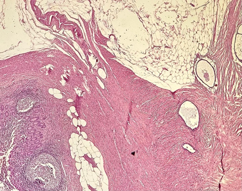
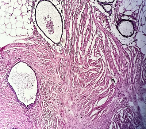
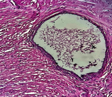
Under the microscope, the examination reveals numerous cystically dilated glands lined by a single layer of epithelium resembling that of the fallopian tube. Within these glands, one can observe the three distinct cell types typical of normal fallopian tube epithelium: pale ciliated columnar cells, non-ciliated secretory (mucous) cells, and dark intercalated/peg cells, albeit in varying proportions. Surrounding the glands is a stroma consisting of dense connective tissue, often containing mononuclear inflammatory cells. Psammoma bodies, characteristic calcific structures, are frequently found within the gland lumina or in the nearby stroma. In contrast, endometriosis displays glands and stroma resembling those of the endometrium, along with hemosiderin-laden macrophages, a feature absents in endosalpingiosis [16].
Diagnosing pelvic endosalpingiosis through imaging is rare, but when detected, it typically appears as a predominantly cystic lesion with either ill or well-defined boundaries. Post-contrast imaging reveals mural and septal enhancement. On MRI, endosalpingiosis typically exhibits a dark T1 signal and a bright T2 signal, with noticeable septal and mural enhancement, which contrasts with the bright T1 appearance characteristic of endometriosis. However, the cystic appearance of endosalpingiosis on MRI is nonspecific and can be confused with neoplasms, particularly ovarian cystadenomas or metastatic disease when affecting the ovaries [17].
Figure 1: HE x 40, Appendiceal endosalpingiosis.
Figure 2: HE x200, Pelvic endosalpingiosis.
Figure 3: HE x400, ectopic benign glands lined by tubal-type epithelium in the peritoneum.
Conclusion
In summary, we present a rare case of appendiceal endosalpingiosis mimicking appendicitis, reflecting the limited cases reported. Given the association of endosalpingiosis with gynecologic malignancies, it's unclear if further investigation or monitoring is warranted for incidental diagnoses. Additional research is needed to understand the link between appendiceal endosalpingiosis and ovarian cancers. Pelvic endosalpingiosis, characterized by tubal epithelial deposits, typically affects older individuals and has an association with ovarian malignancies. Appendiceal endosalpingiosis is exceedingly rare. Our cases presented with right lower quadrant pain.
References
- Prentice L, Stewart A, Mohiuddin S, Johnson NP. (2012). What Is Endosalpingiosis? Fertil Steril. 98(4):942-947.
Publisher | Google Scholor - Pollheimer MJ, Leibl S, Pollheimer VS, Ratschek M, Langner C. (2007). Cystic Endosalpingiosis of The Appendix. Virchows Arch. 450(2):239-241.
Publisher | Google Scholor - Demir MK, Savas Y, Furuncuoglu Y, Cevher T, Demiral S, et al. (2017). Imaging Findings of the Unusual Presentations, Associations and Clinical Mimics of Acute Appendicitis. Eurasian J Med. 49(3):198-203.
Publisher | Google Scholor - John A. Sampson. (1930). Postsalpingectomy Endometriosis (Endosalpingiosis). American Journal of Obstetrics and Gynecology. 20(4):443-480.
Publisher | Google Scholor - Prentice L, Stewart A, Mohiuddin S, Johnson NP. (2012). What Is Endosalpingiosis? Fertil Steril. 98(4):942-947.
Publisher | Google Scholor - Tudor J, Williams TR, Myers DT, Umar B. (2019). Appendiceal Endosalpingiosis: Clinical Presentation and Imaging Appearance of a Rare Condition of The Appendix. Abdom Radiol (NY). 44(10):3246-3251.
Publisher | Google Scholor - Pollheimer MJ, Leibl S, Pollheimer VS, Ratschek M, Langner C. (2007). Cystic endosalpingiosis of the appendix. Virchows Arch. 450(2):239-241.
Publisher | Google Scholor - Cajigas A, Axiotis CA. (1990). Endosalpingiosis of the Vermiform Appendix. Int J Gynecol Pathol. 9(3):291-295.
Publisher | Google Scholor - Demir MK, Savas Y, Furuncuoglu Y, Cevher T, Demiral S, et al. (2017). Imaging Findings of the Unusual Presentations, Associations and Clinical Mimics of Acute Appendicitis. Eurasian J Med. 49(3):198-203.
Publisher | Google Scholor - Tallón-Aguilar L, Olano-Acosta C, López-Porras M, Flores-Cortés M, Pareja-Ciuró F. (2009). Endosalpingiosis of the appendix. Cir Esp. 85(6):383.
Publisher | Google Scholor - Esselen KM, Terry KL, Samuel A, Elias KM, Davis M, et al. (2016). Endosalpingiosis: More Than Just an Incidental Finding at The Time of Gynecologic Surgery? Gynecol Oncol. 142(2):255-260.
Publisher | Google Scholor - Hermens M, van Altena AM, Bulten J, Siebers AG, Bekkers RLM. (2020). Increased Association of Ovarian Cancer in Women with Histological Proven Endosalpingiosis. Cancer Epidemiol. 65:101700.
Publisher | Google Scholor - Sunde J, Wasickanin M, Katz TA, Wickersham EL, Steed DOE, et al. (2020). Prevalence of Endosalpingiosis and Other Benign Gynecologic Lesions. PLoS One. 15(5):e0232487.
Publisher | Google Scholor - Hesseling MH, De Wilde RL. (2000). Endosalpingiosis in laparoscopy. J Am Assoc Gynecol Laparosc. 7(2):215-219.
Publisher | Google Scholor - Djordjevic B, Clement-Kruzel S, Atkinson NE, Malpica A. (2010). Nodal Endosalpingiosis in Ovarian Serous Tumors of Low Malignant Potential with Lymph Node Involvement: A Case for A Precursor Lesion. Am J Surg Pathol. 34(10):1442-1448.
Publisher | Google Scholor - Mills, S. E., Greenson, J. K., Hornick, J. L., Longacre, T. A., Reuter, V. E. (2015). Sternberg's Diagnostic Surgical Pathology. Lippincott Williams & Wilkins.
Publisher | Google Scholor - Kaneda S, Fujii S, Nosaka K, Inoue C, Tanabe Y, et al. (2015). MR Imaging Findings of Mass-Forming Endosalpingiosis in Both Ovaries: A Case Report. Abdom Imaging. 40(3):471-474.
Publisher | Google Scholor


