

Case Report
Aesthetic Rehabilitation of Anterior Malalinged Dentition with E-Max Laminate Veneers-A Case Report
1Post Graduate 3rd year, Department of Prosthodontics, ITS CDSR Muradnagar, India.
2Tutor, Department of Prosthodontics, Dental College, Rajendra Institute of Medical Science, Ranchi, India.
3Senior Lecturer, Department of Prosthodontics, ITS CDSR Muradnagar, India.
*Corresponding Author: Tushar Rishu, Tutor, Department of Prosthodontics, Dental College, Rajendra Institute of Medical Science, Ranchi, India.
Citation: S Idnani, Tushar R, S khan. (2022). Aesthetic Rehabilitation of Anterior Malalinged Dentition with E-Max Laminate Veneers-A Case Report. Dentistry and Oral Health Care. 1(1); DOI: 10.59657/2993-0863.brs.22.001
Copyright: © 2022 Tushar Rishu, this is an open-access article distributed under the terms of the Creative Commons Attribution License, which permits unrestricted use, distribution, and reproduction in any medium, provided the original author and source are credited.
Received: October 06, 2022 | Accepted: October 31, 2022 | Published: November 07, 2022
Abstract
Various different procedures have been advocated in literature to correct dental anomalies specially in the aesthetic region such as discolouration of tooth due to fluorosis or spacing in dentition due to change in shape of tooth like peg laterals. One of the most common treatment modalities to treat such cases are using VENEERS. This case report presents conservative and aesthetic procedure in the management of malalinged anterior teeth with E max lithium disilicate laminate veneers.
Keywords: emax lithium disilicate venners; conservative procedure; malalinged teeth
Introduction
Patients demand for treatment of unaesthetic anterior teeth has been steadily growing. Patients often come with a request for cosmetic improvements in their teeth. Frequently, such an aesthetic improvement consequently uplifts the patient’s sense of well-being and self-esteem [1]. The Glossary of Prosthodontic Terms, drawn up by The Academy of Prosthodontics, defines veneers as a superficial or attractive display in multiple layers, frequently termed as a laminate veneer [2]. Since 1930s dental veneers have been used to improve the smile of the patient. The indications of dental veneers include [3].
1) Discoloured teeth due to many factors such as tetracycline staining, fluorosis and amelogenesis imperfecta
2) Restoring fractured and worn-out teeth.
3) Masking of abnormal tooth morphology.
4) Correction of minor malposition of the teeth.
5) Intra-oral repair of fractured crown and bridge facings.
Ceramic laminate veneers which are also known as “contact lenses,” are capable of providing an extremely accurate reproduction of natural teeth with great colour stability and also have a very conservative treatment approach [4]. As stated by Muller de van in 1952, “the preservation of that which remains is of utmost importance and not the meticulous replacement of that which has been lost”. Respecting this context, veneers are good alternative to restore the aesthetic of patient without doing much changes in existing condition of tooth. Veneers are also biologically compatible with periodontium [5]. The present case report describes the treatment of spaced dentition due to morphological altered tooth in the anterior dentition with thin porcelain laminate veneers, to restore aesthetics and function.
Case Report
45 years old female patient reported to the Department of Prosthodontics with a chief complaint of spacing and malalinged tooth in anterior region and wanted aesthetic correction for the same. A detailed family history, medical history and dental history was obtained. In family history, none of his family members had similar problem. Extra oral examination could elicit no abnormal findings. Intra oral examination reveals spacing and malalinged anterior teeth int 11,12,21,22. All teeth were vital and had no hypersensitivity. No carious teeth were present. Various treatment options like full ceramic crown and veneers were given to patient. Owing to its minimally invasive nature and excellent aesthetic qualities it was decided to enhance her appearance using E max lithium disilicate veneers.
Procedure
Types of preparation for veneers plays an important role in success of veneers. There are 4 types of preparations are present (Figure 1).
a. Window
b. Bevel
c. Feather
d. Incisal overlap
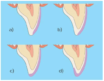
Figure 1: Incisal preparations are possible for veneers: a) window, b) feather, c) bevel or d) incisal overlap
Out of these four types of preparation designs, incised overlap has the highest success rate [6,7].
- Preoperative photos and impression of both arches were obtained. Shade selection was done in natural daylight using a colour scale (VITA Toothguide 3D-MASTER, Zahnfabrik H. Rauter GmbH. & Co.KG Spitalgasse, Germany) (Figure 2).
- Depth orientation grooves were placed on facial surface providing a depth of 0.3mm on gingival region and 0.5mm on incisal half using diamond bur. The remaining tooth structure was removed using round end tapered diamond bur. Chamfer finish line was given at the level of gingival crest.
- Tooth preparation proximally was extended to contact areas without breaking contact to conserve interproximal enamel. An overlapped incisal edge preparation was chosen, which provides a vertical stop to aid in correcting the positioning of the veneer (figure 3).
- Veneers were etched with 5% hydrofluoric acid for 20 seconds. Veneers were washed and dried. The inner surface of veneers was coated with silane-coupling agent (Monobond N; Ivoclar Vivadent, AG Schaan Liechtenstein USA) and allowed to dry for 1min. Then, the etching of the teeth was carried out with 37% phosphoric acid for 15–20s, rinsed thoroughly with water, and dried. A layer of bonding agent (Adper; 3M ESPE, St. Paul, MN, USA) was applied on tooth surface and cured for 20s. Veneers were bonded to teeth with dual cure resin cement (RelyX; 3M ESPE) (figure 4).
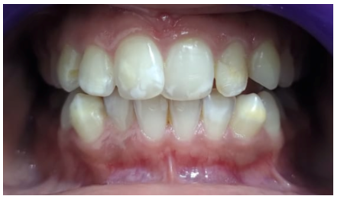
Figure 2: Pre-operative intraoral photo
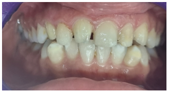
Figure 3: Tooth preparation done int to 12,11,21,22
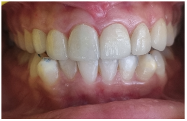
Figure 4: Final cementation of veneers in 21,11,22,12
Discussion
The aim of the treatment in this case was to improve the patient’s smile and aesthetically rehabilitate the teeth. This goal was achieved using porcelain veneers as they can completely mask the spaces present between the teeth with minimal reduction of sound tooth substance.
Porcelain veneers provide both predictable and long-lasting aesthetic rehabilitation [8,9]. The durability and clinical success of porcelain veneers have been widely investigated in the literature. It has been reported that ceramic veneers provide durable and successful restoration with an estimated survival probability of 93.5% over 10 years [10].
The long-term success of ceramic veneers depends on proper case selection and treatment planning procedures such as shade selection, tooth preparation, cementation technique, and patient maintenance. Therefore, the case must be selected carefully and properly planned treatment should be done [11].
Conclusion
Porcelain veneers are considered one of the most popular restorative materials in aesthetic dentistry. They provide excellent aesthetic results when an appropriate treatment plan and protocol are used during the clinical and laboratory fabrication stages. This case report describes the use of porcelain veneers to enhance the appearance of a patient with spaced dentition, thus improving the patient’s smile and, consequently, self-esteem [12].
References
- Lim CC. (1995). Case selection for porcelain veneers. Quintessence International. 26(5):311-315.
Publisher | Google Scholor - The Academy of Prosthodontics. (2017). The glossary of prosthodontic terms. J Prosthet Dent. 117(5S):1-105.
Publisher | Google Scholor - Alothman Y, Bamasoud MS. (2018). The success of dental veneers according to preparation design and material type. Open Access Maced J Med Sci. 6(12):2402-2408.
Publisher | Google Scholor - Silva G, Normandes AC, Barros Júnior E, Gatti J, Maranhão K, Reis AC et al. (2018). Ceramic Laminate Veneers for Reestablishment of Esthetics in Case of Lateral Incisor Agenesis. Case Rep Dent.
Publisher | Google Scholor - Fradeani M, Redemagni M, Corrado M. (2005). Porcelain laminate veneers: 6 to 12 year clinical evaluation-A retrospective study. Int J Periodontics Restorative Dent. 25:9-17.
Publisher | Google Scholor - Castelnuovo J, Tjan AH, Phillips K, Nicholls JI, Kois JC. (2000). Fracture load and mode of failure of ceramic veneers with different preparations. J Prosthet Dent. 83(2):171-180.
Publisher | Google Scholor - Stappert CF, Ozden U, Gerds T, Strub JR. (2005). Longevity and failure load of ceramic veneers with different preparation designs after exposure to masticatory simulation. J Prosthet Dent. 94(2):132-139.
Publisher | Google Scholor - Peumans M, Van Meerbeek B, Lambrechts P, Vanherle G. (2000). Porcelain veneers: a review of the literature. J Dent. 28(3):163-177.
Publisher | Google Scholor - Magne P, Douglas WH. (1999). Porcelain veneers: dentin bonding optimization and biomimetic recovery of the crown. Int J Prosthodont. 12:111-121.
Publisher | Google Scholor - Beier US, Kapferer I, Burtscher D, Dumfahrt H. (2012). Clinical performance of porcelain laminate veneers for up to 20 years. Int J Prosthodont. 25(1):79-85.
Publisher | Google Scholor - Tushar, Kumar KN, Garg S, Vijayan A. (2020). Aesthetic correction of spaced dentition with Emax lithium disilicate veneers: Case report. IJADS. 6(3):648-650.
Publisher | Google Scholor - El Mourad AM. (2018). Aesthetic rehabilitation of a severe dental fluorosis case with ceramic veneers: a step-by-step guide. Case Rep Dent. 1-4.
Publisher | Google Scholor


