

Case Report
Adaptation of the Healthcare Team as The Key to TAVI Implantation
Enfermera/o del Servicio de Hemodinámica del Hospital Universitario Miguel Servet de Zaragoza, Spain.
*Corresponding Author: Virginia Abadía Piquero, Enfermera/o del Servicio de Hemodinámica del Hospital Universitario Miguel Servet de Zaragoza, Spain.
Citation: Susana B. Medina, Consuelo A. Alfageme, Lorena C. Toral,Virginia A. Piquero. (2023). Adaptation of the Healthcare Team as The Key to TAVI Implantation, Journal of BioMed Research and Reports, BioRes Scientia Publishers. 3(1):1-6. DOI: 10.59657/2837-4681.brs.23.052
Copyright: © 2023 Virginia Abadía Piquero, this is an open-access article distributed under the terms of the Creative Commons Attribution License, which permits unrestricted use, distribution, and reproduction in any medium, provided the original author and source are credited.
Received: October 12, 2023 | Accepted: October 26, 2023 | Published: November 02, 2023
Abstract
We present the case of a 79-year-old female patient with a history of ischemic heart disease revascularized with stents and severe aortic stenosis, recently included in the surgical waiting list for treatment. She consulted for syncope and humerus fracture, preceded by chest pain with electrocardiographic changes. During the sequence of diagnostic tests indicated in this context, she suffered a severe clinical worsening, complicating to the point of acute pulmonary edema, cardiogenic shock and requiring mechanical ventilation.
The underlying cause was severe aortic stenosis and, therefore, the ideal treatment was urgent percutaneous aortic valve implantation, since he had a peak peak gradient of 64 mmHg.
The resolution of the case was not easy, since it required the transfer of a hemodynamically unstable patient from her hospital to the center specialized in TAVI implantation, located only 2 km away.
What was new and exceptional was the adaptation of the usual treatment strategy (transfer of the patient to the referral center and scheduled implant) to a patient-centered strategy, which required the transfer of the TAVI expert team (nurses and physicians) together with the implant kit to the hospital where the patient was admitted.
Keywords: syncope; gradient; TAVI; severe aortic stenosis
Introduction
Aortic stenosis is one of the most common heart valve diseases, occurring most frequently in people over 75 years of age. When the aortic valve area is ≤ 1 cm2 it is considered severe and of poor prognosis when the patient presents with symptomatology. Currently, there are no medical therapies to prevent or delay disease progression. Percutaneous aortic valve implantation (TAVI) is an alternative to aortic valve replacement for symptomatic patients at high or intermediate surgical risk. Valve replacement has been proposed as the definitive treatment even for patients with severe, non-symptomatic AS [1,2].
A large percentage of patients with severe aortic stenosis have coronary artery disease, due, in part, to the similar pathogenesis of both. Angina is a common symptom in patients with severe aortic stenosis. The cardiovascular risk profile of the patient will determine the severity of coronary artery disease, the greater the number of risk factors. To reduce the risk of cardiovascular death in patients with severe and complex coronary artery disease undergoing TAVI, both aortic stenosis and coronary artery disease should ideally be resolved [3].
Cardiogenic shock is the primary cardiac disorder characterized by a state of low cardiac output and circulatory failure that triggers target organ hypoperfusion and tissue hypoxia. Acute myocardial infarction is the most common cause of cardiogenic shock. There are other disorders that cause myocardial deterioration such as: the valves, the pericardium or the conduction system. Morbidity and mortality among patients with cardiogenic shock remain high despite advances in mechanical circulatory support therapies [4].
Clinical observation
We present the case of a 79-year-old female patient who came to the hospital emergency department on 27-7-2021 with chest pain radiating to the left upper extremity, dyspnea and syncope, which resulted in a fracture of the head of the left humerus.
Physical examination revealed: BP: 62/31, HR: 78 p.m., oxygen saturation: 95% with facial mask with reservoir, poor general condition, diaphoresis and lip cyanosis.
Diagnostic impression of acute coronary syndrome with ST elevation (STEACS)/cardiogenic shock.
Infarction Code is activated by 061 for transfer to hospital with hemodynamics unit.
Clinical history of: pulmonary hypertension, dyslipidemia, hypothyroidism, chronic ischemic heart disease with percutaneous revascularization (stents in middle and distal LAD, distal DC with proximal PD, PTCA on ostial CX OCT, moderate lesion at distal LMCA level), degenerative aortic valve disease with severe stenosis and hyperuricemia.
Complementary tests
*ECG: ST-segment elevation in V1-V3 and AvR and generalized ST- segment elevation suggestive of ischemia in the context of critical AS (Figures 1 and 2).
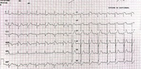
Figure 1: ST segment elevation in V1-V3 and AvR.

Figure 2: ST segment depression.
*X-ray: Cardiomegaly. Signs of heart failure.
*Blood tests: Ultrasensitive Troponin (TnT us) of 220 to 4000 and D-Dimer of 15000.
*Echocardioscopy: hypertrophic left ventricle with mild to moderately depressed LVEF (40%). Severe aortic stenosis, moderate mitral insufficiency.
*Coronary angiography: On arrival at the hemodynamics unit, a diagnostic study was performed through the right radial artery in which TIMI 3 flow was observed throughout the coronary tree with no obstructive lesions in the coronary arteries. Good late results of Everolimus-eluting stents previously implanted in the middle LAD, distal LAD and ostial PD. Good result of PTCA on OCT of ostial CX with distal bed of moderate caliber. Moderate lesion at distal CT level (assessed by IVUS). (Figures 3 and 4)
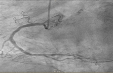
Figure 3: CD without significant lesions.
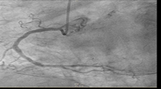
Figure 4: Moderate injury at the distal CT level.
Given the results of the coronary angiography and the presence of Dimero D of 15000, it is of interest to rule out PTE by means of pulmonary CT angiography, the con clusion of which was:
*Pulmonary CT angiography: No signs of PTE. Signs of PHT. Signs of hemodynamic decompensation with probable perihilar cardiogenic edema, bilateral pleural effusion and cardiomegaly (Figures 5 and 6).
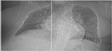
Figure 5: Cardiomegaly. Bilateral pleural effusion.
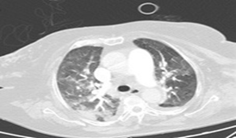
Figure 6: No signs of PE.
After going up to the cardiology ward, the patient worsened with acute pulmonary edema, with sudden onset dyspnea, tachypnea, tachycardia of 120 bpm, sweating, bilateral crackles up to midfields and significant respiratory distress, which resolved with pharmacological treatment of Furosemide 40 mg, Morphic Chloride 3.3 mg and O2 in a reservoir.
The following day and in the morning shift he presented hemodynamic stability but with a tendency to arterial hypotension, O2 saturation 98% with reservoir and a diuresis of 400cc.
The case is discussed with the referral hospital to schedule the TAVI implant in the next 24 hours.
In the afternoon shift, hypotension and oliguria persisted despite diuretic treatment. Dopamine perfusion pump was started at a dose of 0.5 mcgr/kg/min. O2 saturation of 92%, nervousness, but without dyspnea or pain.
Finally, he was transferred to the ICU due to worsening general condition with oliguria, anginal chest pain, profuse sweating, tachypnea, hypotension of 65/38 mmHg, HR 100 bpm; where orotracheal intubation and mechanical ventilation were performed.
Family members were notified due to the patient’s serious condition and poor prognosis.
Given the sudden development of the clinical symptoms, and after ruling out coronary artery disease again, TAVI implantation was prioritized as the treatment of choice.
Due to the poor condition of the patient, her transfer to a hospital specialized in the technique was not considered. Therefore, it was decided to move the healthcare team of hemodynamicists, anesthesiologist, instrumentalist nurse, nurse expert in valve assembly and the necessary material for the implant.
Communication between the units responsible for the patient, ICU and hemodynamics, was continuous, since it varied as the patient’s condition worsened. As a result, the information that reached the hemodynamics unit specialized in the implant was disparate, going from having the procedure scheduled for the day after the notification, to having to do it immediately on the same day, to the transfer of personnel and material to the hospital where the patient was located.
Likewise, the nursing team of both centers coordinated the preparation of the necessary material for the implant successfully.
The transfer approach was uncertain, since there was no means of transport available for this situation. The decision was made to do it in several cabs, with the inconvenience that the valve kit did not fit in any vehicle.
Finally, carrying the bulky box with the valve kit, we arrived at the unit where the implant was to be performed. There, the medical team was waiting for us with great expectation, with everything ready.
The first intention was to perform a palliative aortic valvuloplasty, but after measuring gradients and assessing the patient’s situation, it was decided to definitively implant the TAVI.
TAVI Implantation
Patient included in the surgical demand list for TAVI implant since 07/22/2021.
In the previous study, it appeared that the origin of the left common trunk could be compromised by the TAVI stent, so it was decided to perform left coronary protection with a 3.5 (6F) EBU catheter, Asahi Sion angioplasty guidewire and NC angioplasty balloon through the right brachial artery.
Percutaneous therapeutic access through the right femoral artery, successful implantation of Sapien 3 valve, nº26 (Edwards) (Figure 7 and 8).
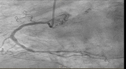
Figure 7: Alignment of valve prosthesis.
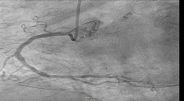
Figure 8: Implantation of aortic prosthesis.
The patient was able to go from an initial gradient of 64 m mHg to 0 mmHg, without regurgitation. The hemodynamic improvement was evident from the first moment (Figure 9 and 10).
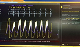
Figure 9: 64 mmHg gradient.
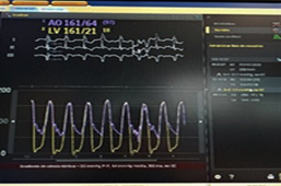
Figure 10: 0 mmHg gradien t.
Nursing care plan
The care plan was based on an assessment framework based on the Virginia Henderson model and NANDA, NIC, NOC Taxonomy.5,6 (Table 1)
Table 1
| Nursing Diagnosis | Related Factors | NOC | NIC | Activities |
| Risk of body temperature imbalance 00005 | Sedative drugs and low operating room temperature | Thermoregulation 000800 | Temperature regulation: intraoperative 3902 | Check ambient temperature |
| Prepare and regulate the corresponding heating devices. | ||||
| Heat or cool all irrigation, i.v. and skin preparation solutions. | ||||
| Cover the patient with reflective | ||||
| blankets. | ||||
| Infection Risk 00004 | invasive procedures | Knowledge: infection control 1842. Immune status 0702 | Infection control: intraoperative 6545 | Limiting and controlling the entry and exit of people in the operating room |
| Ensure that surgical personnel wear the appropriate equipment. | ||||
| Verify antibiotic prophylaxis administered | ||||
| Apply antiseptic as prescribed | ||||
| Wash hands before and after each patient care activity. | ||||
| Open sterile supplies and instruments using aseptic techniques. | ||||
| Verify sterilization indicators | ||||
| Hand and nail brushing, gowns and gloves, according to center standards. | ||||
| Inspect the skin/tissues around the surgical site. | ||||
| Keep the room clean and disinfected. | ||||
| Risk of bleeding 0206 | Interventionism Use of anticoagulant medication | Risk control 4160 | Hemorrhage prevention 4010 | Perform coagulation studies |
| Close monitoring of the patient | ||||
| Observe signs and symptoms | ||||
| Risk of urinary retention 00023 | Volume management Immobility on the stretcher | Urinary elimination 0508 | Bladder catheterization 0580 Urinary catheter care 1876 | -Sterile catheterization |
| -Catheter handling with asepsis | ||||
| -Check the correct placement of the balloon. | ||||
| Fluid volume deficit 27 | Active fluid volume loss | Water balance 0601 | Liquid handling 4120 | -Monitor hydration status (moist mucous membranes, adequate pulse and blood pressure). |
| -Monitor blood pressure, heart rate cardiac and respiratory status | ||||
| -Administer IV fluids at room | ||||
| temperature | ||||
| Ineffective breathing pattern 00032 | hypoventilation syndrome, anxiety, decreased energy and respiratory muscle fatigue. | Respiratory status: airway patency 0410 Respiratory status: ventilation 0403 Vital signs status 0802 | Airway management 3140 Ventilation aid 3390 Monitoring of vital signs 6680 Respiratory monitoring 3350 Airway suction 3270 | -To assess the state of consciousness of the patient |
| -Evaluate skin color and filling | ||||
| capillary | ||||
| -Evaluate the | ||||
| respiratory dynamics and expansion | ||||
| thoracic | ||||
| -Place in position semifowler | ||||
| -Evaluate the need for administration of ventilation mechanics | ||||
| -Participate in the intubation orotracheal |
*Likert scale: 1: Severely compromised; 2: Substantially compromised; 3: Moderately compromised; 4: Slight deviation from the normal range; 5: Normal.
Discussion
Clearly, the nursing team has a fundamental role to play in the management of this type of patient who is so unstable and, in turn, so fragile. They must be specialized in highly versatile, highly precise and continuously updated techniques. The role of the expert nurse in TAVI assembly is particularly important, as it is essential that she or he be properly trained to assume this responsibility, since without her the valve prosthesis implantation could not have been carried out and, therefore, we would be facing a fatal outcome.
We must emphasize the importance of the work performed by the multidisciplinary team involved in the care and treatment of the patient. It is fundamental for making the right decisions and in our case the intervention of the TAVI implant in an emergent manner was the key to success.
Severe aortic stenosis requires immediate treatment at the time of symptoms (syncope, angina and heart failure). Otherwise, the clinical course leads to cardiogenic shock, multiorgan failure and, therefore, death, which in this case had a successful outcome, since the patient evolved favorably and was discharged 10 days after implantation.
The nurse will watch over the patient’s well-being at all times, attending to the vital needs intrinsic to this type of surgery and will work together with the rest of the professionals to ensure that the procedure is successful.
Acknowledgments
None.
Conflicts of interest
Authors declare that there are no conflicts of interest.
References
- SEC Working Group for the 2021. (2022). ESC/EACTS guidelines for the management of valvular heart disease and SEC Guidelines Committee. Comments on the 2021 ESC/EACTS guidelines for the management of valvular heart disease. Rev Esp Cardiol (Engl Ed), 75(6):466-471.
Publisher | Google Scholor - González Saldivar H, Vicent Alaminos L, Rodríguez–Pascual C, et al. (2022). Evolution of patients with severe aortic stenosis after indication for intervention. Rev Esp Cardiol, 72(5):392-397.
Publisher | Google Scholor - Vilacosta I, Vivas D, López J, et al. (2015). Symptomatic severe aortic stenosis: what is severe, what is symptomatic and what do clinical practice guidelines say about its management? Rev Esp Cardiol Supl., 15:3-9.
Publisher | Google Scholor - Vahdatpour C, Collins D, Goldberg S. (2019). Cardiogenic shock. J Am Heart Assoc., 8(8).
Publisher | Google Scholor - Cribier A, Eltchaninoff H, Bash A, et al. (2002). Percutaneous transcatheter implantation of an aortic valve prosthesis for calcific aortic stenosis: first human case description: First human case description. Circulation, 106(24):3006-3008.
Publisher | Google Scholor - Herdman, editor. Kamitsuru S, (2019). NANDA nursing diagnoses definitions and classification 2018-2020. In: Barcelona. Elsevier.
Publisher | Google Scholor


