
Case Report
Xeroderma Pigmentosum: A Case Diagnosed Using the Diagnostic Criteria Proposed in 2022
Pediatric Hospital “Martyrs of Tunas”, Tunas, Cuba.
*Corresponding Author: Yordania Velázquez Avila, Pediatric Hospital “Martyrs of Tunas”, Tunas, Cuba.
Citation: Avila YV, Valenciano CRR. (2026). Xeroderma Pigmentosum: A Case Diagnosed Using the Diagnostic Criteria Proposed in 2022, International Journal of Biomedical and Clinical Research, BioRes Scientia Publishers. 7(3):1-6. DOI: 10.59657/2997-6103.brs.26.150
Copyright: © 2026 Yordania Velázquez Avila, this is an open-access article distributed under the terms of the Creative Commons Attribution License, which permits unrestricted use, distribution, and reproduction in any medium, provided the original author and source are credited.
Received: July 21, 2026 | Accepted: August 24, 2026 | Published: September 02, 2026
Abstract
Xeroderma pigmentosum is an autosomal recessive genodermatosis characterized by a failure in deoxyribonucleic acid repair caused by oxidative damage and ultraviolet radiation, producing skin lesions with photodamage and photoaging from pediatric ages. Diagnosis is guided based on clinical elements and the presence of premalignant or malignant lesions confirmed by dermoscopy. Although molecular studies currently exist that have allowed the detection of eight mutations that cause xeroderma pigmentosum, this technology is unavailable in some countries. In Cuba, histopathological study is used for diagnosis, which is not conclusive for this disease itself, as it may present in other conditions or diseases with chronic actinic damage such as actinic skin, albinism, Rothmund-Thomson syndrome, porphyria, progeroid diseases, among others; and the genetic study called the comet assay in isolated lymphocytes from peripheral blood, which detects deoxyribonucleic acid damage that is not exclusive to xeroderma pigmentosum. In Las Tunas, a province in eastern Cuba, a methodology for comprehensive care of patients with genodermatoses was implemented, which included the proposal of clinical elements for the diagnosis of xeroderma pigmentosum. A case of xeroderma pigmentosum is described that presented clinical manifestations and whose diagnosis was guided using the proposed diagnostic criteria, with the purpose of highlighting them. The importance of the xeroderma pigmentosum diagnostic criteria proposed in the methodology for the care of patients with genodermatoses was demonstrated, which prevents diagnostic error and inappropriate overdiagnosis.
Keywords: genodermatoses; xeroderma pigmentosum; chronic actinic damage; cutaneous genetic diseases; diagnostic
Introduction
Genodermatoses are a group of diseases of genetic origin with phenotypic expression in the skin and its appendages, without environmental factors influencing their expression. Other systems may be affected [1]. Advances in molecular sequencing have made it possible to identify the genes responsible for numerous genetic diseases, among which are genodermatoses related to cancer formation such as xeroderma pigmentosum (XP).
XP is an autosomal recessive genodermatosis characterized by a failure in deoxyribonucleic acid (DNA) repair caused by oxidative damage and short-wavelength ultraviolet (UV) radiation, between 290 and 320 nm, which can affect the skin, increasing susceptibility to skin cancer, and cause damage to other organs such as the eyes and the neurological system. Diagnosis should be suspected in patients with increased photosensitivity and characteristic cutaneous, ophthalmological, and neurological findings [2-5].
It was initially described by the dermatologist Mortiz Kaposi in 1874 based on the study of four patients who shared clinical characteristics such as xerosis, premature aging, hyperpigmentation, and skin carcinomas. In 1926, exposure to ultraviolet rays was identified as an important factor in the progression of the disease. Subsequently, the etiology of the disease was defined as a genetic defect in DNA repair, specifically in nucleotide excision repair [4].
It is estimated to be present in 1 in 1 million people in Europe and the United States. However, it has a higher incidence in regions with high rates of consanguinity. In Japan, it has a frequency of 1 per 22,000 people and 1 per 10,000 people in North Africa. The rate in Latin American countries is lower [2,6].
The authors found no population studies or any characterization showing the prevalence of the disease in Cuba; however, a study on the quality of life of 32 patients with XP in Villa Clara province was found [7]. In this study, the authors demonstrated that the lifestyle that must be implemented and sun damage prevention measures generate limitations in collective human functioning, implying a negative psychosocial impact. Patients with xeroderma pigmentosum tend to experience ridicule, discrimination, abuse, and loneliness to a greater extent than the general population [5,8].
In a population study conducted in Las Tunas, a province in eastern Cuba, it was found that xeroderma has a prevalence of 0.6:100,000 inhabitants [9], which, although it behaves as a rare disease, is present in the population and deserves to be addressed.
Although molecular studies currently exist that have allowed the detection of eight mutations that cause xeroderma pigmentosum, seven of these mutations are associated with a defect in nucleotide excision repair. In some countries, this technology is unavailable [2]. In Cuba, histopathological study is used for diagnosis, which is not conclusive for this disease itself, and the genetic study called the comet assay in isolated lymphocytes from peripheral blood, for the evaluation of DNA repair capacity against damage induced by ultraviolet C light [6].
In Las Tunas, as part of a methodology for the care of patients with genodermatoses, designed using the Delphi method [1,10,11] the proposal of XP diagnostic criteria was included, presented at the International Congress of Community Genetics in 2021 and the International Congress of Dermatology in 2022 [6,12].
Major Criteria
- Premature photoaging (skin hyperplasia, thickening, wrinkles, yellow or reddish hyperpigmentation, solar elastosis) at pediatric ages.
- Family history of xeroderma pigmentosum in consanguineous families.
Minor Criteria
- Skin phototypes I or II.
- Onset from early ages (before 5 years of age) of photosensitivity lesions (actinic dermatitis, actinic cheilitis).
- Skin biopsy with lesions suggestive of premalignant lesions (actinic keratoses).
- Malignant neoplasm at pediatric ages diagnosed by dermoscopy and biopsy.
- Ocular photodamage (photophobia, cataracts, conjunctival melanosis).
- Association with neoplastic tumors in other organs (digestive tract).
- Neurological involvement
Taking as diagnostic criteria the fulfillment of two major criteria or one major criterion and two minor criteria.
The following describes a case of XP that presented clinical manifestations and whose diagnosis was guided using the proposed diagnostic criteria, with the purpose of highlighting them.
Case Presentation
A 6-year-old male patient, urban origin, whose mother noticed that since the first year of life, he burned easily upon sun exposure, and although she protected him from exposure since then, she noticed progressive changes in the skin giving it an aged appearance. With this dermatological condition, he arrived at the specialized genodermatosis consultation at the "Mártires de Las Tunas" Provincial Pediatric Hospital.
Personal Pathological History
None reported.
Relevant Family Pathological History
Great-grandfather who suffered from skin cancer from a young age. The parents were cousins.
Medication Reactions
None reported.
Dermatological Examination
actinic skin (phototype II) with photodamage manifested by cheilitis on the lips (Figure 1), ephelides and lentigines in photo exposed areas with yellowish hyperpigmentation (Figure 2), elastosis on the neck and dorsum of the hands (Figure 3), and presence of two small pearly nodules on the auricular pavilions (Figure 4).
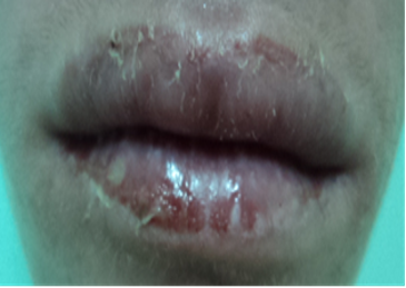
Figure 1: Cheilitis on lips. Authors' image, taken with prior informed consent.
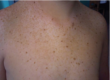
Figure 2: Presence of multiple ephelides and solar lentigines and hyperpigmentation in photo exposed areas typical of chronic actinic damage. Authors' image, taken with prior informed consent.
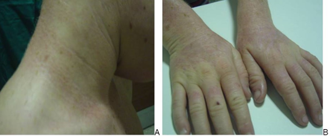
Figure 3: Premature photoaging manifested by solar elastosis and hyperpigmentation on the neck (A) and dorsum of the hands (B). Authors' image, taken with prior informed consent.
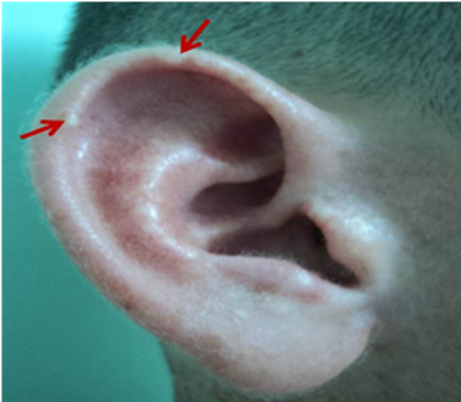
Figure 4: Pearly nodules on the auricular pavilions of the patient with XP (indicated by red arrows). Authors' image, taken with prior informed consent.
On dermoscopy of the pearly nodular lesions, areas of shiny whitish-red color and the presence of comma-shaped vessels were observed. With these elements, a skin biopsy was indicated.
Histopathological Study
Hyperkeratosis, irregular acanthosis with areas of spongiosis and basal layer hyperpigmentation. In the dermis, presence of basophilic degeneration of collagen and elastosis. Undifferentiated basaloid cells with a micronodular pattern compatible with non-invasive micronodular basal cell carcinoma were observed.
Ophthalmology
Presence of keratitis was found. A pedigree (Figure 5) was constructed, determining an autosomal recessive (AR) inheritance pattern.
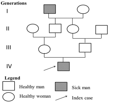
Figure 5: Pedigree showing AR inheritance pattern. Authors' image.
With this clinical presentation, the possibility of XP was considered, concordant with the proposed diagnostic criteria (XP criteria), so a molecular study "Comet assay" was performed, resulting positive.
Management
Genetic counseling was provided to the family, along with multidisciplinary care from dermatologists, geneticists, pediatricians, and psychologists. Surgical excision of the lesions suggestive of basal cell carcinoma was performed. Extreme photoprotection was prescribed, both physical (clothing covering as much skin surface as possible, such as long-sleeved shirts and long pants, glasses, caps and hats, use of umbrellas), and chemical (use of sunscreen applied 30 minutes before sun exposure); daily use of vitamin D, both in the diet (fish, dairy, eggs) and as a supplement; monthly periodic skin self-examination to detect the appearance of possible premalignant lesions.
Currently, the patient is followed up every 6 months with dermoscopy, has not presented malignant lesions, and premalignant lesions have been removed.
Discussion
Xeroderma pigmentosum is a genetic disease transmitted with an autosomal recessive inheritance pattern, so it is observed more frequently in consanguineous families [13], a characteristic present in the case presented.
The photosensitivity that characterizes it produces UV-induced cutaneous pigmentary changes of early onset, an increased risk of developing skin tumors, and, in some cases, progressive neurological degeneration [2]. Many of these manifestations were present in the case, beginning with acute actinic damage and rapidly evolving into chronic actinic damage with basal cell carcinoma formation.
The damage is produced because the nucleotide excision repair and translesion synthesis (NER) system, induced by ultraviolet light, does not act correctly as a result of an abnormality in the related genes (XPA, XPB (ERCC3), XPC, XPD (ERCC2), XPE (DDB2), XPF (ERCC4), XPG (ERCC5), and XPV (POLH)), affecting DNA damage recognition, DNA strand opening around the lesion, lesion excision by endonucleases, and the synthesis of new complementary DNA strands [14]. The accumulation of pyrimidine-pyrimidine 6-4 dimers and cyclobutene-pyrimidine dimers from UV radiation exposure is crucial in cutaneous carcinogenesis. Patients with XP develop numerous precancerous actinic keratoses and skin carcinomas in childhood [5].
Also guiding in the case was the presence of keratitis, given that in cases with chronic actinic damage, photophobia, keratitis, ectropion, and even neoplastic changes in the retina due to sun exposure may occur; dermoscopy is a useful non-invasive tool for differentiating between benign and malignant lesions, and histopathological study corroborates their presence, in addition to showing typical changes of chronic photodamage manifested by hyperkeratosis, irregular acanthosis with areas of spongiosis, and basal layer hyperpigmentation. In the superficial dermis, signs of photoaging such as basophilic degeneration of collagen and elastosis are found [13].
Other dermatoses with which differential diagnosis should be made are those that produce premature aging, such as progeroid syndromes [13], in which from birth the skin has an aged, atrophic appearance, and if exposed to the sun it will suffer photodamage, but it does not present premature photoaging like XP.
In this sense, the diagnostic criteria proposed in 2022, as part of a methodology for comprehensive care of patients with genodermatoses, are crucial for guiding diagnosis. In the case presented, the presence of both major criteria was fulfilled (premature photoaging and family history of consanguinity) in addition to more than two minor criteria (skin phototype II, early onset of photosensitivity, malignant neoplasm at pediatric ages diagnosed by dermoscopy and biopsy, and presence of ocular photodamage).
Definitive diagnosis can be established through the identification of biallelic pathogenic variants in one of the causative genes [5]. The Comet assay, or alkaline single-cell gel electrophoresis, has become an established method for the study of DNA damage, with multiple applications in genotoxicity assays, human biomonitoring studies, molecular epidemiology, ecotoxicology, as well as a fundamental tool for research on DNA damage and repair, although it is not specific to XP, it indicates the presence of DNA breakage [12].
Currently, there is no cure for xeroderma pigmentosum (XP); extreme photoprotection is essential, vitamin D administration to prevent rickets since its synthesis through cutaneous phototherapy is not possible, periodic skin self-examination to detect premalignant lesions from early stages, and multidisciplinary follow-up both for patient and family education with genetic counseling, as well as to prevent and treat possible complications [13].
In other research, foreign authors [2-4], have emphasized the importance of establishing an early diagnosis of XP to prevent complications in this disease that is disfiguring and potentially fatal.
Conclusion
In the case presented, the importance of the XP diagnostic criteria proposed in the methodology for the care of patients with genodermatoses was demonstrated, which prevents diagnostic error and inappropriate overdiagnosis.
Abbreviations
DNA: deoxyribonucleic acid; AR: autosomal recessive inheritance; XP: xeroderma pigmentosum
Declarations
Ethical Responsibilities
An informed consent form has been completed and signed by the legal representative (mother), and the research committee of the institution where the clinical history was taken has granted authorization, thereby ensuring the patient's right to privacy and guaranteeing the protection of his or her identity in accordance with international regulations.
Economic Financing
No financial support was received.
Author Contributions
Yordania Velázquez Avila: Investigation, and writing - original draft
Carmen Rosa Rodríguez Valenciano: Investigation.
Conflicts of Interest
The authors declare no conflicts of interest.
References
- Avila, Y. V., Romagosa, M. B., Reyes, D. E., Castillo, M. O., Bruzón, D. P. (2022). Metodología para la atención integral a pacientes con genodermatosis. Multimed, 26(6):e2668.
Publisher | Google Scholor - Sánchez Cadena, A. M. (2025). Métodos diagnósticos de xeroderma pigmentoso (Bachelor's thesis).
Publisher | Google Scholor - Leung, A. K., Barankin, B., Lam, J. M., Leong, K. F., Hon, K. L. (2022). Xeroderma pigmentosum: an updated review. Drugs in Context, 11.
Publisher | Google Scholor - Molina, F. M. W. (2024). Xerodermia pigmentosa: la importancia del diagnóstico precoz. Revista Médica Sinergia, 9(8):15.
Publisher | Google Scholor - Rivera Peñaranda, S., Ortuño Gil, C., Vera Lorente, M., Iniesta Mompeán, R., Cascales Barceló, I. (2003). Xeroderma pigmentoso. Dos casos más en España. An. pediatr. 58(4):402-403.
Publisher | Google Scholor - Avila, Y. V., Jiménez, M. U., Valenciano, C. R. R., Solís, M. M. (2022). Propuesta de elementos clínicos para establecer criterios diagnósticos del xeroderma pigmentoso. Folia Dermatológica Cubana, 16(1).
Publisher | Google Scholor - Gutiérrez, N. C., Navarro, J. L. T., Pedraja, N. A., Fleites, E. A. E., Fleites, D. P., et al. (2010). Calidad de vida de los pacientes con xeroderma pigmentoso según la percepción del médico y los familiares. Medicentro Electrónica, 14(3).
Publisher | Google Scholor - Velázquez, Y. (2026). Calidad de vida en adolescentes con genodermatosis evaluados en consulta especializada en Las Tunas.
Publisher | Google Scholor - Velázquez-Ávila, Y., Valenciano-Rodríguez, C. R. (2021). Genodermatoses in Las Tunas Province, Cuba, 1989-2019. MEDICC Review, 23(2):34-41.
Publisher | Google Scholor - Avila, Y. V., Rodriguez, C. R. V., Enamorado, P. B. P., Rojas, Y. F. (2024). Validación de una metodología para la atención a pacientes con genodermatosis mediante el método Delphi. Folia Dermatológica Cubana, 18(2).
Publisher | Google Scholor - Velázquez Y, Rodríguez CR, Orraca M, Morales M, Llamosa AM. (2025). Contribuciones al perfeccionamiento de la atención a pacientes con genodermatosis. An Acad Cienc Cuba. 15(3).
Publisher | Google Scholor - Avila, Y. V., Jiménez, M. U., Valenciano, C. R. R., Solís, M. M. (2022). Propuesta de elementos clínicos para establecer criterios diagnósticos del xeroderma pigmentoso. Folia Dermatológica Cubana, 16(1).
Publisher | Google Scholor - Velázquez Y. (2025). Dermatología clínica y su relación con la genética. Ed. Editorial Académica Española, 387-392.
Publisher | Google Scholor - Prada, C., Liendo, J., Suarez-Mattos, A., Gnecco, J. P., Melo, M., et al. (2022). Angiosarcoma cutáneo en una adolescente con Xeroderma Pigmentoso: Reporte de caso. Revista Colombiana de Cancerología, 26(1):111-116.
Publisher | Google Scholor