
Research Article
Unraveling Risk Factors: Investigating Triggers for Recoarctation in Patients with Coarctation of the Aorta
1 Department of cardiac surgery, Tashkent Pediatric Medical Institute Hospital, Uzbekistan.
2 Department of Congenital Heart Defects, Republican specialized scientific and practical medical center of surgery named after academician V. Vakhidov, Uzbekistan.
*Corresponding Author: Turaev B.B., Department of cardiac surgery, Tashkent Pediatric Medical Institute Hospital, Uzbekistan.
Citation: Turaev B.B, Abralov Kh.K., Ibragimov N.Sh. (2026). Unraveling Risk Factors: Investigating Triggers for Recoarctation in Patients with Coarctation of the Aorta, International Clinical Case Reports and Reviews, BioRes Scientia Publishers. 4(2):1-9. DOI: 10.59657/2993-0855.brs.26.046
Copyright: © 2026 Turaev B.B, this is an open-access article distributed under the terms of the Creative Commons Attribution License, which permits unrestricted use, distribution, and reproduction in any medium, provided the original author and source are credited.
Received: April 10, 2026 | Accepted: May 20, 2026 | Published: August 07, 2026
Abstract
Introduction: Coarctation of the aorta (CoA) is a congenital heart defect characterized by a narrowing of the aorta, often necessitating surgical repair to restore normal blood flow. Despite successful initial interventions, a significant subset of patient’s experiences recoarctation, the reoccurrence of aortic narrowing, presenting a considerable clinical challenge. This study aims to investigate the triggers or contributing factors associated with the development of recoarctation (reCoA) following the initial repair of coarctation of the aorta (CoA), to identify potential strategies for its prevention and management.
Materials and methods: A retrospective cohort study includes information about 120 patients, who underwent 4 different types of surgical repairs of coarctation of aorta through left thoracotomy between 2012-2022. Recoarctation was evaluated using the pressure gradient on the coarctation site measured by echocardiography (echoCG). A threshold of more than 20mmHg was employed to define recoarctation. All statistical analysis was performed using SPSS and Jamovi applications.
Results: The study revealed that 30 patients (25%) experienced early recoarctation, while 52 patients (43.7%) encountered late recoarctation. Patient-related variables such as age, height, weight, gender, and BMI were not correlated with early or late recoarctation. Among the 28 patients (23.3%) who had arch hypoplasia, 12 of them experienced early recoarctation, and 22 of them exhibited late recoarctation. Correlation tests demonstrated a strong negative correlation of the z-score of the arch size with both early recoarctation (r=-0.229, p=0.013) and late recoarctation (r=-0.421, p<0.001). Resection and end-to-end anastomosis (EEA) displayed the highest proportions of early (59%) and late (77%) recoarctation. Prosthetic patch aortoplasty (PPA) showed a relatively higher rate of recoarctation, with 27% of patients experiencing early recoarctation and 44% exhibiting late recoarctation. Resection and extended end-to-end anastomosis displayed a comparatively lower rate, with 0% experiencing early recoarctation and 23% exhibiting late recoarctation.
Conclusion: In conclusion, patient-related variables like age, height, weight, gender, and BMI may not directly cause early or late recoarctation. However, aortic arch hypoplasia emerges as a significant risk factor for both early and late recoarctation. Additionally, while all coarctation repair methods carry some risk of recoarctation, resection and end-to-end anastomosis and prosthetic patch aortoplasty may pose a higher risk compared to extended end-to-end anastomosis. Recognizing these factors is crucial for optimizing surgical outcomes and reducing recoarctation incidence in patients with coarctation of the aorta.
Keywords: congenital heart defects; coarctation of aorta; recoarctation, aortic arch hypoplasia; surgical repair of coarctation of aorta
Introduction
Coarctation of the aorta (CoA) is a congenital heart defect characterized by a narrowing of the aorta, typically near the insertion of the ductus arteriosus. While significant strides have been made in improving the outcomes of CoA repair, the occurrence of recoarctation poses a formidable challenge, warranting a meticulous exploration of its prevalence, associated risk factors, and subsequent management. Understanding the prevalence of recoarctation is essential for gauging the success of surgical interventions and developing targeted postoperative care strategies. Current literature suggests a variable incidence of recoarctation, ranging from 5% to 30% across different cohorts [1] [2]. This broad range underscores the complexity of factors influencing recoarctation dynamics, prompting the need for a nuanced examination of contributing variables. In this study, we embark on a comprehensive exploration of recoarctation after CoA repair, delving into the intricate interplay of factors contributing to its occurrence. This study seeks to unravel the prevalence of recoarctation, identify significant risk factors influencing its development, and assess the impact of different surgical techniques on its incidence.
Materials and Methods
A retrospective cohort investigation was conducted wherein medical records and CT scans were retrospectively analyzed. Pertinent data, including intraoperative procedures, intraoperative and postoperative complications, and CT scan measurements, were gathered and recorded in an Excel spreadsheet for subsequent statistical analysis. Patient information and CT scan measurements were separately documented in distinct Excel sheets to minimize performance and detection bias. Given that clinical audits entail no deviation from standard clinical management, patient consent or formal ethical review/approval was not required; thus, the present study was registered as a clinical audit, and all data were de-identified.
The study included 120 patients diagnosed with isolated Coarctation of the Aorta (CoA) who underwent elective surgical repair over the past decade. Patient data encompassed preoperative medical examination results, including EchoCG and MSCT findings, postoperative hospital status with EchoCG results, and follow-up examination findings at 1 month and 1-year post-operation with EchoCG. All echocardiography (EchoCG) examinations were conducted in accordance with established guidelines [3]. In our investigation, recoarctation was evaluated using the pressure gradient on the coarctation site measured by echocardiography (echoCG). A threshold of more than 20mmHg was employed to define recoarctation, aligning with the recommendations of various authors and guidelines [1] [4]. Early recoarctation was characterized by a pressure gradient exceeding 20 mmHg during the initial examination after the operation, typically conducted during the hospital stay. Late recoarctation, on the other hand, was identified when a pressure gradient beyond 20 mmHg was observed in follow-up examinations conducted one-year post-operation.
Preoperative CT scans were scrutinized, and measurements were conducted utilizing "Syngo.via ProtoNeo" software (Siemens Healthcare GmbH/Siemens Medical Solutions USA, 2018), adhering to the Society for Vascular Surgery guidelines and reporting standards [5]. Statistical analyses were conducted using SPSS Statistics 22.0 software, checking for homogeneity (Levene's test) and normal distribution (QQ-plot). Mean and Standard Deviation (SD) summarized symmetrically distributed numerical variables, while Median and Inter-quantile range (IQR) described non-symmetric numerical variables. Chi-square test used for categorical (nominal and ordinal) variables, the comparing of means was performed using paired t-test or Wilcoxon signed-rank test for paired variables and independent t-test or Mann-Whitney U test for independent variables according to normality and homogeneity. Pearson correlation coefficient was utilized for correlation tests. A significance level of P < 0.05 was employed in this study.
Results
The study revealed that 30 patients (25%) experienced early recoarctation, while 52 patients (43.7%) encountered late recoarctation. A correlation test conducted between early and late recoarctation demonstrated a robust positive correlation (r=0.644, p<0>
Table 1: The investigation into patient-related factors.
| Information | Patients with early recoarctation (30 patients) | Patients without early recoarctation (90 patients) | p-value | Patients with late recoarctation (52 patients) | Patients without late recoarctation (68 patients) | p-value |
| Age (months) | 116 ±102 | 81.4 ±125 | 0.177 | 77.4 ±94.6 | 100 ±137 | 0.336 |
| Gender (males) | 21 (70%) | 64 (71%) | 0.908 | 37 (71%) | 47 (69%) | 0.905 |
| Height (m) | 1.20 ±0.401 | 0.985 ±0.415 | 0.053 | 1.04 ±0.398 | 1.04 ±0.444 | 0.936 |
| Weight (kg) | 29.0 ±23.4 | 20.7 ±22.8 | 0.088 | 21.7 ±21.2 | 23.8 ±24.8 | 0.629 |
| BMI | 16.5 ±4.33 | 15.6 ±4.7 | 0.325 | 15.7 ±3.88 | 16 ±5.14 | 0.724 |
Upon examining the occurrence of early and re-coarctation status within different age groups, the following results were observed (table 2).
Table 2: The occurrence of early and re-coarctation status within different age groups
| Age groups (120 patients) | Patients with early recoarctation | Patients with late recoarctation |
| Group 1 (<1> | 4 (8.7%) | 19 (41%) |
| Group 2 (1-3 years) (15 patients) | 4 (26.7%) | 7 (46.6%) |
| Group 3 (3-10 tears) (25 patients) | 11 (44%) | 14 (56%) |
| Group 4 (>10 years) (34 patients) | 11 (32.3%) | 12 (35%) |
| p-value | P=0.006 | P=0.358 |
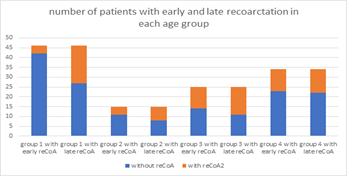
Figure 1: number of patients with early and late recoarctation in each age group
From the table (table 2) and figure (figure 1) above, it is evident that the early recoarctation rate was higher in older age groups compared to younger groups, and the chi-squared test showed a significantly different distribution (p=0.006). However, the number of patients with recoarctation changed significantly in younger age groups, especially in patients less than one year of age, and the percentage of patients with late recoarctation did not differ significantly between age groups (p=0.358). Overall, in older patients, the risk of recoarctation is higher than in younger patients, but this proportion remained stable with time. Meanwhile, the proportion of patients with early recoarctation can be lower in younger patients, but this tends to increase over time. This finding requires further investigation to assess the factors that affect younger patients in developing recoarctation later. Analyzing the influence of pre-operative echocardiographic findings on the development of early and late recoarctation yielded the following results (table 3).
Table 3: Analyzing the influence of pre-operative echocardiographic findings on the development of early and late recoarctation
| Information | Early recoarctation (n = 30) | No early recoarctation (n = 90) | p-value | Late recoarctation (n = 52) | No late recoarctation (n = 68) | p-value |
| Pressure gradient at coarctation site (mmHg) | 57.3 ± 19.3 | 52.8 ± 15.0 | 0.196 | 55.0 ± 17.0 | 52.9 ± 15.7 | 0.492 |
| Mitral regurgitation | 0.160 | 0.100 | ||||
| Without | 19 (63.3%) | 71 (78.9%) | 35 (67.3%) | 55 (80.1%) | ||
| Mild | 6 (20.0%) | 10 (11.1%) | 9 (17.3%) | 7 (10.3%) | ||
| Moderate | 5 (16.7%) | 8 (8.9%) | 8 (15.4%) | 1 (1.5%) | ||
| Severe | 0 | 1 (1.1%) | 0 | 4 (5.9%) | ||
| Tricuspid regurgitation | 0.442 | 0.565 | ||||
| Without | 24 (80.0%) | 75 (83.3%) | 43 (82.3%) | 56 (82.4%) | ||
| Mild | 3 (10.0%) | 11 (12.2%) | 5 (9.6%) | 9 (13.2%) | ||
| Moderate | 3 (10.0%) | 4 (4.5%) | 4 (7.7%) | 2 (2.9%) | ||
| Severe | 0 | 0 | 0 | 0 | ||
| Aortic regurgitation | 0.318 | 0.713 | ||||
| Without | 24 (80.0%) | 77 (85.5%) | 43 (82.7%) | 57 (83.8%) | ||
| Mild | 4 (13.3%) | 11 (12.2%) | 7 (13.4%) | 8 (11.8%) | ||
| Moderate | 2 (6.7%) | 2 (2.3%) | 2 (3.9%) | 2 (2.9%) | ||
| Severe | 0 | 0 | 0 | 0 | ||
| LV dilation | 19 (63.0%) | 32 (35.5%) | 0.007 ** | 27 (51.9%) | 23 (33.8%) | 0.054 |
| End-diastolic volume of LV | 80.4 ± 49.9 | 64.2 ± 33.1 | 0.045* | 67.5 ± 43.7 | 68.0 ± 33.8 | 0.943 |
| Ejection fraction of LV | 56.1 ± 16.2 | 44.4 ± 23.0 | 0.013* | 48.4 ± 22.2 | 46.9 ± 22.0 | 0.721 |
Our initial hypothesis suggesting that the severity of aortic coarctation influences the development of early and late recoarctation was not confirmed by statistical tests. The analyses revealed that only patients with left ventricular (LV) dysfunction, characterized by LV dilation, higher end-diastolic volume of the left ventricle (EDVLV), and lower ejection fraction of the left ventricle (EFLV), are at risk of early recoarctation. Additional comparison tests of CT scan findings could provide further insights into this topic (table 4).
Table 4: Analyzing the influence of pre-operative CT scan findings on the development of early and late recoarctation
| Information | Patients with early recoarctation (30 patients) | Patients without early recoarctation (90 patients) | p-value | Patients with late recoarctation (52 patients) | Patients without late recoarctation (68 patients) | p-value |
| Diameter of ascending aorta | 21.7 ±6.83 | 17.0 ±8.17 | 0.008** | 18.3 ±7.72 | 18.2 ±8.48 | 0.968 |
| z-score of ascending aorta | +2.13 ±1.13 | +1.69 ±1.21 | 0.098 | +1.76 ±1.153 | +1.83 ±1.25 | 0.750 |
| Diameter of aortic arch | 14.2 ±4.46 | 12.6 ±5.86 | 0.215 | 11.9 ±5.02 | 13.8 ±5.87 | 0.073 |
| z-score of aortic arch | -1.06 ±1.91 | -0.183 ±1.54 | 0.013* | -1.20 ±1.74 | +0.216 ±1.35 | <0> |
| Diameter of isthmus (coarctation site) | 4.66 ±2.20 | 4.48 ±2.82 | 0.766 | 4.23 ±2.24 | 4.73 ±2.96 | 0.348 |
| z-score of isthmus (coarctation site) | -6.11 ±2.44 | -4.64 ±2.14 | 0.004** | -5.82 ±2.54 | -4.44 ±1.93 | 0.002** |
| Diameter of descending aorta | 14.5 ±4.80 | 11.6 ±5.71 | 0.018* | 12.3 ±4.99 | 12.4 ±6.12 | 0.886 |
| z-score of descending aorta | +1.30 ±1.19 | +1.01 ±1.47 | 0.362 | +1.00 ±1.12 | +1.16 ±1.59 | 0.576 |
| Diameter of aorta at diaphragm level | 14.2 ±5.19 | 10.3 ±4.25 | 0.002** | 12.0 ±5.31 | 10.7 ±4.39 | 0.192 |
| z-score of aorta at diaphragm level | +1.45 ±1.42 | +0.715 ±1.42 | 0.022* | +1.15 ±1.38 | +0.731 ±1.49 | 0.144 |
Several factors, such as the sizes of the ascending and descending aorta, and the size and z-score of the aorta at the diaphragm level, were initially considered to be influenced by age. However, upon closer examination, early recoarctation was found to occur more frequently in older age groups, and these factors became non-significantly different in the late recoarctation check. Only two variables remained significantly different, namely the z-score of the aortic arch and the z-score of the isthmus. Consequently, the z-score of the aortic arch and the z-score of the isthmus were significantly lower in patients with early and late recoarctation. Many authors have reported [1,2,4] that one of the main risk factors for recoarctation is the surgical technique employed. To assess the impact of different operation types on recoarctation, we compared the incidence of recoarctation among four distinct surgical groups.
Table 5: Impact of different operation types on recoarctation
| Operation type groups (120 patients): | Patients with early recoarctation | Patients with late recoarctation |
| Group I (EEA) (27 patients) | 16 (59%) | 21 (77%) |
| Group II (PPA) (52 patients) | 14 (26.9%) | 23 (44%) |
| Group III (EEEA) (35 patients) | 0 | 8 (23%) |
| Group IV (PIG) (6 patients) | 0 | 0 |
| p-value | P<0.001*** | P<0.001*** |
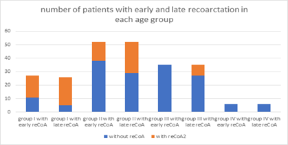
Figure 2: Number of patients with early and late recoarctation in each age group
The analysis revealed notable variations in the rates of recoarctation among different surgical techniques. Patients who underwent prosthetic interposition graft (PIG) did not exhibit any instances of early or late recoarctation. In contrast, resection and end-to-end anastomosis (EEA) displayed the highest proportions of early (59%) and late (77%) recoarctation. Prosthetic patch aortoplasty (PPA) showed a relatively higher rate of recoarctation, with 27% of patients experiencing early recoarctation and 44% exhibiting late recoarctation. Resection and extended end-to-end anastomosis displayed a comparatively lower rate, with 0% experiencing early recoarctation and 23% exhibiting late recoarctation.
Discussion
The data in the table reveals that the mean age of patients with early recoarctation was higher compared to patients without early recoarctation. Intriguingly, the mean age at the time of the operation was lower in patients who later developed late recoarctation. However, a comparison test (t-test) indicated no statistical significance (p=0.177). This implies that patient-related variables such as age, height, weight, gender, and BMI may not be causative factors in the development of early or late recoarctation. The clinical significance of age at the time of repair in relation to recoarctation has been underscored in several studies [6] [7]. Additionally, investigations encompassing various factors, such as weight before surgery, have yielded mixed findings [6-9]. Some studies have suggested a noteworthy association between lower weight at the time of repair and arch restenosis [9].
However, the complexity of these associations becomes apparent when considering multivariable models. Notably, Gorbatykh et al. [8] observed that weight did not emerge as a significant risk factor when included in a multivariable model alongside different surgical strategy. Furthermore, contrasting perspectives have been presented regarding birthweight and body length at surgery as potential risk factors. While some studies posit lower birthweight [10] and smaller body length at surgery [11] as risk factors, our series challenges these assertions, suggesting that these factors may not be conclusive indicators of susceptibility to early or late recoarctation.
Among the 28 patients (23.3%) who had arch hypoplasia, 12 of them experienced early recoarctation, and 22 of them exhibited late recoarctation. Interestingly, only 6 patients (21.4% of patients with arch hypoplasia) did not experience recoarctation. Correlation tests demonstrated a strong negative correlation of the z-score of the arch size with both early recoarctation (r=-0.229, p=0.013) and late recoarctation (r=-0.421, p<0.001). Therefore, it can be concluded that arch hypoplasia is one of the main risk factors for the development of both early and late recoarctation.
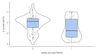
Figure 3: Statistics of z-score of aortic arch size in patients with and without early recoarctation
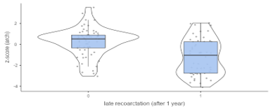
Figure 4: Statistics of z-score of aortic arch size in patients with and without late recoarctation
Numerous studies [6,7,8,10,11,12] have explored the relationship between recoarctation and aortic arch morphometry. Conflicting results have been reported, with some studies identifying a hypoplastic aortic arch as a significant risk factor. Hager et al. [11] reported that the presence of a hypoplastic arch increased the odds of developing recoarctation or experiencing mortality by 2.9 to 1. However, Gorbatykh et al. [8] demonstrated that a hypoplastic arch did not remain a determinant factor when incorporated into a multivariable regression model alongside different types of surgical strategies. Intriguingly, McElhinney et al. [7] found that a smaller transverse arch diameter was associated with an elevated risk of recoarctation, and this effect was more pronounced when indexed to weight. Additionally, Burch et al. [12] concluded that for every 1-mm increase in the transverse arch diameter, the risk for recoarctation decreased by 43%. This collective evidence underscores the importance of considering hypoplastic aortic arch as a crucial risk factor for recoarctation when deciding on the optimal repair strategy.
While assessing the impact of different surgical methods on developing recoarctation, it's important to note that prosthetic interposition graft (PIG) was considered a less risky group. However, this observation should be interpreted cautiously, as PIG was primarily performed in older patients whose growth had nearly concluded. Consequently, the lack of recoarctation in the PIG group may be attributed to limited patient growth, preventing the graft size from becoming insufficient. Therefore, while PIG demonstrated a lower risk in this specific context, it cannot be conclusively deemed a universally safe method for all patients. In summary, all methods of coarctation repair carry some risk of recoarctation, but resection and end-to-end anastomosis (EEA) and prosthetic patch aortoplasty (PPA) appear to have a higher risk than extended end-to-end anastomosis (EEEA).
Concerning the choice of surgical technique, the literature has emphasized that extended end-to-end anastomosis is considered a superior alternative for preventing recoarctation. This preference is attributed to the method's advantages, including a more extensive resection, preservation of the subclavian artery, and the use of an oblique anastomosis [13,14,15]. To assess the impact of operation type within distinct age groups, the obtained results are as follows (table 6).
Table 6: The impact of operation type within distinct age groups
| Age groups | Operation type groups | |||||||
| Group I (EEA) | Group II (PPA) | Group III (EEEA) | Group IV (PIG) | |||||
| Early reCoA | Late reCoA | Early reCoA | Late reCoA | Early reCoA | Late reCoA | Early reCoA | Late reCoA | |
| Group 1 (<1> | 4 (33%) | 8 (66.7%) | 0 | 3 (60%) | 0 | 8 (27.6%) | 0 | 0 |
| Group 2 (1-3 years) | 4 (80%) | 4 (80%) | 0 | 3 (75%) | 0 | 0 | 0 | 0 |
| Group 3 (3-10 years) | 5 (71%) | 6 (100%) | 6 (37.5%) | 8 (50%) | 0 | 0 | 0 | 0 |
| Group 4 (>10 years) | 3 (100%) | 3 (100%) | 8 (29.6%) | 9 (33.3%) | 0 | 0 | 0 | 0 |
The table illustrates that early recoarctation predominantly occurred in older age groups, particularly in cases involving EEA and PPA. EEA demonstrated a higher rate of early recoarctation across all age groups, with a more pronounced increase in older groups from an early stage, while in younger groups, it exhibited a tendency to rise over time. PPA exhibited more favorable short-term results in younger groups; however, the proportion of recoarctation in these groups significantly escalated, reaching up to 75%. In contrast, in older groups, although short-term results might be less favorable, they did not worsen significantly over time. EEEA exhibited the best short-term results with 0
Conclusion
The analysis of patient-related variables, including age, height, weight, gender, and BMI, suggests that these factors may not directly contribute to the development of early or late recoarctation. However, our findings indicate that aortic arch hypoplasia emerges as a significant risk factor for both early and late recoarctation, highlighting the importance of this anatomical characteristic in patient risk assessment and surgical planning.
Furthermore, while all methods of coarctation repair carry inherent risks of recoarctation, our study suggests that certain surgical techniques may pose a higher risk than others. Specifically, resection and end-to-end anastomosis (EEA) and prosthetic patch aortoplasty (PPA) appear to be associated with a greater likelihood of recoarctation compared to extended end-to-end anastomosis (EEEA).
In summary, our findings underscore the complexity of recoarctation development and its multifactorial nature. Recognizing the significance of aortic arch hypoplasia and considering the differential risks associated with various surgical techniques are essential for optimizing patient outcomes and reducing the incidence of recoarctation in individuals with coarctation of the aorta. Further research and ongoing surveillance are warranted to refine risk stratification strategies and improve the long-term management of patients undergoing surgical repair for coarctation of the aorta.
References
- Backer, C. L., & Mavroudis, C. (2014). Coarctation of the aorta. In C. Mavroudis & C. L. Backer (Eds.):Pediatric cardiac surgery. Mosby, 4th ed.:840–844.
Publisher | Google Scholor - Dias, M., Barros, A., Leite Moreira, A., & Miranda, J. (2020). Risk factors for recoarctation and mortality in infants submitted to aortic coarctation repair: A systematic review. Pediatric Cardiology, 41(4):561–575.
Publisher | Google Scholor - Baumgartner, H., Bonhoeffer, P., De Groot, N. M. S., de Haan, F., Deanfield, J. E., et al. (2010). ESC Guidelines for the management of grown-up congenital heart disease (new version 2010). European Heart Journal, 31(23):2915–2957.
Publisher | Google Scholor - Dodge-Khatami, A., Backer, C. L., & Mavroudis, C. (2000). Risk factors for recoarctation and results of reoperation: A 40-year review. Journal of Cardiac Surgery, 15(5):369–377.
Publisher | Google Scholor - Chaikof, E. L., Dalman, R. L., Eskandari, M. K., Jackson, B. M., Lee, W. A., Mansour, et al. (2018). The Society for Vascular Surgery practice guidelines on the care of patients with an abdominal aortic aneurysm. Journal of Vascular Surgery, 67(1):2–77.e2.
Publisher | Google Scholor - Lehnert, A., Villemain, O., Gaudin, R., Meot, M., Raisky, O., & Bonnet, D. (2019). Risk factors of mortality and recoarctation after coarctation repair in infancy. Interactive CardioVascular and Thoracic Surgery, 29(4):562–567.
Publisher | Google Scholor - McElhinney, D. B., Yang, S. G., Hogarty, A. N., Rychik, J., Gleason, M. M., et al. (2001). Recurrent arch obstruction after repair of isolated coarctation of the aorta in neonates and young infants: Is low weight a risk factor? The Journal of Thoracic and Cardiovascular Surgery, 122(5):883–890.
Publisher | Google Scholor - Gorbatykh, A., Nichay, N., Ivantsov, S., Voitov, A., Kulyabin, Y., Gorbatykh, Y., & Bogachev-Prokophiev, A. (2017). Risk factors for aortic coarctation development in young children. Pediatria, 96(3):118–124.
Publisher | Google Scholor - Soynov, I., Sinelnikov, Y., Gorbatykh, Y., Omelchenko, A., Kiseliov, I., Nichay, N., Bogachev-Prokophiev, A., & Karaskov, A. (2018). Modified reverse aortoplasty versus extended anastomosis in patients with coarctation of the aorta and distal arch hypoplasia. European Journal of Cardio-Thoracic Surgery, 53(1):254–261.
Publisher | Google Scholor - Truong, D. T., Tani, L. Y., Minich, L. L., Burch, P. T., Bardsley, T. R., & Menon, S. C. (2014). Factors associated with recoarctation after surgical repair of coarctation of the aorta by way of thoracotomy in young infants. Pediatric Cardiology, 35(1):164–170.
Publisher | Google Scholor - Hager, A., Schreiber, C., Nutzl, S., & Hess, J. (2009). Mortality and restenosis rate of surgical coarctation repair in infancy: A study of 191 patients. Cardiology, 112(1):36–41.
Publisher | Google Scholor - Burch, P. T., Cowley, C. G., Holubkov, R., Null, D., Lambert, L. M., Kouretas, P. C., & Hawkins, J. A. (2009). Coarctation repair in neonates and young infants: Is small size or low weight still a risk factor? The Journal of Thoracic and Cardiovascular Surgery, 138(3):547–552.
Publisher | Google Scholor - Lansman, S., Shapiro, A. J., & Schiller, M. S. (1986). Extended aortic arch anastomosis for repair of coarctation in infancy. Circulation, 74(Suppl. I):I137–I141.
Publisher | Google Scholor - Kaushal, S., Backer, C. L., Patel, J. N., et al. (2009). Coarctation of the aorta: Midterm outcomes of resection with extended end-to-end anastomosis. The Annals of Thoracic Surgery, 88(6):1932–1938.
Publisher | Google Scholor - Thomson, J. D., Mulpur, A., & Guerrero, R. (2006). Outcome after extended arch repair for aortic coarctation. Heart, 92(1):90–94.
Publisher | Google Scholor - Backer, C. L., Paape, K., & Zales, V. R. (1995). Coarctation of the aorta: Repair with polytetrafluoroethylene patch aortoplasty. Circulation, 92(Suppl. II):II132–II136.
Publisher | Google Scholor - Elzenga, N. J., Gittenberger-de Groot, A. C., & Groot, A. C. (1983). Localised coarctation of the aorta. British Heart Journal, 49(4):317–323.
Publisher | Google Scholor - Minotti, C., Scioni, M., Castaldi, B., Guariento, A., Biffanti, R., Di Salvo, G., Vida, V., & Padalino, M. (2022). Effectiveness of repair of aortic coarctation in neonates: A long-term experience. Pediatric Cardiology, 43(1):17–26.
Publisher | Google Scholor - Kotani, Y., Anggriawan, S., Chetan, D., et al. (2014). Fate of the hypoplastic proximal aortic arch in infants undergoing repair for coarctation of the aorta through a left thoracotomy. The Annals of Thoracic Surgery, 98(4):1386–1393.
Publisher | Google Scholor