
Case Report
Supra-Cardiac Partial Anomalous Pulmonary Venous Return in A 2-Month-Old Infant: A Case Report of Diagnostic Challenge
1 Department of Diagnostic Radiology, College of Medical Radiologic Sciences, Karary University, Khartoum, Sudan.
2 Department of Diagnostic Radiology, Kassala Advanced Diagnostic Center, Kassala, Sudan.
3 Department of Radiology and Medical Imaging, college of Medicine, Omdurman Islamic University, Khartoum, Sudan.
*Corresponding Author: Alhassan Hamed Alhassan,Department of Diagnostic Radiology, College of Medical Radiologic Sciences, Karary University, Khartoum, Sudan.
Citation: Alhassan H Alhassan, Awad M, Elshafie A (2026). Supra-Cardiac Partial Anomalous Pulmonary Venous Return in A 2-Month-Old Infant: A Case Report of Diagnostic Challenge. International Journal of Medical Case Reports and Reviews. BioRes Scientia Publishers. 6(2):1-6. DOI: 10.59657/2837-8172.brs.26.092
Copyright: © 2026 Alhassan H Alhassan, this is an open-access article distributed under the terms of the Creative Commons Attribution License, which permits unrestricted use, distribution, and reproduction in any medium, provided the original author and source are credited.
Received: July 15, 2026 | Accepted: July 29, 2026 | Published: August 25, 2026
Abstract
Introduction: Partial anomalous pulmonary venous return (PAPVR) is a rare congenital cardiovascular anomaly in which one or more pulmonary veins drain into the systemic venous circulation instead of the left atrium. Supra-cardiac PAPVR is an uncommon subtype and may present with nonspecific symptoms in infancy, resulting in delayed diagnosis or misdiagnosis, particularly in low-resource settings.
Case Presentation: We report the case of a 2-month-old male infant who presented with a 3-day history of progressively worsening shortness of breath, cough, poor feeding, and excessive sweating during breastfeeding. The infant had initially been treated for malaria because of an associated febrile illness. Physical examination revealed tachycardia, tachypnea, pallor, an active precordium, and a systolic murmur without cyanosis. Chest radiography demonstrated cardiomegaly with pulmonary vascular congestion. Transthoracic echocardiography showed anomalous pulmonary venous drainage, a moderate secundum atrial septal defect with bidirectional shunting, right-sided cardiac chamber dilatation, and preserved ventricular systolic function. Computed tomography angiography further delineated the anatomy, demonstrating a large vertical vein collecting the left pulmonary veins and draining into the left brachiocephalic vein and subsequently the superior vena cava, while the right pulmonary veins drained normally into the left atrium, confirming a diagnosis of supra-cardiac partial anomalous pulmonary venous return. The aortic arch and major branches were normal.
Conclusion: This case highlights the diagnostic challenges of supra-cardiac PAPVR in infancy, particularly in settings where infectious etiologies are more commonly suspected. Persistent respiratory distress, feeding difficulties, and cardiac murmurs in infants should prompt early cardiovascular evaluation. Echocardiography and computed tomography angiography play complementary roles in establishing the diagnosis and guiding management.
Keywords: partial anomalous pulmonary venous return; PAPVR; supra-cardiac anomalous pulmonary venous return; congenital heart disease; infant; computed tomography angiography
Introduction
Total Anomalous Pulmonary Venous Return (TAPVR), also referred to as total anomalous pulmonary venous connection (TAPVC), is a rare congenital cardiovascular anomaly characterized by the absence of a direct connection between the pulmonary veins and the left atrium. Instead, all pulmonary venous blood returns anomalously to the systemic venous circulation, ultimately draining into the right atrium. TAPVR accounts for approximately 1–3% of all congenital heart diseases and frequently presents during infancy with cyanosis, respiratory distress, feeding difficulties, and congestive heart failure, particularly in obstructed forms [1].
TAPVR is traditionally classified into four anatomical subtypes according to the site of anomalous pulmonary venous drainage: supra-cardiac (type I), cardiac (type II), infra-cardiac (type III), and mixed type (type IV). Among these, the supra-cardiac variant (type I) is the most common subtype, accounting for nearly 45–50% of cases. In supra-cardiac TAPVR, the pulmonary venous confluence typically drains through a vertical vein into the innominate (brachiocephalic) vein, superior vena cava (SVC), and eventually the right atrium. Less common anatomical variants include direct drainage into the right SVC [2].
The pathophysiology of supra-cardiac TAPVR involves complete mixing of oxygenated and deoxygenated blood in the right atrium, making survival dependent on the presence of an interatrial communication, usually an atrial septal defect or patent foramen ovale. Clinical severity varies according to the presence or absence of pulmonary venous obstruction. Obstructed TAPVR often presents as a neonatal emergency associated with severe pulmonary hypertension, profound cyanosis, and hemodynamic instability requiring urgent surgical intervention [3].
Early diagnosis using transthoracic echocardiography is essential; however, advanced imaging modalities such as computed tomography angiography (CTA) and magnetic resonance imaging (MRI) are increasingly utilized to delineate complex pulmonary venous anatomy and assist in preoperative planning, especially in atypical or rare anatomical variants. Surgical correction remains the definitive treatment and aims to establish unobstructed pulmonary venous drainage into the left atrium while ligating the anomalous venous pathway. Despite improvements in surgical outcomes, postoperative pulmonary venous obstruction remains an important complication influencing long-term prognosis [4].
Given the rarity of supra-cardiac TAPVR and its diverse anatomical presentations, reporting individual cases remains valuable for expanding the current understanding of imaging findings, diagnostic challenges, surgical management, and clinical outcomes. Therefore, we present a case of supra-cardiac type I TAPVR to highlight its radiological characteristics and clinical significance [5].
Case Presentation
A 2-month-old male infant, born at term via normal vaginal delivery in a hospital setting, presented with respiratory and feeding difficulties. The infant cried immediately after birth and had an unremarkable immediate neonatal period, including passage of urine and meconium within the first 24 hours. He was apparently well until approximately three days prior to presentation, when he developed progressive shortness of breath associated with cough and intermittent low-grade fever. The caregiver reported poor feeding, with breastfeeding lasting less than five minutes and accompanied by excessive sweating. The symptoms gradually worsened over the three-day period before presentation. There was no history of cyanosis, seizures, or loss of consciousness.
The infant had a recent febrile illness for which he was treated as a case of malaria and reportedly received two doses of artesunate. There was no history of previous hospitalizations, surgeries, or blood transfusions. He was exclusively breastfed without supplemental feeding, and developmental milestones were appropriate for age. There was no family history of congenital anomalies or chronic illnesses. Although the family belonged to a low socioeconomic background, they had access to adequate housing and water supply. No known drug or food allergies were reported. Review of systems was unremarkable except for respiratory symptoms, including cough and shortness of breath, and cardiovascular symptoms suggestive of cardiac involvement, including feeding difficulty and excessive sweating. No neurological symptoms such as seizures or abnormal movements were reported.
On examination, the infant appeared irritable, mildly febrile, and mildly distressed but clinically stable. General examination revealed tachycardia, tachypnea, pallor, and dysmorphic facial features suggestive of Down syndrome. No cyanosis or jaundice was observed. Anthropometric assessment showed a weight of 4.3 kg and a length of 53 cm. Vital signs demonstrated a heart rate of 125 beats/min, blood pressure of 75/50 mmHg, and oxygen saturation of 84% on room air, with normal peripheral pulses and capillary refill time of less than 2 seconds.
Cardiovascular examination revealed an active precordium with the apex beat localized at the fifth intercostal space and a grade 3/6 systolic murmur best heard over the left upper sternal border, raising suspicion of an underlying structural cardiac defect. Respiratory examination demonstrated equal bilateral air entry with a clear chest on auscultation and no added sounds. Abdominal examination revealed mild hepatomegaly (approximately 2 cm below the costal margin) with a soft and lax abdomen. Neurological examination was unremarkable, with the infant conscious, alert, active, and exhibiting normal tone, power, and pupillary responses.
Based on the clinical presentation and examination findings, congenital heart disease was initially suspected, with ventricular septal defect (VSD) considered as a provisional diagnosis. Chest radiography demonstrated cardiomegaly with pulmonary vascular congestion. figure [1] Transthoracic echocardiography revealed situs solitus with levo-cardia and normal atrioventricular and ventriculoarterial concordance. All four pulmonary veins formed a common confluence draining through an ascending vertical vein into the innominate vein, superior vena cava, and subsequently the right atrium, consistent with supra-cardiac total anomalous pulmonary venous return (TAPVR) without evidence of pulmonary venous obstruction. A moderate-sized secundum atrial septal defect (ASD II) with bidirectional shunting was identified. Additional echocardiographic findings included dilation of the superior vena cava, right atrium, and right ventricle, a mildly small left atrium, mild tricuspid regurgitation, preserved left ventricular systolic function with an ejection fraction of 73%, and absence of pulmonary hypertension. No patent ductus arteriosus, pericardial effusion, or significant valvular abnormalities were noted.
Computed tomography angiography (CTA) of the chest was subsequently performed for further anatomical delineation and surgical planning. CTA demonstrated a large vertical vein collecting the left pulmonary veins and draining into the left brachiocephalic vein and subsequently into the superior vena cava figure [2, 3], while the right pulmonary veins drained normally into the left atrium figure [4], consistent with a supra-cardiac anomalous pulmonary venous return pattern. The aortic arch and its major branches were normal, with no associated vascular abnormalities identified figure [5].
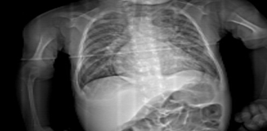
Figure 1: chest x-ray showed: cardiomegaly with pulmonary vascular congestion.
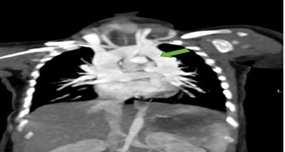
Figure 2: Coronal MIP images of heart great vessels, showed: green arrow representing large vertical vein draining the superior and inferior pulmonary veins of the left side. Which drains in to left brachiocephalic to superior vena cava.
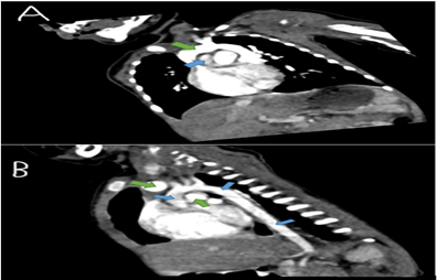
Figure 3: A, B sagittal MIP images showed: green arrow large vertical vein that drain in brachiocephalic vein, blue arrow normal (ascending, arch, descending) aorta.
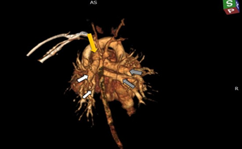
Figure 4: A, B 3D image of great vessel heart showed:(white arrow left, gray arrow right) pulmonary veins, main pulmonary vein (yellow arrow main).
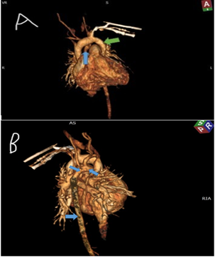
Figure 5: A, B 3D image showed: green arrow large vertical vein, blue arrow normal (ascending, arch, descending) aorta.
Discussion
Partial anomalous pulmonary venous return (PAPVR) is an uncommon congenital cardiovascular anomaly characterized by anomalous drainage of one or more, but not all, pulmonary veins into the systemic venous circulation rather than the left atrium. In contrast to total anomalous pulmonary venous return (TAPVR), where all pulmonary veins drain abnormally, PAPVR preserves normal drainage of at least one pulmonary venous system into the left atrium. PAPVR is rare, accounting for approximately 0.4–0.7% of congenital cardiac anomalies, and may remain clinically silent or present with symptoms depending on the degree of left-to-right shunting and associated cardiac abnormalities, particularly atrial septal defects (ASDs) [6,7].
The present case describes a 2-month-old infant who presented with progressive respiratory distress, poor feeding, diaphoresis during breastfeeding, and recurrent symptoms suggestive of congestive heart failure. These clinical manifestations are consistent with increased pulmonary blood flow and right heart volume overload, commonly observed in anomalous pulmonary venous return anomalies [8]. Feeding difficulty and excessive sweating in infancy are well-recognized indicators of cardiac insufficiency and increased metabolic demand in congenital heart disease [9].
An important feature of this case was the absence of obvious cyanosis despite low oxygen saturation and the presence of congenital cardiac pathology. In PAPVR, cyanosis is generally less prominent than in TAPVR because part of the pulmonary venous return reaches the left atrium normally, thereby preserving a degree of systemic oxygenation [10]. This may contribute to delayed diagnosis, particularly in infants presenting predominantly with respiratory symptoms that mimic infectious conditions such as pneumonia or bronchiolitis.
In the present case, the infant had initially received treatment for malaria because of fever, highlighting a diagnostic challenge in resource-limited and endemic settings where infectious diseases are often considered before congenital cardiac disorders. Persistent respiratory symptoms, poor feeding, sweating, cardiomegaly, and the presence of a systolic murmur should prompt consideration of an underlying congenital cardiac lesion, particularly when clinical improvement is not achieved with standard treatment for presumed infections [11].
Echocardiography played a crucial role in the initial diagnosis by demonstrating anomalous pulmonary venous drainage, a moderate secundum atrial septal defect with bidirectional shunting, right-sided chamber dilatation, and preserved ventricular systolic function. However, computed tomography angiography (CTA) provided superior anatomical characterization and was instrumental in confirming the diagnosis and defining the drainage pathway for surgical planning. In this patient, CTA demonstrated a large vertical vein collecting the left pulmonary veins and draining into the left brachiocephalic vein and subsequently the superior vena cava, while the right pulmonary veins drained normally into the left atrium. This drainage pattern is consistent with a supra-cardiac form of PAPVR involving the left pulmonary venous system. Furthermore, the aortic arch and major branches were normal, with no additional vascular anomalies identified. Cross-sectional imaging modalities such as CTA are particularly valuable in defining complex pulmonary venous anatomy and avoiding misclassification of pulmonary venous anomalies [12].
The coexistence of a moderate secundum ASD in this patient is clinically important, as ASDs are frequently associated with PAPVR and may contribute to right-sided volume overload and symptom severity [7,10]. The observed dilation of the right atrium and right ventricle on echocardiography likely reflects increased pulmonary venous return to the systemic venous circulation and chronic right-sided hemodynamic burden. Despite the absence of pulmonary hypertension or pulmonary venous obstruction in our patient, early surgical intervention remains necessary to prevent long-term complications, including progressive right ventricular dysfunction, pulmonary vascular disease, recurrent respiratory infections, and failure to thrive [13].
This case highlights the importance of maintaining a high index of suspicion for congenital cardiovascular anomalies in infants presenting with respiratory distress, feeding intolerance, and recurrent hospitalization, particularly in low-resource settings. It also emphasizes the complementary role of echocardiography and CTA in accurately delineating pulmonary venous anatomy and guiding definitive surgical management.
Declarations
Acknowledgements
The authors would like to thank Kassala advance diagnostic center for their support in preparing this case report.
Conflicts of interest
The authors declare no conflicts of interest.
Funding
No funding was received for this work
Ethics approval
Not required for this anonymised clinical image.
Consent
Written informed consent for publication of the images and clinical details was obtained from the patient.
Guarantor
Dr Alhassan Hamed Alhassan accepts full responsibility for the integrity of the content of this submission.
References
- Haas NA, Driscoll DJ, Rickert-Sperling S (2024). Clinical presentation and therapy of total anomalous pulmonary venous return. Adv Exp Med Biol.,1441:587–591.
Publisher | Google Scholor - Hirsch JC, Bove EL (2007). Total anomalous pulmonary venous connection. Multimed Man Cardiothorac Surg.,
Publisher | Google Scholor - Liu X, Liufu R, Liu T, et al., (2023). Supracardiac total anomalous pulmonary venous connection type Ib: morphology and outcomes. J Thorac Cardiovasc Surg.,166(1):193–200.e1.
Publisher | Google Scholor - Tucker BL, Lindesmith GG, Stiles QR, Meyer BW (1976). The superior approach for correction of the supracardiac type of total anomalous pulmonary venous return. Ann Thorac Surg.,22(4):374–377.
Publisher | Google Scholor - Zhu Y, et al., (2017). Prenatal diagnoses of an uncommon isolated obstructed supracardiac total anomalous pulmonary venous connection: Case report and review of the literature. Medicine (Baltimore).96(5): e5897.
Publisher | Google Scholor - Ho ML, Bhalla S, Bierhals A, Gutierrez F (2009). MDCT of partial anomalous pulmonary venous return (PAPVR) in adults. J Thorac Imaging.24(2):89–95.
Publisher | Google Scholor - Healey JE Jr. (1952). An anatomic survey of anomalous pulmonary veins: their clinical significance. J Thorac Surg.,23(5):433–444.
Publisher | Google Scholor - Kliegman RM, St Geme JW (2023). Nelson Textbook of Pediatrics. 22nd ed. Philadelphia: Elsevier.
Publisher | Google Scholor - Allen HD, Driscoll DJ, Shaddy RE, Feltes TF (2016). Moss & Adams’ Heart Disease in Infants, Children, and Adolescents. 9th ed. Philadelphia: Wolters Kluwer.
Publisher | Google Scholor - Dillman JR, Yarram SG, Hernandez RJ (2009). Imaging of pulmonary venous developmental anomalies. AJR Am J Roentgenol.,192(5):1272–1285.
Publisher | Google Scholor - Seale AN, et al., (2009). Delay in diagnosis of congenital heart disease: a systematic review. Arch Dis Child.,94(6):468–473.
Publisher | Google Scholor - Haramati LB, Moche IE, Rivera VT, Patel PV, Heyneman L, et al., (2003). Computed tomography of partial anomalous pulmonary venous connection in adults. J Comput Assist Tomogr.,27(5):743–749.
Publisher | Google Scholor - Alsoufi B, Cai S, Van Arsdell GS, et al., (2007). Outcomes after surgical treatment of partial anomalous pulmonary venous connection. Ann Thorac Surg.,84(6):2020–2026.
Publisher | Google Scholor