
Case Report
Small Bowel Obstruction Secondary to Band Adhesions from An Unruptured Ectopic Pregnancy-A Case Report
- Asfaw Kibret Msc, IESO 1*
- Getachew Worku MD 2
- Talaksew Fasika MD 3
- Chernet Lema Msc 4
- Thomas Ameno Msc 5
1 Department of emergency surgery, Leku general hospital, Sidama, Ethiopia.
2 Department of ECCM, Leku general hospital, sidama, Ethiopia.
3 Department of Surgery, Leku general hospital, sidama, Ethiopia.
4 Department of Emergency surgery, Aletawondo general hospital, sidama, Ethiopia.
5 Department of Anesthesia, Leku general hospital, sidama, Ethiopia.
*Corresponding Author: Asfaw Kibret Msc, IESO, Department of emergency surgery, Leku general hospital, Sidama, Ethiopia.
Citation: Kibret A., Worku G., Fasika T, Lema C., Ameno T. (2026). Small Bowel Obstruction Secondary to Band Adhesions from An Unruptured Ectopic Pregnancy. A Case Report, Clinical Case Reports and Studies, BioRes Scientia Publishers. 13(2):1-6. DOI: 10.59657/2837-2565.brs.26.344
Copyright: © 2026 Asfaw Kibret, this is an open-access article distributed under the terms of the Creative Commons Attribution License, which permits unrestricted use, distribution, and reproduction in any medium, provided the original author and source are credited.
Received: July 27, 2026 | Accepted: August 10, 2026 | Published: August 17, 2026
Abstract
Background: Small bowel obstruction (SBO) in a "virgin abdomen" is rare, typically caused by congenital bands, internal hernias, or malignancies. SBO complicating pregnancy is exceptionally rare (0.001% to 0.003%). While hemoperitoneum from ruptured ectopics can cause paralytic ileus, an unruptured tubal pregnancy causing mechanical SBO via localized inflammatory adhesions is an extreme clinical rarity. This paper presents such a case with a review of its integrated pathophysiology.
Case Presentation: A 25-year-old female with no surgical history presented with acute bowel obstruction and hemodynamic compromise. Evaluation confirmed a positive urine hCG, while ultrasound showed dilated small bowel loops and a right adnexal mass. During emergency exploratory laparotomy, an unruptured right tubal ectopic pregnancy was found anchoring the small bowel via a dense inflammatory band. The band was released, bowel viability returned, and a right salpingectomy was performed. Recovery was uneventful.
Conclusion: An unruptured tubal pregnancy is a vital differential diagnosis for acute mechanical SBO in reproductive-aged females without prior surgery. In resource-limited settings, qualitative hCG and basic ultrasound are critical diagnostic tools. Additionally, it validates the clinical impact of the Integrated emergency surgical officers (IESOs) task-shifting model, showing how integrated surgical capabilities can bypass resource barriers to provide timely, definitive, and life-saving dual-specialty emergency care.
Keywords: small bowel obstruction; unruptured ectopic pregnancy; band adhesions; virgin abdomen; integrated emergency surgical officer (ieso); low-resource setting
Introduction
Small bowel obstruction (SBO) is a critical surgical emergency, accounting for roughly 20% of acute surgical admissions globally [1]. The vast majority of SBO cases (approximately 70% to 80%) result from intra-abdominal postoperative adhesions [2]. Conver-sely, a mechanical bowel obstruction developing in a "virgin abdomen"—where no prior laparotomy, trauma, pelvic inflammatory disease, or clinical hernia exists—is relatively rare, often caused by congenital bands, internal hernias, intussusception, or malignan-cies [4]. When mechanical bowel obstruction occurs during pregnancy or alongside gynecological emergen-cies, it presents a life-threatening diagnostic and therapeutic challenge. The incidence of SBO complicating pregnancy is estimated to be exceedingly low, between 0.001% and 0.003% [1, 2]. Delayed diagnosis in these cases drastically increases both maternal mortality and fetal loss [2]. While ruptured ectopic pregnancies have occasionally been reported to cause localized peritonitis, chemical irritation, or paralytic ileus due to massive hemoperitoneum [5], an unruptured tubal ectopic pregnancy inducing an acute mechanical SBO via localized band adhesions is an exceptionally rare clinical phenomenon.
An unruptured ectopic pregnancy typically presents with localized pelvic pain, amenorrhea, and abnormal vaginal bleeding. However, when the fallopian tube or adjacent inflammatory tissue forms an adhesive band that constricts or loops around the small intestine, the classic gynecological presentation is completely masked by aggressive acute gastrointestinal symptoms such as bilious vomiting, severe abdominal distention, and obstipation. This case report documents a rare presentation of an acute mechanical small bowel obstruction secondary to localized band adhesions caused by an unruptured tubal ectopic pregnancy in a 25-year-old female. We highlight the clinical presentation, resource-limited diagnostic challenges, and successful surgical management of this condition to remind clinicians to consider pelvic pathologies when managing mechanical bowel obstructions in reproductive-aged females presenting with an acute abdomen.
Case Presentation
A 25-year-old female patient presented to our resource-limited emergency department with a chief complaint of severe abdominal pain of one day’s duration. The pain was acute, generalized, and characterized by a distinct crampy, colicky nature. Associated with the pain, she experienced repeated episodes of bilious vomiting, obstipation and rapidly progressive abdominal distention. She had no history of prior abdominal or pelvic surgeries, no documented chronic medical conditions, and no reported history of external hernias. No hx of recent vaginal bleeding. She discontinued injectable family planning method 6months back and doesn’t see her regular menstrual bleeding yet after the Depo-Provera injection. Upon physical examination, the patient was acutely sick-looking, dehydrated, presenting with sunken eyes, a dry buccal mucosa, and a dry tongue. Her vital signs revealed hemodynamic compromise: her blood pressure (BP) was 90/60 mmHg, her pulse rate (PR) was tachycardic at 115 beats per minute, her oxygen saturation (SpO2) was 94% on room air, and she was hypothermic with a temperature of 35.8°C. Abdominal examination demonstrated moderate abdominal distention with hyper-tympanic percussion notes. Digital rectal examination revealed an empty rectum.
Aggressive fluid resuscitation was immediately initiated using intravenous crystalloids, and a nasogastric tube was placed for gastrointestinal decompression. Emergency available laboratory investigations were ordered (cbc-8000, hgb-12gm/dl, PLT-234000), and a qualitative urine Human Chorionic Gonadotropin (hCG) test was performed, which yielded a positive result [7], confirming an early pregnancy of unknown location. Due to the lack of advanced cross-sectional imaging in our resource-limited facility, a bedside transabdominal abdominopelvic ultrasound was performed. The ultrasound demonstrated markedly dilated, fluid-filled small bowel loops with a distinct distal collapse, though the precise mechanical transition point could not be demonstrated due to extensive bowel gas artifacts. Crucially, the pelvic view revealed an empty uterus with a right-sided, complex cystic adnexal lesion located extrauterine. The radiologist concluded that the patient had an acute small bowel obstruction of undetermined mechanical cause, alongside a right-sided complex cystic mass highly suspicious of an extrauterine gestational sac, likely representing an ectopic pregnancy. Immediate exploratory laparotomy was recommended.
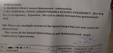
Figure 1: abdomenopelvic U/S report
Following stabilization of her blood pressure and heart rate with fluid resuscitation, the patient was taken to the operating theater for an emergency open abdominal exploration, performed by an Integrated Emergency Surgical Officer (IESO) [6]. Upon entering the peritoneal cavity via a lower midline laparotomy, there was marked dilation of the proximal small bowel. Deep pelvic exploration revealed a dense, inflammatory band adhesion arising from the rt adnexal complex extending to the small bowel mesentery loops, obstructing the bowel loops and anchoring firmly to the pelvic wall. This complex adnexal mass was subsequently identified as an intact, unruptured right-sided tubal ectopic pregnancy.
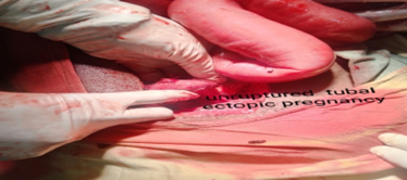
Figure 2: Intra-operative image of unraptured tubal ectopic pregnancy
The entrapped small bowel loop demonstrated a clear "band tag sign" at the site of constriction, with a localized, slight duskiness and color change indicative of early ischemic stress [8]. The inflammatory band was sharply cut and released. Following decompression and the application of warm saline-soaked laparotomy sponges for several minutes, the compromised segment of the small bowel rapidly regained its normal pink color, active peristalsis, and palpable mesenteric pulsations, confirming excellent bowel viability and eliminating the need for intestinal resection.
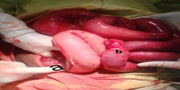
Figure 3: (a) Intra-operative images of unruptured tubal ectopic pregnancy and (b) dialated small bowel loops with clear band tag sign after release of the band.
Definitive management of the pelvic pathology was completed by performing a right salpingectomy to completely excise the unruptured ectopic pregnancy. Thorough warm saline peritoneal lavage was executed to clear any inflammatory debris. The abdomen was closed in layers. The patient’s postoperative course was smooth and completely uneventful, with a rapid return of bowel function, and she was successfully discharged home in stable condition.
Discussion & Review of Literature
Contextualizing the Case within Existing Literature
Mechanical small bowel obstruction secondary to pelvic pathology poses a formidable challenge to general surgeons and gynecologists alike. While postoperative adhesions remain the leading cause of SBO overall [2], the development of a mechanical adhesive band originating from an intact, unruptured fallopian tube hosting an ectopic gestation represents an extreme clinical outlier [3]. The intersection of SBO and pregnancy is broadly categorized in literature as a high-mortality clinical scenario. Historically, maternal mortality from SBO during pregnancy reached as high as 20% to 30%, though modern advancements in surgical resuscitation have lowered this to roughly 5% [1]. However, the vast majority of these reported obstructions occur during the second and third trimesters of a viable intrauterine pregnancy [1, 2]. During these later stages, mechanical obstruction is physically triggered by the rapidly expanding gravid uterus, which shifts positions, causing direct extrinsic compression or traction on pre-existing postoperative adhesive bands, or trapping a loop of the small bowel in the pelvic basin [2].
In contrast, intestinal complications resulting from extrauterine/ectopic pregnancies follow a completely different pathophysiological timeline and display a distinct scarcity in surgical registries. The predominant body of literature surrounding bowel issues caused by ectopic gestations features paralytic ileus or subacute adynamic obstructions secondary to ruptured tubal pregnancies [5]. In those scenarios, the driving force is chemical peritonitis and massive hemoperitoneum. Blood accumulation within the peritoneal cavity acts as an irritant to the myenteric plexus of the intestines, paralyzing smooth muscle peristalsis and presenting clinically as a pseudo-obstruction. Saha et al. documented a rare instance where a ruptured tubal mass formed a massive, organized pelvic hematoma over several weeks, which subsequently compressed the rectum and terminal ileum [5]. True mechanical luminal occlusion caused by an unruptured ectopic pregnancy in a patient without prior operations is almost entirely missing from standard surgical literature. A sparse number of historical case reports describe unruptured tubal pregnancies causing mechanical SBO, but these are almost universally associated with a history of prior pelvic surgeries, such as previous cesarean deliveries or cystectomies, where old adhesions acted as the physical bridge [3]. Al-Wadaani documented a baseline vulnerability where the fallopian tube itself, suffering from severe chronic pelvic inflammatory disease (PID) or endometriosis, became abnormally elongated and mobile, creating an internal hernia loop that ensnared the bowel [3]. The presentation of a completely unruptured, early first-trimester tubal mass independently synthesizing an active, de novo inflammatory band of sufficient strength to crimp and occlude the small bowel in a strictly "virgin abdomen" represents an isolated anomaly [4, 8].
Cellular and Biochemical Pathophysiology of Adhesion Formation
The biological mechanism behind the rapid formation of a mechanical band adhesion by an intact, unruptured fallopian tube hosting an ectopic gestation involves an aggressive local cellular cascade. Under normal physiologic conditions, the pelvic peritoneum maintaining the fallopian tube is coated with a single layer of mesothelial cells that secrete lubricants to ensure smooth, unhindered movement of adjacent small bowel loops. However, during a tubal ectopic pregnancy, the extrauterine blastocyst aggressively invades the highly vascularized, thin walls of the fallopian tube. This invasive trophoblastic migration, coupled with the progressive mechanical stretching of the muscularis layer, induces a profound localized, transmural inflammatory response. This microenvironment generates high concentrations of localized pro-inflammatory cytokines, specifically tumor necrosis factor-alpha (TNF-alpha), interleukin-1 (IL-1), and interleukin-6 (IL-6), alongside potent angiogenic signaling molecules like vascular endothelial growth factor (VEGF).
This focal chemical peritonitis alters local vascular permeability, triggering the exudation of a protein-rich fluid high in fibrinogen into the immediate pelvic basin. This fluid rapidly polymorphs into a sticky, gelatinous fibrin matrix. Under normal circumstances, the body’s endogenous peritoneal fibrinolytic system—primarily driven by tissue plasminogen activator (tPA)—destroys these temporary fibrin gels within a few days. However, the continuous ischemic and inflammatory stress exerted by the expanding unruptured tubal tissue suppresses tPA production while overexpressing plasminogen activator inhibitor-1 (PAI-1). This shifts the physiological balance toward permanent adhesion formation. This hyper-coagulable fibrin gel acts as a physical mesh that recruits the highly mobile omentum or low-hanging loops of the terminal ileum and its mesentery. Within a remarkably brief window often less than 24 to 48 hours circulating fibroblasts migrate into this persistent fibrin network, laying down collagen fibrils that mature into a taut, vascularized, and inelastic fibrotic band. When this band anchors tightly between the adnexal complex and the rigid pelvic wall, any subsequent movement of the small intestine can cause the bowel loop to slip beneath or twist around the band. This results in the "band tag sign" seen intraoperatively: a focal point of intense mechanical constriction leading to immediate localized venous congestion, luminal occlusion, and early ischemic duskiness [8].
Diagnostic Resolution Amid Substantial Structural Constraints
This case underscores a monumental diagnostic dilemma. The classic diagnostic triad of an ectopic pregnancy captioned as amenorrhea, localized unilateral pelvic pain, and abnormal vaginal bleeding was entirely absent or completely overshadowed by the hyper-acute, dramatic presentation of a mechanical proximal SBO. The patient's clinical picture was dominated by severe colicky abdominal pain, repetitive bilious emesis, absolute obstipation, and progressive abdominal distention. Furthermore, her advanced state of dehydration (sunken eyes, dry mucosa, tachycardia of 115 bpm, and hypotension of 90/60 mmHg) demanded immediate, aggressive resuscitation, leaving little initial window for complex diagnostic reflection. In high-resource tertiary healthcare systems, this diagnostic ambiguity is quickly resolved through an array of advanced technologies. Quantitative serum beta -hCG tracking provides an exact biochemical window into gestational status, while contrast-enhanced Computed Tomography (CT) or Magnetic Resonance Imaging (MRI) effortlessly displays the mechanical transition point of the bowel alongside the exact dimensions of the adnexal pathology.
In our resource-limited hospital setup, the complete absence of these modalities forced an absolute reliance on basic, universally available point-of-care tools: a qualitative urine hCG strip and a transabdominal ultrasound. While a qualitative urine hCG is frequently dismissed in modern Western protocols as inadequate due to its inability to monitor trend lines or confirm viability, it remains an invaluable tool in low-resource environments [7]. Its positive status instantly alters the clinical paradigm for any female patient of reproductive age presenting with an acute abdomen, mandating that the clinician evaluate for a pregnancy-related complication regardless of the prominence of gastrointestinal signs.
However, interpreting a pelvic ultrasound in the face of an active SBO introduces massive technical challenges. The immense volume of trapped intraluminal gas and fluid within the dilated small bowel loops creates a massive acoustic barrier, generating widespread gas artifacts that blind the transabdominal probe. We bypassed this by utilizing a targeted pelvic window, confirming an empty uterine cavity alongside a right-sided, complex extrauterine cystic mass. This combination—a positive pregnancy test, an empty uterus, a complex adnexal lesion, and concurrent mechanical bowel dilation—allowed us to safely bypass the need for an advanced CT scan and confidently proceed directly to life-saving surgical intervention.
Task-Shifting and the Clinical Value of the IESO Framework
Laparoscopic management is globally recognized as the gold standard for treating both early, unruptured ectopic pregnancies and mechanical small bowel obstructions due to minimized tissue manipulation, lower rates of secondary adhesion formation, and shorter hospital stays. However, in our setting, an open lower midline laparotomy was chosen. In a resource-restricted facility, open surgery provides a critical safety buffer: it permits rapid manual exploration of the entire small bowel from the ligament of Treitz to the ileocecal valve, allows the operator to physically feel changes in bowel wall compliance, and facilitates local resuscitation using warm-saline packs without relying on complex laparoscopic gas insufflators or specialized video towers. The most vital systemic lesson from this case lies in the human resource framework that executed the operation: the Integrated Emergency Surgical Officer (IESO) model [6]. IESOs are clinical professionals dully known in Ethiopia explicitly trained through an intensive, integrated postgraduate curriculum to execute emergency general surgery, obstetrics, and gynecology in rural, underserved, and low-resource healthcare ecosystems [6,10]. This task-shifting strategy is a cornerstone response to the profound deficit of specialized surgeons and gynecologists in developing regions.
A standard, highly segregated healthcare delivery model would struggle significantly with a case of this nature. Because the pathology simultaneously spanned two entirely distinct surgical fields—general emergency surgery (requiring SBO band identification, lysis, and bowel viability assessment) and gynecology (requiring salpingectomy for an ectopic mass)—it would traditionally demand multiple specialty consultations or a highly dangerous, prolonged patient transfer to a tertiary institution. Such a delay would inevitably culminate in bowel necrosis, tubal rupture, massive internal hemorrhage, and maternal mortality. The integrated scope of the IESO completely removed these institutional barriers [6]. Because a single clinician possessed verified competency in both abdominal emergency surgery and pelvic gynecology, they were able to seamlessly transition from lysing the strangulating inflammatory band to performing a definitive right salpingectomy in a single, uninterrupted operative window. This case serves as a powerful validation of the IESO framework, proving that integrated surgical training directly optimizes clinical throughput, bypasses structural specialist shortages, and actively saves lives when managing complex, overlapping multi-system emergencies [9,10].
Conclusion
An unruptured tubal ectopic pregnancy is an exceptional but highly critical cause of mechanical small bowel obstruction via band adhesions in young women with a virgin abdomen. This case underscores the necessity of maintaining a high index of suspicion and routinely testing pregnancy status via qualitative urine hCG in reproductive-aged females presenting with an acute abdomen. Furthermore, it validates the clinical impact of the IESO task-shifting model, showing how integrated surgical capabilities can bypass resource barriers to provide timely, definitive, and life-saving dual-specialty emergency care.
Declarations and Statements
Ethics Approval and Consent to Participate
Institutional review board approval was waived as this text contains a single anonymized case report with no direct identifying details.
Consent for Publication
Written informed consent was obtained from the patient for publication of this case report and accompanying clinical/radiological findings. A copy of the written consent form is available for review by the Editor-in-Chief of this journal.
Availability of Data and Materials
All data supporting our findings are contained entirely within the body of this manuscript.
Competing Interests
The authors declare that they have no competing financial or personal interests.
Funding
This work received no external funding or financial support from any public or commercial entity.
Authors' Contributions
AK- operated on the patient and designed the clinical treatment plan, collected the clinical/ultrasound data and drafted the initial Case Presentation, Conceptualize, reviewed the paper. And GW, CL, TF, TA, reviewed, heavily edited, and approved the final manuscript for submission.
Acknowledgments
The authors would like to acknowledge the emergency nursing staff, ward nursing staffs and operation theater teams for their dedicated patient care under resource-limited hospital conditions.
References
- Goussous, N., Nightingale, C. E., Das, K., et al. (2016). Small bowel obstruction in pregnancy is a complex surgical and obstetric challenge: A review of contemporary literature. BMC Surgery, 16(1):70.
Publisher | Google Scholor - Okafor, C., Sharma, D. G., & Al-Khaffaf, H. (2021). Adhesive small bowel obstruction in pregnancy: Case report and a review of the literature. The Journal of Maternal-Fetal & Neonatal Medicine, 34(11):1850–1856.
Publisher | Google Scholor - Al-Wadaani, H. (2018). Fallopian tube as an unusual cause of intestinal obstruction: A rare case report with review of the literature. International Journal of Surgery Case Reports, 49:124–127.
Publisher | Google Scholor - Ramos-Sosa, J., Marín-Méndez, S., et al. (2024). Band adhesions in a virgin abdomen: A case report of mechanical bowel obstruction. Annals of Medicine and Surgery, 86(4):2100–2104.
Publisher | Google Scholor - Saha, P. K., Gupta, P., et al. (2009). Ruptured ectopic pregnancy presenting primarily as subacute mechanical intestinal obstruction. Journal of Obstetrics and Gynaecology, 29(5):442–443.
Publisher | Google Scholor - Lisanu, M., Tadesse, A., et al. (2022). Task-shifting in emergency surgical care: Outcomes of surgeries performed by Integrated Emergency Surgical Officers (IESOs) in resource-limited settings. Ethiopian Journal of Health Sciences, 32(4):789–796.
Publisher | Google Scholor - Gessessew, A. (2019). Ectopic pregnancy in a rural hospital of a low-income country: Spectrum of presentation and diagnostic challenges using qualitative urine hCG. East African Medical Journal, 96(3):210–217.
Publisher | Google Scholor - Singh, R., Deoraj, A., Gupta, M., & Pathi, D. (2025). Band adhesion causing intestinal obstruction without previous surgery in pregnancy: A rare case. Cureus, 17(4):e81873.
Publisher | Google Scholor - Kebede, M. A., Beyene, A. D., Ifeanyichi, M., Bognini, M., Takele, M., Hargest, R., & Friebel, R. (2025). Political economy analysis of Ethiopia’s Integrated Emergency Surgical Officer programme. BJS, 112(Supplement_15):xv41–xv42.
Publisher | Google Scholor - Gobeze, A. A., Kebede, Z., Berhan, Y., & Ghosh, B. (2016). Clinical performance of Emergency Surgical Officers in Southern Ethiopia. Ethiopian Journal of Health Sciences, 26(5):463–470.
Publisher | Google Scholor