

Case Report
Progression of Disease with Multiple Metastases in Patients with Adenocarcinoma of The Lung-Case Report
- Raden Mirjana *
- Dobrinka Božović-Ćosović
- Vladičić Ana
Univerzitetska bolnica Foča, Bosnia and Herzegovina.
*Corresponding Author: Raden Mirjana, Univerzitetska bolnica Foča, Bosnia and Herzegovina.
Citation: Mirjana R, Dobrinka B. Ćosović, Ana V. (2025). Progression of Disease with Multiple Metastases in Patients with Adenocarcinoma of The Lung - Case Report, Journal of BioMed Research and Reports, BioRes Scientia Publishers. 8(5):1-4. DOI: 10.59657/2837-4681.brs.25.202
Copyright: © 2025 Rađen Mirjana, this is an open-access article distributed under the terms of the Creative Commons Attribution License, which permits unrestricted use, distribution, and reproduction in any medium, provided the original author and source are credited.
Received: September 04, 2025 | Accepted: September 18, 2025 | Published: September 25, 2025
Abstract
Lung cancer, of which non-small-cell lung cancer comprises the majority, is one of the most common malignancies in the world and in our country and is the leading cause of death among all cancers. Adenocarcinoma of the lung is a major subtype of NSCLC. Compared to other cancers, lung cancer in men is in the first place. Most patients are diagnosed at an advanced stage of the disease because there are no screening methods for early detection of lung carcinoma. We present a patient who has proven adenocarcinoma of the lung with rapid disease progression in multiple metastases occurred in a short period of time, and very fast lethal outcome. The patient admitted to our department for diagnostic processing shadows on the lungs, the patient is a good general condition except fever and occasional pain in the chest has no other symptoms. The patient underwent bronchoscopy and proven ph findings of adenocarcinoma of the lung, abdominal sonography done-multiple secondary deposits in the liver. In the following days, the patient's general condition is worse, there are lymph glands in the neck and nodules on the back, which were diagnosed as metastatic. After that, the diagnosis of bone metastasis and expressed anemia. 17th day of hospitalization comes to the fatal outcome of the disease.
Keywords: adenocarcinoma of the lung; disease progression; metastasis
Introduction
Lung cancer, of which non-small cell lung cancer (NSCLC) is the majority, is one of the most common malignant diseases in the world and in our country and is the leading cause of death among all cancers. 1.2 million new cases of lung cancer are registered annually in the world. Lung adenocarcinoma is the main subtype of non-small cell lung cancer and is increasing in both men and women, smokers and non-smokers [1,2]. In men, lung cancer is the most common cancer among other cancers, and in women, it is the third most common. In addition to smoking (active and passive), which is the main risk factor for the development of lung cancer, other factors also influence its development, such as: air pollution, environmental factors, increase in inorganic and organic substances in the atmosphere, occupational exposure to harmful radiation, genetic predisposition. [3,6]. Most patients are detected in a clinically advanced stage of the disease because there are no screening methods for early detection of lung cancer. High mortality from lung cancer is also caused by concomitant diseases caused by cigarette smoking (COPD, ischemic heart disease). Depending on the stage of the disease in patients with NSCLC, surgical treatment is applied only for early stages, and for advanced disease combined chemotherapy and radiotherapy.
Goal of The Work
The aim of this paper is to present a patient diagnosed with lung adenocarcinoma with rapid disease progression and the development of multiple metastases, and a very rapid fatal outcome.
Materials and Methods
This paper used medical documentation from the pneumophthisiology and oncology departments of the University Hospital Foča.
Case Report
This is a patient born in 1954. P.M. who was hospitalized in the pneumophthisiology department of our institution in July 2015. due to high fever and occasional pain in the right half of the chest. The symptoms began 5 days before admission. He cites appendectomy as a previous illness. From personal history: long-time smoker, denies allergies to drugs and food.
From family history: younger sister died of lung cancer; she fell ill and was diagnosed with lung cancer - died two months later.
On admission, the patient was conscious, oriented in time and space, febrile 38.2 C, eupnoic at rest, acyanotic, moderately OMG, nourished, mobile, giving the impression of a mildly ill patient.
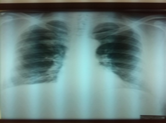
Picture 1
Lab findings on admission: SE 75/100, Le 14.2, Er 3.82, Hgb 122, Hct0.36m, Tr317, urea 9.1, creatinine 103.5, GGT 121.0, AST 31.2 ALT 71.7, ECG: sinus rhythm, fr 90/min, rs in D2, D3, aVF individual VES without changes in ST/T. An inhomogeneous round shadow can be seen on the X-ray image of the right subclavicular lung/picture 1/.
Antibiotic therapy, infusion solutions are prescribed. For technical reasons, we were not able to do a chest CT. Abdominal echo performed: The liver is of unchanged localization, diameter in MCL 154 mm, completely hyperechoic structure with several circular changes of diameter up to 20 mm, hypo to anechoic. /sec. deposits/. Bronchoscopy findings: The findings in the larynx and trachea are normal. The carina of the trachea is slightly dilated. On the left, the findings in the bronchial tree are normal. On the right, the upper interlobar carina is dilated. BB is taken from the upper interlobar carina on the right, KB and BRAŠ from the right upper one for PH diagnosis.
After 2 days, the patient complains of swelling of the neck and the appearance of nodules on the back, physical examination reveals an enlarged lymph gland on the left supraclavicular vel. 2 cm, minor swellings are palpated in several places on the back. A needle puncture of the gland in the neck is performed, as well as a puncture of the change on the back, the sample is sent to pathology.
The same day we receive the pathology findings from the bronchological examination: Cytological findings of the right upper lung: cylindrical cells, mucus, rare metaplastic cells, lymphocytes, macrophages and granulocytes and rare larger groups of malignant epithelial cells - non-microcellular carcinoma, in the differential diagnosis in the first-place adenocarcinoma.
In the following days, the patient's general condition suddenly worsened, with pronounced weakness and malaise along with constant high fever. The patient complained of pain throughout the body, especially severe pain in the left upper arm with the inability to move the arm and assume a forced position. An X-ray of the left upper arm was performed/ Picture2/. and an orthopedist was consulted. Dg. Meta humeri sin. Th: bisphosphonates were recommended.
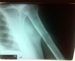
Picture 2
The general condition of the patient is constantly worsening with pronounced pains in the whole body, suffocation, pronounced general weakness. Lab tests are again: Le 20.3, Er 2.40, Hgb 70, Hct 0.23, Tr 153, uk Ca 1.85, ion. Ca 1.25, urea 10.7, creatinine 70.7, due to severe anemia in the following days, the patient was prescribed deplasmatized Er, fresh frozen plasma on several occasions (in addition to the already prescribed th-bronchodilator, antibiotics, analgesics and other symptomatic therapies).
We also get a needle puncture cytology finding of a gland on the left neck and a needle puncture finding of changes on the back: Numerous single and in groups of atypical cells characteristic of non-microcellular carcinoma, in the differential diagnosis the first place is adenocarcinoma.
On the prescribed therapy, the patient's general condition improved slightly, laboratory tests showed: Le 21.7, Er 3.08, Hgb 86, Hct 0.30, Tr 115, he was afebrile, presented to the oncology council for malignant diseases of the respiratory tract of our institution. The council's decision: Apply bisphosphonates (amp Arediae). The application of chemotherapy according to the Cis-Gemzar protocol in the second cycle is indicated with monitoring of blood count elements and renal function parameters. The patient is transferred to the oncology department for the application of th according to the council's decision. The patient was prescribed Aredia, HT planned for use after improvement of the CBC.
But after two days, on the 17th day of admission to our institution, there is a sudden deterioration of the general condition and a lethal outcome of the diseases.
Discussion
Lung adenocarcinoma is the main subtype of non-small cell lung cancer and the number of patients is constantly increasing in most countries in the world [5]. The paper presents a patient whose disease began suddenly, with daily deterioration of the general condition and in a short period of time a lethal outcome occurred. He was admitted to our department for the diagnosis of a shadow on the right lung. The patient is in good general condition, and among the complaints he reports high fever and occasional chest pain, the complaints that occurred a few days before admission, he denies other complaints. The patient underwent bronchoscopy, the Ph finding proved lung adenocarcinoma, and an abdominal echo was performed/multiple sec. deposits in the liver/. A few days after hospitalization, the patient's general condition suddenly worsened, multiple nodules appeared on the back, supraclavicular lymph nodes on the right, the biopsy of the described changes proved expanded disease. In the following days, the patient's general condition worsened, he complained of severe pain in the whole body, bones, joints, especially the left upper arm, inability to move it, he also mentioned general weakness, malaise. An orthopedist was consulted -/dg.meta humeri/, the blood count showed severe anemia, deplasmatic ER, fresh frozen plasma was prescribed. The patient was presented to the oncology council, according to the council's decision, the patient was prescribed Aredia, and HT was planned after the blood count improved. Two days after the administration of Aredia, on the 17th day from admission to our institution, the patient's general condition deteriorated and the disease was fatal.
Conclusion
Lung cancer is one of the most common malignant diseases in the world and in our country and is the leading cause of death among all cancers. In men, it is in first place in terms of frequency compared to other cancers. Smoking is still the main risk factor for the development of lung cancer, but other factors are not excluded /air pollution, environmental factors, increase in inorganic and organic substances in the atmosphere, occupational exposure to harmful radiation, genetic predisposition [6]. Most patients are detected in a clinically advanced stage of the disease because there are no screening methods for early detection of lung cancer. In some patients, such as in our case, the disease progresses very quickly and in a short time the outcome is fatal. The only way to reduce the number of patients is prevention - the fight against smoking, reducing air pollution and improving working and living conditions [3,4].
References
- Jemal, A., Bray, F., Center, M. M., Ferlay, J., Ward, E., & Forman, D. (2011). Global cancer statistics. CA: A Cancer Journal for Clinicians, 61(2):134.
Publisher | Google Scholor - Kadara, H., Kabbout, M., & Wistuba, I. I. (2012). Pulmonary adenocarcinoma: A renewed entity in 2011. Respirology, 17(1):50–65.
Publisher | Google Scholor - Alberg, A. J., Brock, M. V., Ford, J. G., Samet, J. M., & Spivack, S. D. (2013). Epidemiology of lung cancer. Chest, 143(5, Suppl.):e1S–e29S.
Publisher | Google Scholor - Dela Cruz, C. S., Tanoue, L. T., & Matthay, R. A. (2011). Lung cancer: Epidemiology, etiology, and prevention. Clinics in Chest Medicine, 32(4):605–644.
Publisher | Google Scholor - Janssen-Heijnen, M. L., & Coebergh, J. W. (2003). The changing epidemiology of lung cancer in Europe. Lung Cancer, 41(3):245–258.
Publisher | Google Scholor - McCarthy, W. J., Meza, R., Jeon, J., & Moolgavkar, S. (2012). Lung cancer in never smokers: Epidemiology and risk prediction models. Cancer, 32(Suppl. 1):S69–S84.
Publisher | Google Scholor


