
Case Report
Occipital Wormian Bone: What Are Its Implications? A Case Report
- Josphat Kimani *
- Wallace Chege
- Roy Olunga
- Oliver Kinoti
- John Kuany
Department of Human Anatomy and Medical Physiology, University of Nairobi, Kenya.
*Corresponding Author: Josphat Kimani, Department of Human Anatomy and Medical Physiology, University of Nairobi, Kenya.
Citation: Kimani J., Chege W, Olunga R., Kinoti O., Kuany J. (2026). Occipital Wormian Bone: What Are Its Implications? A Case Report, Clinical Case Reports and Studies, BioRes Scientia Publishers. 13(2):1-4. DOI: 10.59657/2837-2565.brs.26.345
Copyright: © 2026 Josphat Kimani, this is an open-access article distributed under the terms of the Creative Commons Attribution License, which permits unrestricted use, distribution, and reproduction in any medium, provided the original author and source are credited.
Received: August 11, 2026 | Accepted: August 25, 2026 | Published: September 03, 2026
Abstract
Peculiar bone ossicles called Wormian bones may form during skull development around the cranial bones, inside the fontanelles, or along the cranial sutures of the skull. These bones, which are also known as intrasutural or sutural bones, have been linked to a number of conditions such as osteogenesis imperfecta, rickets, and cleidocranial dysostosis. They have been considered as innocuous anatomical variations of the skull bones. The occipital region of the skull is less likely to have Wormian bones. However, when present, the midline ossicles are referred to as "Inca bones." In this case study, the presence of a Wormian bone within the occipital bone of an adult human skull was identified during a research study visit to an archaeological museum. A review of the literature on Wormian bones, including their developmental anatomy, therapeutic and forensic implications, and anthropological significance, follows a detailed description of the skull under study. The study findings highlight the importance of recognizing Wormian bones as both diagnostic characteristics and structural variations for diagnostic and forensic practice.
Keywords: wormian bone; occipital bone; sutural ossicle
Introduction
The intricate structure of the human skull is formed by a combination of several ossification centres during embryological development [1]. The development of the brain throughout infancy and early childhood depends on sutures, which are fibrous joints between cranial bones of the skull [2]. Sometimes extra ossification centres grow inside these sutures, resulting in the formation of small, irregular bones called Wormian bones or sutural bones [3]. Ole Worm, a Danish physician from the 17th century, is credited with being the first to provide a detailed description of these ossicles, thus the term "Wormian" for these bones [3,4]. Wormian bones are most commonly found at the lambdoid suture, perhaps because of their complex structure and mechanical vulnerability [5]. However, they may also be present in the sagittal, coronal, and metopic sutures, as well as in cranial fontanelles such as the anterior fontanelle, posterior fontanelle (lambda), and pterion regions [6]. When Wormian bones are found in the midline of the occipital bone at the lambda, they are frequently called Inca bones because they are so prevalent in Andean societies, where they were once thought to be a racial marker [7,8].
The reported frequency of Wormian bones varies greatly in various populations, ranging from 8 to 80%, according to anthropological studies (9,10). Several studies have shown that genetic and environmental factors, including cranial deformation processes, influence their incidence [7,10]. Importantly, many Wormian bones, which are strongly associated with pathological disorders such as osteogenesis imperfecta, may be present in up to 80-90% of patients [10]. They may also be seen in patients with rickets, hypothyroidism, and cleidocranial dysostosis [6]. From a medical viewpoint, Wormian are significant clinically because they may be mistaken for skull fractures on radiographic imaging, particularly in young patients [9]. In addition, their presence may help differentiate traumatic lesions from developmental variations and provide useful information for skeletal identification in forensic medicine [7]. Studies on Wormian bones have been used to give data on human variations, demographic linkages, and cultural patterns of skull deformation [7]. In this case study, we discuss the morphological characteristics, clinical and forensic significance, anthropological distribution, and embryological origin of a Wormian bone discovered in the occipital region of an adult skull at the Kenya National Museum.
Case Presentation
Morphological features of the Wormian bone
The specimen under study in this case report was a dried adult human skull from the Kenya National Museum's anatomical collection. The available morphological traits of the skull that were present did not allow for a definitive determination of the sex for the specimen. Abnormal thickness of cranial bones, porosity, or craniosynostosis were among the pathological abnormalities that were not evident in the skull. An analysis of the occipital region led to the identification of a Wormian bone. The bone was formed at the posterior fontanelle, where the lambda is, where the sagittal and lambdoid sutures converge. Its location inside the midline occipital region led to its classification as an Inca bone variety. The Wormian bone was quadrilateral in shape and had uneven serrated edges on the sides. It was calculated to be around 2.1 cm tall and 4.8 cm broad using digital vernier callipers. The margins of the bone interdigitated with the surrounding occipital and parietal bones, forming a tight articulation with the skull, as seen in Figures 1 and 2. The surface and cortical bone quality of the Wormian bone matched that of the surrounding cranial bone, and there was no porosity or pathological remodelling.
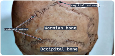
Figure 1
Figure 1 shows the posterior aspect of the skull. Note the Wormian bone on the occipital region. The other sutures, such as the squamosal, coronal, sagittal, and lambdoid sutures, were all undamaged, as shown in Figure 2. There were no other Wormian bones in the cranium.
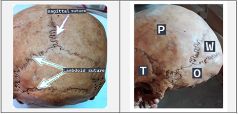
Figure 2&3
Figure 2 shows the lambdoid and sagittal sutures. The Wormian bone is interdigitated by the parietal and occipital bones. Figure 3 shows the lateral aspect of the skull. The various bones of the skull have been shown. O - occipital bone, P - parietal bone, T - temporal bone. The Wormian bone (W) can be seen on the right side of the image.
Furthermore, there were no indications of skull traumatic damage, congenital deformity, or trepanation. Based on our findings during skull examination, the Wormian bone appeared to be a developmental variant rather than a pathological manifestation.
Discussion
Embryological basis of wormian bones
During development, accessory ossification centres form within the cranial sutures, leading to the formation of Wormian bones. Sutures serve as sites for the intramembranous ossification, enabling the formation of the cranial bones. During the embryological development of the skull, in the sutural mesenchyme, ectopic ossification centres can occasionally form. Failure to merge with surrounding developing bones may lead to the formation of distinct, separate ossicles (6). The formation of the Wormian bones is believed to be influenced by genetic predisposition, mechanical stress, and delayed suture closure [3].
For the occipital bone, given that it develops from many ossification centres that are derived from both membranous and cartilaginous origins, its formation is especially complicated (5). A possible Wormian bone that develops in the midline occipital region is provided by this case report.
Incidence and distribution
There are significant differences in the occurrence of Wormian bones amongst populations. Midline occipital Wormian bones, which are also known as Inca bones, can occur in up to 27-36% of people in Andean cultures, but their prevalence in the European population varies from 8-15% [7]. According to a study done on Turkish skulls, 52.7% of specimens had Wormian bones, with the majority being at the lambdoid suture (43.1%), while others were in the lambda (10.3%) and occipitomastoid area (12.1%) [7,8]. The overall presence of Wormian bones in Central Anatolia was 27.7%, with the lambda accounting for 7.6% of the bones [7,8]. Despite not being the most common site for formation of the Wormian bones, these results show that the occipital region is a recognized site with population-specific variation for the bones.
Anthropological and pathological significance
Wormian bones have long been employed as markers in anthropological research. Because of their great prevalence in Andean people, midline occipital bones have been dubbed "Inca bones" (7). Although they were regarded as racial markers by early anthropologists, current studies have revealed that they are present in a variety of populations, albeit with different prevalences. Cranial deformation in some populations is one cultural practice that influences their prevalence. Wormian bones in forensic anthropology help population affinity estimates, but they are not diagnostic markers [7]. Other skeletal and cranial indicators must be taken into consideration when interpreting their existence.
Pathological diseases, especially those involving abnormal bone formation, are significantly linked to multiple Wormian bones. Up to 80-90% of individuals with osteogenesis imperfecta (OI) have Wormian bones, which are frequently more than 10 in number [3]. The appearance of multiple Wormian bones in young populations is considered a diagnostic characteristic of OI (16). Rickets, hypothyroidism, cleidocranial dysostosis, hypophosphatasia, and Menke's kinky hair syndrome are significant disorders linked to Wormian bones [6]. However, a benign developmental and structural variant rather than pathology is suggested in this case by the isolated occurrence of a single Wormian bone in the absence of other skeletal abnormalities in the skull.
Radiological and forensic clinical implications
Wormian bones can be mistaken for fractures in clinical radiography, particularly in the imaging of pediatric patients. They may be distinguished from fractures due to traumatic injury as the bone fractures often have sharp, linear borders, while Wormian bones have their borders being irregular and interdigitated borders [10]. Potentially serious consequences may result from misinterpretation of the variant bones, especially when child abuse is suspected. Therefore, radiologists and physicians must be aware of their normal position and features. Wormian bones can make surgical techniques more complicated in neurosurgery and craniofacial surgery, especially when they are present in the posterior cerebral fossa [3]. Identifying them preoperatively can help avoid inadvertent complications.
Distinguishing Wormian bones from traumatic lesions is crucial in cases involving forensics. They might resemble postmortem bone breaks or comminuted fractures. Understanding their morphological characteristics, such as serrated edges and characteristic sutural positions, is necessary for their identification. Furthermore, their presence can lead to a better understanding of the skeletal system by providing information on developmental variance and ancestry.
Conclusion
In this case study, a Wormian bone was found in the occipital area of an adult skull. Despite being sometimes dismissed as of no significance, Wormian bones are important in anthropology, forensics, and clinical practice. To prevent diagnostic failures, especially in radiology and forensic investigations, it is essential to recognize them. Furthermore, information about human cranium development and diversity is still provided by their varying occurrence among groups. This case study highlights the necessity for interdisciplinary knowledge and leads to a better understanding of cranial anatomical variations.
References
- Callisthenics Association. (n.d.). The human cranium: Structure, function, and clinical relevance. Retrieved October 4, 2025.
Publisher | Google Scholor - Gray’s anatomy: The anatomical basis of clinical practice. (2008). Churchill Livingstone/Elsevier.
Publisher | Google Scholor - Bellary, S. S., Steinberg, A., Mirzayan, N., Shirak, M., Tubbs, R. S., Cohen-Gadol, A. A., et al. (2013). Wormian bones: A review. Clinical Anatomy, 26(8):922–927.
Publisher | Google Scholor - White, T. D. (2005). The human bone manual. Elsevier Academic Press.
Publisher | Google Scholor - Scheuer, L. (2000). Developmental juvenile osteology. Academic Press.
Publisher | Google Scholor - Sanchez-Lara, P. A., Graham, J. M., Hing, A. V., Lee, J., & Cunningham, M. (2007). The morphogenesis of wormian bones: A study of craniosynostosis and purposeful cranial deformation. American Journal of Medical Genetics Part A, 143A(24):3243–3251.
Publisher | Google Scholor - Hanihara, T., & Ishida, H. (2001). Os incae: Variation in frequency in major human population groups. Journal of Anatomy, 198(2):137–152.
Publisher | Google Scholor - Movsesian, A. (2024). Does the distribution of Wormian bone frequencies across different world regions reflect genetic affinity between populations? Egyptian Journal of Forensic Sciences, 14(1):33.
Publisher | Google Scholor - Kumar, R. P., & Gaikwad, M. (2021). Topographical variations of Wormian bones in Eastern-Indian dry human skulls: Current perspective and review of literature. Journal of Human Anatomy, 5.
Publisher | Google Scholor - Gaillard, F. (n.d.). (2025). Wormian bone. Radiopaedia.
Publisher | Google Scholor