
Case Report
Neonatal Acute Liver Failure: A Diagnostic Challenge in The Intensive Care Unit
- Vallejo Chaves Saul Hernando ID 1,2*
- Quevedo Juana María 1,3
- Portilla Fanny 1,2
- Caro Jesica 1,2
- Gómez Martina 1,2
- Barrios Juan Camilo 1,2
1Neonatal Intensive Care Unit, UROS Clinic. Neiva, Huila, Colombia.
2Pediatric Service, UROS Clinic. Neiva, Huila, Colombia.
3Pediatric Gastroenterology Service, UROS Clinic. Neiva, Huila, Colombia.
*Corresponding Author: Vallejo Chaves Saul Hernando, Neonatal Intensive Care Unit, UROS Clinic. Neiva, Huila, Colombia.
Citation: Hernando VCS, María QJ, Fanny P, Jesica C, Martina G, et al. (2026). Neonatal Acute Liver Failure: A Diagnostic Challenge in The Intensive Care Unit, International Clinical Case Reports and Reviews, BioRes Scientia Publishers. 4(1):1-6. DOI: 10.59657/2993-0855.brs.26.042
Copyright: © 2026 Vallejo Chaves Saul Hernando, this is an open-access article distributed under the terms of the Creative Commons Attribution License, which permits unrestricted use, distribution, and reproduction in any medium, provided the original author and source are credited.
Received: January 16, 2026 | Accepted: July 03, 2026 | Published: July 14, 2026
Abstract
We present the case of a full-term newborn admitted to the neonatal intensive care unit at 15 days of age with persistent hypoglycemia, multisystem involvement, severe hepatomegaly, refractory coagulopathy, and acute liver failure with encephalopathy. Initial studies suggested a severe hepatic etiology with vascular compromise and hepatic hypervascularization, with hereditary metabolic diseases and hepatic neoplasia considered as differential diagnoses. This case highlights the diagnostic and therapeutic complexity of neonatal liver failure.
Keywords: acute liver failure; neonates; congenital alloimmune hepatitis; metabolic diseases; liver transplantation
Introduction
Acute liver failure during the neonatal period is a rare, very serious disease with high mortality. Coagulopathy, with an International Normalized Ratio ≥ 3, is the key parameter for defining it. The most frequent causes are fetal alloimmune hepatitis, previously known as neonatal hemochromatosis, viral infections, metabolic disorders, and lymphohistiocytosis. Hemophagocytic. There is a group of treatable diseases that need to be diagnosed very quickly in order to provide the appropriate treatment.
The presentation of a baby-led weaning syndrome (BLWS) is variable and includes feeding refusal, growth retardation, hypoglycemia, coagulopathy unresponsive to vitamin K administration, and cholestatic jaundice. It is essential to quickly identify treatable causes, such as galactosemia, fatty acid oxidation defects, hereditary fructose intolerance, herpes virus infection, etc [1-2].
The patient should be referred early to a specialized center with pediatric liver transplant availability in order to provide this therapeutic alternative when indicated.
Clinical Case Report
Prenatal and Perinatal History
Newborn female, product of the second pregnancy, born at term (38 weeks), daughter of a 41-year-old mother, G2 C2 V2, with seven adequate prenatal checkups. Previous pregnancy complicated by preeclampsia. No family consanguinity. Family history of a brother with surgically corrected craniosynostosis, associated with moderate cognitive impairment, without genetic testing.
Maternal infectious screening (STORCH): Treponema pallidum negative, HIV non-reactive, Toxoplasma IgG and IgM negative, Cytomegalovirus (24/04/25) IgG positive with IgM negative, Hepatitis B negative, Rubella IgG positive, Hemoparasites negative.
Birth by repeat cesarean section. Apgar score 8/10 at one minute and 9/10 at five minutes. Spontaneous neonatal adaptation, Silverman-Andersen score 0. Birth weight 2,830 g, length 47 cm. Received vitamin K at birth.
Current Illness
From birth, she experienced recurrent episodes of hypoglycemia and progressive deterioration of her general condition. At 15 days old, she was transferred as a life-threatening emergency from Mocoa (Putumayo) to the Neonatal Intensive Care Unit of the Uros Clinic.
On admission, she was in poor general condition, lethargic, and pale yellow. She had good bilateral ventilation, with no initial signs of respiratory distress. Her abdomen was distended with marked hepatomegaly and a palpable liver edge. She rapidly progressed to respiratory compromise, requiring high-frequency mechanical ventilation.
Hemodynamic and Cardiovascular Compromise
He presented with cardiovascular collapse refractory to inotropic support. Echocardiogram: good systolic -diastolic function of the left ventricle, physiological tricuspid and pulmonary regurgitation, estimated pulmonary systolic pressure of 34 mmHg, foramen ovale of 3 mm, TAPSE 11 mm, no vegetations.
Renal Involvement
She developed multifactorial acute kidney injury, oliguric and anuric, in the context of hepatorenal syndrome. Despite normal initial renal function tests, she remained anuric, requiring peritoneal dialysis. She did not respond to continuous infusion of furosemide and albumin. Renal ultrasound showed right perirenal fluid.
Liver and Gastrointestinal Involvement
The patient presented with acute liver failure of undetermined etiology, associated with encephalopathy. Severe abdominal distension, collateral circulation, and massive hepatomegaly were noted. Peritoneal drainage was performed using a multipurpose catheter, yielding scant serosanguineous fluid.
Hepatobiliary ultrasound with Doppler: enlarged liver with diffuse areas of decreased echogenicity. Right hepatic lobe 96 mm and left lobe 61 mm. Globally increased hepatic vascularity. Portal vein 4.5 mm with hepatopeteric flow and elevated pulsatility index (0.46), suggestive of portal hypertension. Common bile duct 2 mm. Gallbladder 32 × 10 × 10 mm, with homogeneous anechoic contents. Pancreas not evaluable.
Ultrasound Conclusion: hepatomegaly with parenchymal changes suggestive of neoplastic etiology hypervascular. Contrast-enhanced magnetic resonance imaging was recommended.
Laboratory Studies
Liver function tests: total bilirubin 16.7 mg/ dL (indirect 14.4 mg/ dL, direct 3.7 mg/ dL), cholestatic pattern. AST 845 U/L, ALT 143 U/L, alkaline phosphatase 280 U/L (elevated), LDH 1,808 U/L (elevated). Total protein 3.7 g/ dL and albumin 2.0 g/ dL. Triglycerides and cholesterol within normal ranges.
Severe coagulopathy with prolonged unresponsive times to vitamin K; fibrinogen 40 mg/ dL. Required transfusion of fresh frozen plasma, platelets, and tranexamic acid.
Tumor Markers: markedly elevated alpha-fetoprotein (≈498,000), negative beta-HCG. elevated ferritin 2,990 ng/ mL, transferrin saturation 14%. Normal ammonia.
Blood Count: anemia (Hb 7.7 g/ dL) and severe thrombocytopenia (47,000/µL), red blood cell and platelet transfusion was indicated.
Infectious and Metabolic Studies
Expanded infectious disease screening: toxoplasma IgM negative, CMV IgG positive with pending IgM, hepatitis An IgM negative, hepatitis C negative, herpes, parvovirus and toxoplasma IgA negative. Gamma glutamyl transferase within normal range.
An expanded metabolic workup was initiated to rule out galactosemia and tyrosinemia. Enteral and parenteral nutrition were discontinued until the metabolic etiology was clarified. Management consisted of intravenous fluids and strict blood glucose monitoring due to the high risk of hypoglycemia.
Antimicrobial Management
cefepime was initiated; vancomycin was not added due to nephrotoxicity. Acyclovir was administered given the possibility of a viral etiology. Pediatric infectious diseases deemed the regimen appropriate and recommended further investigation.
Neurological Evaluation
Patient with hypoactive encephalopathy, weak sucking for gestational age, and neurological examination with apparent deficit. Transfontanellar ultrasound without pathological abnormalities. Pediatric neurology classified the patient as high neurological risk and ordered a non-contrast brain MRI.
Discussion
This case represents severe neonatal liver failure with multisystem involvement. The findings of massive hepatomegaly, refractory coagulopathy, and markedly elevated ferritin and alpha-fetoprotein suggest a hereditary metabolic disorder, fulminant neonatal hepatitis, or a liver neoplasm such as hepatoblastoma as the main differential diagnoses. The absence of a clear infectious etiology and normal ammonia levels point toward non-infectious causes.
It is a serious disease that causes high fetal and neonatal mortality, and is currently being recognized as one of the most important causes of perinatal cirrhosis and liver failure. The pattern of hepatic fibrosis and the abundant iron deposition in the liver and other organs distinguish this entity from other etiologies that cause neonatal liver failure [7-9]
The high mortality rate of acute neonatal liver failure justifies the re-evaluation of diagnostic tests and clinical history in cases of patients with progressive cytopenias, sepsis with poor response to antibiotic therapy, or persistent hypoglycemia along with clinical and laboratory data of acute liver failure, in order to clarify the underlying cause and provide appropriate treatment [1,3]. Hypoglycemia is present in around 40% of cases of acute liver failure, probably related to decreased gluconeogenesis and decreased plasma insulin levels [4].
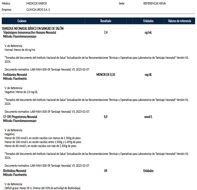
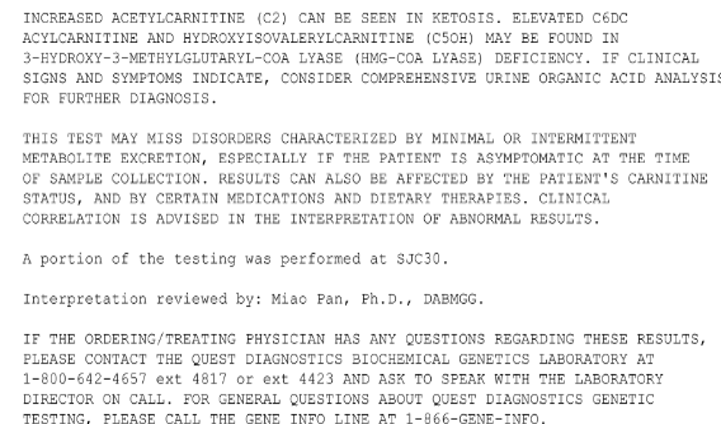
Inborn errors of metabolism were a central focus of the differential diagnosis. The acylcarnitine profile showed elevated levels of C2, C6DC, and C5OH, findings that have been described in HMG- CoA lyase deficiency, a condition that can manifest in the neonatal period with hypoketotic hypoglycemia, metabolic acidosis, variable hyperammonemia, and severe liver involvement.
However, the interpretation of these results is limited by the context of critical illness, prolonged fasting, and hepatic dysfunction, conditions that can lead to secondary elevations in acylcarnitines. Additionally, the report cautions that the screening test may be falsely negative or nonspecific in disorders with minimal or intermittent metabolic excretion. Therefore, it was deemed essential to supplement the study with urinary organic acids, lactate/pyruvate ratio, and targeted genetic testing, although these results were unavailable at the time of the clinical outcome.
Regarding treatment, prior to definitive etiological diagnosis and specific treatment, general measures should be taken to control liver failure and empirically treat the most frequent specific causes, as the causal diagnosis is usually delayed. In this regard, admission to the ICU is necessary to ensure support of vital functions, as well as continuous monitoring and the appropriate administration of supportive treatments (blood replacement, gastric protection, vasoactive amines, vitamin K). To control possible infectious causes, broad-spectrum antibiotic coverage (ampicillin and gentamicin/cefotaxime) is recommended, as well as acyclovir (60 mg/kg/day) to cover herpes simplex virus, since this treatment has been shown not only to improve survival but also the long-term neurological development of these patients [5-6].
Conclusion
Figure 1The patient meets the diagnostic criteria for acute neonatal liver failure, defined as impaired hepatic synthesis within the first four weeks of life, evidenced by severe coagulopathy and hypoalbuminemia, associated with hepatic encephalopathy and hepatorenal syndrome. Based on the clinical, biochemical, and imaging findings, the following differential diagnostic approach was performed:
Infectious Causes
These were initially considered due to their frequency in this age group. Viral infections such as herpes simplex virus and parvovirus B19 are under investigation, with serological tests already ordered and empirical management with intravenous acyclovir. Bacterial causes were also considered, and the patient is currently receiving broad-spectrum antibiotic coverage while awaiting culture results, including those for Escherichia coli. E. coli, a pathogen described in association with galactosemia, was present. The TORCH test was negative (toxoplasma, HIV, CMV, HCV), and hepatitis A was negative. Taken together, these findings make a primary infectious etiology unlikely.
Gestational Alloimmune Liver Disease (GALD)
It represents one of the main diagnostic possibilities, given the clinical and paraclinical characteristics: fulminant onset in the first weeks of life, hepatomegaly, severe coagulopathy, hypoalbuminemia, generalized edema, ascites, hypoglycemia on admission, jaundice, multiple organ failure, serum ferritin >1000 ng/ mL, low serum iron with decreased transferrin saturation, moderately elevated transaminases, as well as elevated LDH and alkaline phosphatase.
Normal ammonia levels and non-persistent, non-refractory hypoglycemia were identified as contraindications. Nevertheless, the diagnostic probability remains high, which is why intravenous immunoglobulin (IVIG) was initiated, and a double-volume exchange transfusion is being considered depending on clinical evolution. Once relative stability is achieved, abdominal magnetic resonance imaging (MRI) is recommended to look for extrahepatic siderosis, or an oral mucosal biopsy with Perls' stain is recommended. A viral gastrointestinal panel, especially for enterovirus, was also suggested as a differential diagnosis, but it was negative.
Inborn Errors of Metabolism
Given the presence of hypoglycemia on admission, metabolic acidosis, and hyperlactatemia, several entities were considered:
- Tyrosinemia type 1, due to its presentation in the first weeks of life and association with hepatorenal syndrome. Although alpha-fetoprotein is normal for age (usually >100,000 in this condition), a diagnostic approach will be performed because succinylacetone is the key biomarker, as it constitutes a relevant differential diagnosis from hepatorenal syndrome.
- Galactosemia was considered unlikely due to the absence of reducing sugars in urine and lack of clinical response to fasting; however, a genetic study was requested.
- Other conditions such as urea cycle disorders or protein metabolism disorders are considered unlikely due to the absence of significant hyperammonemia.
- Additionally included as differential diagnoses are mitochondrial respiratory chain defects and fatty acid oxidation defects.
- Wolman disease is considered unlikely based on abdominal ultrasound findings.
Vascular and Neoplastic Causes
Vascular etiologies are considered unlikely: echocardiogram without pathological findings, Doppler with data suggestive of portal hypertension and secondary vascular changes, without evidence of congenital malformations. If suspicion persists, abdominal venous and arterial magnetic resonance angiography is recommended. There are no data to suggest a neoplastic etiology, with negative tumor markers (normal alpha-fetoprotein and β- hCG).
Anemia
It is interpreted as multifactorial, secondary to hemolysis, systemic inflammation, consumption, and both macroscopic and microscopic losses.
Finally, early referral to a reference center is a priority in order to have specific second-line tests and make an accurate etiological diagnosis, as well as to perform a liver or hematopoietic stem cell transplant if necessary.
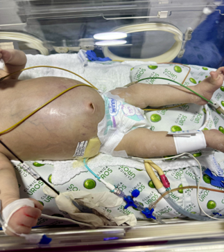
Figure 1
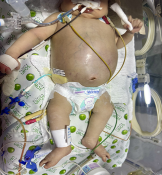
Figure 2
References
- Shanmugam, N. P., Bansal, S., Greenough, A., Verma, A., Dhawan, A. (2011). Neonatal liver failure: aetiologies and management-state of the art. European Journal of Pediatrics, 170(5):573-581.
Publisher | Google Scholor - Sundaram, S. S., Alonso, E. M., Narkewicz, M. R., Zhang, S., Squires, R. H., et al. (2011). Characterization and outcomes of young infants with acute liver failure. The Journal of Pediatrics, 159(5):813-818.
Publisher | Google Scholor - Dhawan, A., Mieli-Vergani, G. (2005). Acute liver failure in neonates. Early Human Development, 81(12):1005-1010.
Publisher | Google Scholor - Whitington, P. F., Alonso, E. M., Squires, R. H. (2008). Acute Liver Failure. Diseases of The Liver and Biliary System in Children, 169-188.
Publisher | Google Scholor - Kimberlin, D. W., Lin, C. Y., Jacobs, R. F., Powell, D. A., Corey, L., et al. (2001). Safety and efficacy of high-dose intravenous acyclovir in the management of neonatal herpes simplex virus infections. Pediatrics, 108(2):230-238.
Publisher | Google Scholor - Verma, A., Dhawan, A., Zuckerman, M., Hadzic, N., Baker, A. J., et al. (2006). Neonatal herpes simplex virus infection presenting as acute liver failure: prevalent role of herpes simplex virus type I. Journal of Pediatric Gastroenterology and Nutrition, 42(3):282-286.
Publisher | Google Scholor - Whitington, P. F. (2012). Gestational alloimmune liver disease and neonatal hemochromatosis. In Seminars in liver disease. Thieme Medical Publishers. 32(4):325-332.
Publisher | Google Scholor - Vohra, P., Haller, C., Emre, S., Magid, M., Holzman, I., et al. (2000). Neonatal hemochromatosis: the importance of early recognition of liver failure. The Journal of pediatrics, 136(4):537-541.
Publisher | Google Scholor - Whitington, P. F., Malladi, P. (2005). Neonatal hemochromatosis: is it an alloimmune disease? Journal of Pediatric Gastroenterology and Nutrition, 40(5):544-549.
Publisher | Google Scholor