
Case Report
Marked Eosinophilia as an Initial Diagnostic Clue to Advanced Gastric Adenocarcinoma: Clinical Implications of a Paraneoplastic Presentation
- Sinem Ülke *
- Pelin Yumuşak
- Sevil Uygun İlikhan
Department of Internal Medicine, Ankara Bilkent City Hospital, Ankara, Turkey.
*Corresponding Author: Sinem Ülke, Department of Internal Medicine, Ankara Bilkent City Hospital, Ankara, Turkey.
Citation: Ulke S., Yumuşak P., Sevil U. Ilikhan. (2026). Marked Eosinophilia as an Initial Diagnostic Clue to Advanced Gastric Adenocarcinoma: Clinical Implications of a Paraneoplastic Presentation, Clinical Case Reports and Studies, BioRes Scientia Publishers. 13(1):1-4. DOI: 10.59657/2837-2565.brs.26.341
Copyright: © 2026 Sinem Ülke, this is an open-access article distributed under the terms of the Creative Commons Attribution License, which permits unrestricted use, distribution, and reproduction in any medium, provided the original author and source are credited.
Received: July 20, 2026 | Accepted: July 31, 2026 | Published: August 10, 2026
Abstract
Eosinophilia is commonly associated with allergic diseases, parasitic infections, and hematologic disorders; however, it may rarely occur as a paraneoplastic manifestation of solid tumors. We report the case of an 87-year-old woman who presented with abdominal pain, weakness, and marked eosinophilia. Laboratory evaluation revealed leukocytosis with an absolute eosinophil count of 4,260/mm³. Extensive investigations excluded common secondary causes of eosinophilia, including parasitic, allergic, infectious, and hematologic disorders. Imaging studies demonstrated a gastric cardia mass with hepatic metastasis, and histopathological examination confirmed poorly differentiated gastric adenocarcinoma. Positron emission tomography-computed tomography showed widespread metastatic disease. In the absence of other identifiable causes, eosinophilia was considered paraneoplastic in origin. This case highlights that unexplained eosinophilia, particularly in elderly patients, may represent an important clue to underlying malignancy. Early recognition of this association may facilitate timely diagnosis and management.
Keywords: eosinophilia; gastric adenocarcinoma; paraneoplastic syndrome
Introduction
Eosinophilia, defined as an absolute eosinophil count exceeding 500 cells/mm³, is a relatively common laboratory finding in clinical practice. While mild eosinophilia is frequently associated with benign conditions such as allergic disorders and parasitic infections, marked or persistent eosinophilia warrants a more comprehensive evaluation. Hypereosinophilia (>1,500 cells/mm³) may lead to end-organ damage and is occasionally associated with underlying malignancy [1].
Paraneoplastic eosinophilia is an uncommon but well-recognized phenomenon, most frequently described in hematologic malignancies. In contrast, its occurrence in solid tumors is rare and often under-recognized [2]. The proposed mechanism involves cytokine-mediated eosinophil proliferation, particularly through mediators such as IL-5 and GM-CSF [3].
Gastric adenocarcinoma associated with paraneoplastic eosinophilia has been reported only in a limited number of cases and is typically linked to advanced-stage disease[4,5]. In such cases, eosinophilia may precede or accompany nonspecific clinical symptoms, posing a diagnostic challenge.
Herein, we present a case of advanced gastric adenocarcinoma in an elderly patient who initially presented with unexplained marked eosinophilia. This report aims to emphasize the diagnostic value of eosinophilia as a potential early indicator of malignancy and to discuss its clinical implications in the context of solid tumors.
Case Presentation
An 87-year-old woman with a history of essential hypertension was admitted with a one-week history of abdominal pain, decreased appetite, and generalized weakness. She denied fever, night sweats, weight loss, pruritus, skin rash, or recent medication use. There was no history suggestive of allergic disease or recent travel associated with parasitic exposure.
On physical examination, vital signs were stable. Pulmonary auscultation revealed decreased breath sounds at the right lung base. Abdominal examination demonstrated tenderness in the epigastric and right upper quadrant regions. A palpable, approximately 3×4 cm soft mass was noted in the epigastric area, with mobility increasing during inspiration. No peripheral lymphadenopathy or hepatosplenomegaly was detected.
Initial laboratory investigations revealed leukocytosis (white blood cell count: 22,300/mm³) with marked eosinophilia (absolute eosinophil count: 4,260/mm³). Hemoglobin level was 12.3 g/dL, and platelet count was within normal limits. Liver and renal function tests were unremarkable. Serum immunoglobulin E (IgE) levels were within normal range. Peripheral blood smear did not demonstrate atypical cells, dysplasia, or blast forms.
Given the marked eosinophilia, an extensive evaluation for secondary causes was undertaken. Repeated stool examinations for parasitic infections were negative, and serologic tests did not indicate parasitic or infectious etiologies. There was no clinical or laboratory evidence of autoimmune or allergic disease. A detailed drug history excluded medication-related eosinophilia. Bone marrow examination was performed to evaluate for primary hematologic disorders, including hypereosinophilic syndrome, and revealed no evidence of malignancy or clonal eosinophilia.
Chest radiography demonstrated right-sided opacity with hilar prominence. Thoracic computed tomography revealed multiple small mediastinal lymph nodes and a loculated pleural effusion in the right hemithorax measuring approximately 25 mm (Figure 1). Diagnostic thoracentesis revealed an exudative effusion according to Light’s criteria.
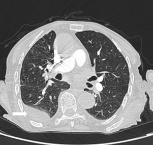
Figure 1: Thoracic CT showing loculated pleural effusion in the right hemithorax and mediastinal lymphadenopathy.
Further evaluation with abdominal dynamic magnetic resonance imaging revealed a mass lesion involving the gastric cardia and an approximately 8 cm metastatic lesion in hepatic segments II–III (Figure 2). Upper gastrointestinal endoscopy identified a mass extending from the distal esophagus into the stomach, causing luminal narrowing. Histopathological examination of endoscopic biopsy specimens demonstrated poorly differentiated gastric adenocarcinoma. Positron emission tomography–computed tomography (PET-CT) confirmed widespread metastatic involvement. Based on clinical, laboratory, and imaging findings, the patient was diagnosed with stage IV metastatic gastric adenocarcinoma. In the absence of identifiable secondary causes, the marked eosinophilia was considered to be paraneoplastic in origin.
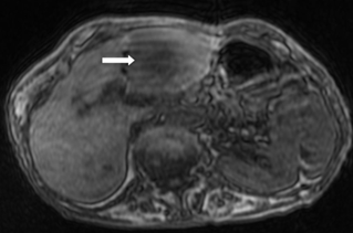
Figure 2: Abdominal MRI showing metastatic lesion in the left hepatic lobe.
Clinical Course and Eosinophil Trend
During hospitalization, eosinophil counts remained persistently elevated, ranging between 3,800–4,500/mm³ on serial measurements. No acute organ damage attributable to hypereosinophilia (such as cardiac, pulmonary, or neurologic involvement) was observed.
Given the absence of symptomatic eosinophil-related organ dysfunction, specific anti-eosinophilic therapy was not initiated. Instead, management was primarily directed toward the underlying malignancy. The patient was referred to the oncology department for further evaluation and treatment planning.
The persistence of eosinophilia in parallel with extensive metastatic disease further supported a paraneoplastic etiology. Follow-up data regarding eosinophil response to oncologic treatment were not available at the time of reporting. This temporal association between persistent eosinophilia and advanced disease supports a paraneoplastic mechanism.
Discussion
Paraneoplastic eosinophilia is a rare but clinically significant finding in patients with solid malignancies [6]. Although it is more commonly associated with hematologic cancers, it has also been reported in a limited number of solid tumors, including gastric adenocarcinoma [4–6]. Paraneoplastic eosinophilia has also been reported in other solid malignancies, including pancreatic and bladder cancers [7,8].
In the present case, marked eosinophilia was identified in the absence of common secondary causes such as parasitic infections, allergic conditions, drug reactions, or primary hematologic disorders. Comprehensive diagnostic evaluation, including repeated microbiological and hematologic investigations, failed to reveal an alternative etiology, thereby supporting a paraneoplastic mechanism.
The mechanism of paraneoplastic eosinophilia is not fully understood, but it is thought to be mediated by cytokines that promote eosinophil proliferation and survival [9,10]. Although cytokine levels were not directly measured in this patient, the clinical findings are consistent with a cytokine-mediated mechanism.
Notably, paraneoplastic eosinophilia has been associated with advanced-stage disease [6]. Consistent with this observation, our patient was diagnosed with metastatic gastric adenocarcinoma at presentation. Previous reports suggest that paraneoplastic eosinophilia may serve as a diagnostic clue and may be associated with poorer clinical outcomes in solid tumors [6].
From a clinical perspective, unexplained eosinophilia—particularly in elderly patients—should prompt a systematic and guideline-based evaluation [9,10]. After exclusion of common etiologies such as parasitic infections, allergic diseases, and medication-related causes, clinicians should consider both hematologic and solid malignancies in the differential diagnosis.
A structured, stepwise diagnostic approach may facilitate earlier recognition of underlying malignancy, especially in patients presenting with nonspecific symptoms. Failure to recognize paraneoplastic eosinophilia may lead to diagnostic delay and suboptimal management.
Management of paraneoplastic eosinophilia primarily involves treatment of the underlying malignancy [9,10]. In cases where eosinophil-mediated organ damage is suspected, corticosteroids or cytoreductive therapies may be considered; however, evidence guiding optimal management in solid tumor–associated eosinophilia remains limited. Further studies are needed to better define prognostic and therapeutic implications in solid tumor–associated eosinophilia.
Conclusion
Unexplained marked eosinophilia should not be regarded as a benign laboratory abnormality, particularly in elderly patients. Instead, it may represent an important clinical indicator of underlying malignancy. In our case, persistent eosinophilia in the absence of identifiable secondary causes preceded the diagnosis of advanced gastric adenocarcinoma, supporting its role as a paraneoplastic manifestation. Recognition of this association is essential to avoid delays in diagnosis, especially when clinical symptoms are nonspecific. Incorporating malignancy screening into the diagnostic workup of persistent eosinophilia may facilitate earlier diagnosis and more timely management. Furthermore, eosinophilia in this context may reflect advanced disease burden and carry potential prognostic significance.
Declarations
Informed Consent
Written informed consent was obtained from the patient for the publication of this case report.
Ethical Consent
Written informed consent was obtained from the patient for publication of this case report and accompanying images.
Conflict of Interest
The authors declare no conflict of interest.
Financial Support
The authors received no financial support for this study.
Author Contributions
Concept: SU, SUI; Design: SU, PY; Data Collection and/or Processing: SU, PY; Literature Review: SU, SUI; Writing: SU; Critical Review: SUI.
Acknowledgments
The authors would like to thank all healthcare professionals involved in the diagnosis and management of the patient.
References
- Rothenberg, M. E. (1998). Eosinophilia. The New England Journal of Medicine, 338(22):1592–1600.
Publisher | Google Scholor - Gleich, G. J. (2000). Mechanisms of eosinophil-associated inflammation. The Journal of Allergy and Clinical Immunology, 105(4):651–663.
Publisher | Google Scholor - Ogbogu, P. U., Bochner, B. S., Butterfield, J. H., et al. (2009). Hypereosinophilic syndrome: A multicenter, retrospective analysis. The Journal of Allergy and Clinical Immunology, 124(6):1319–1325.e3.
Publisher | Google Scholor - Wang, S., Zhang, Y., Chen, L., et al. (2022). Gastric signet-ring cell carcinoma with paraneoplastic eosinophilia: A case report and literature review. Oncology and Translational Medicine, 8(5):264–266.
Publisher | Google Scholor - Teoh, S. C., Siow, W. Y., & Tan, H. T. (2000). Severe eosinophilia in disseminated gastric carcinoma. Singapore Medical Journal, 41(5):232–234.
Publisher | Google Scholor - Zalewska, E., Krawczyk, P., Powrózek, T., et al. (2021). Hypereosinophilia in solid tumors: Case report and clinical review. Frontiers in Oncology, 11, Article 639395.
Publisher | Google Scholor - Shomura, Y., Sato, T., Satoh, M., et al. (2017). Paraneoplastic eosinophilia associated with advanced pancreatic carcinoma. Internal Medicine, 56(10):1273–1276.
Publisher | Google Scholor - Tanaka, T., Takeda, M., Haratake, N., et al. (2020). Severe eosinophilia associated with metastatic bladder cancer: A case report and literature review. BMC Cancer, 20(1):1–5.
Publisher | Google Scholor - Roufosse, F., & Weller, P. F. (2010). Practical approach to the patient with hypereosinophilia. The Journal of Allergy and Clinical Immunology, 126(1):39–44.
Publisher | Google Scholor - Valent, P., Klion, A. D., Horny, H. P., et al. (2020). Contemporary consensus proposal on criteria and classification of eosinophilic disorders and related syndromes. The Journal of Allergy and Clinical Immunology, 145(1):1–17.
Publisher | Google Scholor