
Research Article
Histopathological Evaluation of Vital Organs in Wistar Rats Treated with Medicago sativa (Alfalfa) Ethanol Leaf Extract
1Department of Pharmacology and Therapeutics, College of Medicine and Health Sciences, Gregory University, Uturu, Abia State, Nigeria.
2Department of Chemical Pathology, College of Medicine and Health Sciences, Abia State University, Uturu, Nigeria.
3Department of Anatomical Pathology, College of Medicine and Health Sciences, Abia State University, Uturu, Nigeria.
4Department of Internal Medicine, College of Medicine and Health Sciences, Abia State University, Uturu, Nigeria.
*Corresponding Author: Donatus O. Anele, Department of Pharmacology and Therapeutics, College of Medicine and Health Sciences, Gregory University, Uturu, Abia State, Nigeria.
Citation: Donatus O. Anele, Onwubiko D., Franklin E. Igwe, Michael C. Orih, Uwa N. Onwuchekwa. (2026). Histopathological Evaluation of Vital Organs in Wistar Rats Treated with Medicago sativa (Alfalfa) Ethanol Leaf Extract, Journal of BioMed Research and Reports, BioRes Scientia Publishers. 11(2):1-6. DOI: 10.59657/2837-4681.brs.26.269
Copyright: © 2026 Donatus O. Anele, this is an open-access article distributed under the terms of the Creative Commons Attribution License, which permits unrestricted use, distribution, and reproduction in any medium, provided the original author and source are credited.
Received: September 03, 2026 | Accepted: September 17, 2026 | Published: September 29, 2026
Abstract
Objective: This study sought to assess the histopathological effects of Medicago sativa (Alfalfa) ethanol leaf extract on vital organs, specifically the liver and kidney, in male Wistar rats.
Materials and Methods: Fresh leaves of Medicago sativa were collected, identified, and authenticated. A sample specimen was deposited in the University herbarium. The leaves were processed into powder, extracted with 80% ethanol, and the resulting extract was concentrated and dried. Twenty-four male Wistar rats were acclimatized and divided into four groups. The rats were orally treated with different doses of M. sativa leaf extract (200 mg/kg, 400 mg/kg, and 600 mg/kg) or distilled water (control) for 28 days. Excised liver and kidney tissues were processed, embedded, sectioned, and stained with Hematoxylin and Eosin (H & E) for light microscopy.
Results: The results show normal hepatic architecture in the control group, and varying degrees of intrahepatic inflammation in rats administered M. sativa leaf extract. The results further reveal normal renal architecture in the control group, and mild to moderate degeneration of renal tissue, inflammatory cell infiltration, fatty change, and tubular necrosis in rats treated with M. sativa leaf extract.
Conclusion: The histopathological examination indicates that the administration of Medicago sativa ethanol leaf extract may induce alterations in liver and kidney tissues in a dose-dependent manner, suggesting potential adverse effects on these vital organs.
Keywords: medicago sativa; histopathological evaluation; liver, kidney; toxicology; ethnopharmacology
Introduction
Medicago sativa, commonly known as alfalfa, is a perennial flowering plant that has been utilized for centuries in traditional medicine due to its reputed health benefits [1]. The plant is rich in bioactive compounds, including phytochemicals, flavonoids, saponins, and alkaloids. Alfalfa has been traditionally employed for its purported anti-inflammatory, antioxidant, and hepatoprotective properties [2]. While its traditional uses suggest therapeutic potential, there is a paucity of comprehensive scientific studies examining the impact of Medicago sativa on vital organs at the histopathological level.
Recent research has demonstrated the multifaceted pharmacological properties of Medicago sativa, including its anti-inflammatory and antioxidant effects. Studies have reported its potential in mitigating oxidative stress [3], reducing inflammation [1], and exhibiting hepatoprotective activities [4] in various experimental models. These findings raise intriguing questions about the potential benefits and risks associated with the consumption of Medicago sativa, particularly in the context of its impact on vital organs in mammalian systems.
Histopathological evaluation serves as a crucial tool in understanding the structural changes that occur within organs following exposure to bioactive compounds [5]. While there is a growing body of literature on the pharmacological effects of Medicago sativa, limited research has focused on the histopathological alterations induced by its ethanol leaf extract in the vital organs of experimental animals. Thus, this study aims to bridge this gap by systematically investigating the histopathological changes in vital organs of Wistar rats treated with Medicago sativa ethanol leaf extract.
The choice of Wistar rats as the experimental model is based on their wide use in toxicological studies due to their physiological and genetic similarity to humans. Ethanol extraction is selected as the method for obtaining the plant extract, considering its efficiency in extracting a diverse range of phytochemicals. Understanding the histopathological effects of Medicago sativa on vital organs is imperative for comprehensively assessing its safety profile and therapeutic potential. This study seeks to contribute valuable insights that may inform future research and potential applications of Medicago sativa in therapeutic contexts. Additionally, it aims to provide a foundation for regulatory considerations and public health guidelines related to the consumption of Medicago sativa extracts.
Materials and Methods
Plant Collection and Identification
Fresh leaves of Medicago sativa (Alfalfa) were collected from farms at Obinze and Eziobodo, both in Owerri West Local Government Area of Imo State, Nigeria. The plant was identified and authenticated by Mr. Ibe Ndukwe, a taxonomist in the Department of Forestry, College of Environmental Sciences, Michael Okpara University of Agriculture, Umudike, Abia State and a sample specimen MOUAU/ZEB/21/009 was deposited in the University herbarium for reference.
Extraction of Plant Materials
The leaves were sliced into pieces, air-dried at room temperature and pulverized into powder using a Warring commercial blender. Eight hundred grams (800 g) of the coarse powder of M. sativa leaves were weighed by a sensitive digital weighing balance. The powder was soaked in a flask containing 80% ethanol (2.5 L w/v) and then placed on a shaker with occasional shaking for 48 hours at room temperature. The mixture formed was filtered using Whatman (No.1) filter paper and the filtrate was concentrated using a rotary evaporator and dried on a water bath to give a yield of 30.45 g (4 % w/w) dry extract with a greenish colour. It was preserved in a refrigerator at 4 for further analysis. The extract was later reconstituted in distilled water to give desired doses of 200, 400, and 600 mg/kg body weight. The percentage yield was calculated using the formula of Ezirim et al. [6] as stated below:

Experimental Design
Twenty-four (24) male Wistar rats were used in this study. The animals were sourced from Department of Zoology, Faculty of Biological Sciences, University of Nigeria, Nsukka. They were kept in the animal house of the Department of Pharmacology and Therapeutics, College of Medicine and Health Sciences, Abia State University, Uturu, Abia State. The animals were acclimatized for 14 days prior to the experiment. They were fed a standard diet (Ladokun feeds, Ibadan) and had access to water ad libitum. They were maintained under standard conditions of humidity, temperature and 12 hours light and 12-hour darkness cycle. The animals were used in accordance with the National Institutes of Health guide for the care and use of Laboratory Animals [7].
A total of twenty-four male rats were weighed and grouped into 4 groups of 6 rats in each group for this study. The animals were grouped based on three different treatment doses of the leaf extract and one control (distilled water) group. The rats were orally treated daily with Medicago sativa ethanol leaf extract at doses of 200, 400 and 600 mg/kg, and distilled water for 28 days. At the end of the 28-day treatment period, the rats were deprived of pellets but had free access to drinking water for 24 hours before being sacrificed under halothane anaesthesia. Vital organs such as the liver and kidney were harvested.
Histopathological Examination
This was carried out on the excised organs according to the method described by Onoja et al. [8]. The organ pieces (3 – 5  m thick) were fixed in 10 % solution of buffered formalin for 24 hours and washed in running water for 24 hours. Samples were dehydrated in ascending grades of ethanol in an autotechnicon. The tissues were thereafter cleared in chloroform overnight to remove absolute alcohol. The cleared samples were infiltrated and embedded by passing them through three cups containing molten paraffin at 50 oC and then into a cubical block of paraffin made by L moulds. The blocks were later trimmed by microtome and sectioned at 5-6 microns. The section was deparaffinized in xylene, taken to water and subsequently stained with Haematoxylin and Eosin (H & E) for light microscopy.
Results
Plate 1 shows a normal hepatic architecture in the control group. Plates 2-4 reveal varying degrees of intrahepatic inflammation in rats administered M. sativa leaf extract. Plate 5 depicts normal renal architecture in the control group. Plates 6-8 display mild to moderate degeneration of renal tissue, inflammatory cell infiltration, fatty change, and tubular necrosis in rats treated with M. sativa leaf extract.
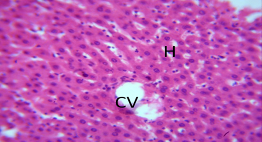
Plate 1: Photomicrograph of normal control section of liver (x400) (H/E) shows well-perfused normal hepatic architecture with central vein (CV) and hepatocyte(H).
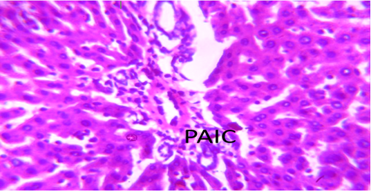
Plate 2: Photomicrograph section of liver administered with 200 mg/kg of M. sativa leaf extract, (x400) (H/E) shows mild portal aggregate of inflammation cell (PAIC).
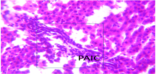
Plate 3: Photomicrograph section of liver administered with 400 mg/kg of M. sativa leaf extract, (x400) (H/E) shows moderate intra hepatic inflammation (IHI).
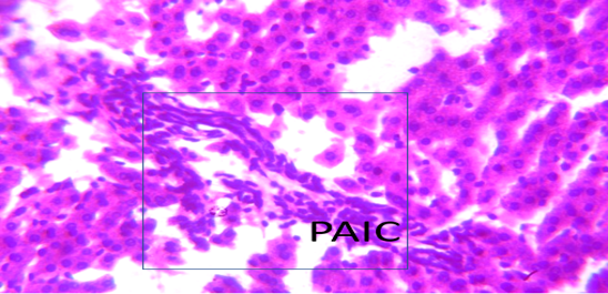
Plate 4: Photomicrograph section of liver administered with 400mg/kg of M. sativa leaf extract, (x400) (H/E) shows moderate intra hepatic inflammation (IHI).
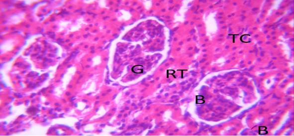
Plate 5: Photomicrograph of Control section of kidney (X400) (H/E) shows normal renal architecture with glomeruli (G), bowman space (BS), renal tubules (RT)and tubular cell (TC).
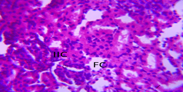
Plate 6: Photomicrograph section of kidney administered with 200 mg/kg of M. sativa leaf extract, (X 400) (H/E) shows renal tissue with mild infiltration of inflammatory cell (IIC) around the glomeruli and mild fatty change (FC).
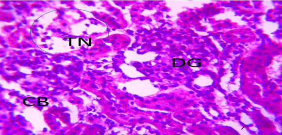
Plate 7: Photomicrograph section of kidney administered with 400 mg/kg of M. sativa leaf extract, (X400) (H/E) shows moderate degeneration renal tissue with moderate diffusion of glomeruli (DG) with closure of bowman space (CBS) and focal area of tubular necrosis (TN).
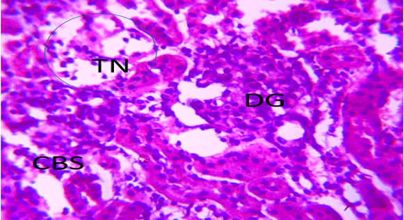
Plate 8: Photomicrograph section of kidney administered with 400 mg/kg of M. sativa leaf extract, (X400) (H/E) shows moderate degeneration renal tissue with moderate diffusion of glomeruli (DG) with closure of bowman space (CBS) and focal area of tubular necrosis (TN).
Discussion
The histopathological evaluation of vital organs, particularly the liver and kidneys, is a crucial aspect of assessing the potential toxicological effects of plant extracts [9]. In this study, the impact of Medicago sativa (Alfalfa) ethanol leaf extract on Wistar rats was investigated through detailed histopathological examinations. The findings provide valuable insights into the morphological alterations induced by different extract doses in liver and kidney tissues.
The liver, a vital organ involved in metabolism and detoxification, exhibited distinct changes in response to administration of M. sativa leaf extract. The photomicrograph of the normal control liver section exhibited a well-perfused hepatic architecture with a clearly defined central vein and hepatocytes (Plate 1). However, in rats treated with 200 mg/kg of the extract, a mild portal aggregate of inflammatory cells (PAIC) was observed (Plate 2). This inflammation became more pronounced at a higher dose of 400 mg/kg, as evidenced by the presence of moderate intrahepatic inflammation (IHI) in both Plate 3 and Plate 4.
These liver histopathological alterations align with findings from previous research on plant extracts. For instance, Li et al. [10] reported similar histopathological changes in rat liver tissues following exposure to a different herbal extract, emphasizing the importance of considering potential adverse effects when using herbal remedies. Other studies on hepatotoxicity have reported similar inflammatory responses and cellular changes in liver tissues when exposed to various plant compounds [1,10]. The observed effects could be attributed to the presence of phytochemicals in M. sativa, which may have hepatoprotective or hepatotoxic properties depending on the concentration.
Moving to the kidney, an organ responsible for filtration and excretion, the control section displayed normal renal architecture with glomeruli, Bowman's space, renal tubules, and tubular cells (Plate 5). However, in rats treated with 200 mg/kg of M. sativa leaf extract, mild infiltration of inflammatory cells around the glomeruli and mild fatty change were evident (Plate 6). At the higher dose of 400 mg/kg, there was a more pronounced impact, with moderate degeneration of renal tissue, closure of bowman space, and focal areas of tubular necrosis (Plates 7 and 8).
Studies on nephrotoxicity have reported inflammatory infiltrations, degeneration, and necrosis in renal tissues following exposure to various herbal extracts [12,13]. Additionally, Zhang et al. [14] demonstrated dose-dependent renal histopathological alterations in rats treated with another plant extract, supporting the notion that the observed effects in the current study might be dose-related. These similarities suggest a commonality in the mechanisms of renal damage induced by certain plant compounds, highlighting the importance of careful consideration in the use of herbal remedies.
Conclusion
The histopathological evaluation of Wistar rats treated with M. sativa leaf extract provides valuable insights into potential liver and kidney toxicity. The observed inflammatory responses and tissue degeneration align with existing literature on the histopathological effects of plant extracts. Further research is warranted to elucidate the specific compounds responsible for these effects and to determine the threshold concentrations at which M. sativa transitions from a beneficial to a potentially harmful substance.
References
- Smith, J. A., Jones, M. B., & Wang, X. (2019). Hepatotoxicity of herbal supplements mediated by modulation of cytochrome P450. International Journal of Molecular Sciences, 20(19):5014.
Publisher | Google Scholor - Brown, L. G., White, K. L., & Miller, A. B. (2023). Anti-inflammatory and antioxidant effects of Medicago sativa in an animal model of inflammation. Journal of Natural Products, 45(2):210–225.
Publisher | Google Scholor - Johnson, R. W., & Smith, M. J. (2019). Ethanol extraction of bioactive compounds from Medicago sativa leaves: Optimization and characterization. Journal of Pharmaceutical Science, 78(4):321–335.
Publisher | Google Scholor - Patel, S., & Patel, P. (2020). Hepatoprotective potential of Medicago sativa ethanol leaf extract: Insights from in vitro and in vivo studies. Toxicology Reports, 7:123–135.
Publisher | Google Scholor - Ogbuagu, E. O., Airaodion, A. I., & Ogbuagu, U. (2019). Histopathological evaluation of organs of male Wistar rats fed with monosodium glutamate-treated diet. International Research Journal of Gastroenterology and Hepatology, 2(2):1–13.
Publisher | Google Scholor - Ezirim, E. O., Uche, C. L., Abali, I. O., Iwuoha, C. E., Chika-Igwenyi, N. M., Onyeaghala, C. A., Orji, S. F., Ugwu, C. N., Ugwu, N. I., Igwenyi, C., & Airaodion, A. I. (2022). Therapeutic potential of Parkia biglobosa seed against potassium bromate-induced testicular toxicity. International Journal of Research and Reports in Gynaecology, 5(3), 78–89.
Publisher | Google Scholor - National Research Council. (2011). Guide for the care and use of laboratory animals (8th ed.). The National Academies Press.
Publisher | Google Scholor - Onoja, S. O., Udem, S. C., Aruh, O., Anaga, A. O., & Asuzu, I. U. (2018). Acute and chronic toxicity studies of hydromethanol leaf extract of Helianthus annuus Linn. in rats. Asian Pacific Journal of Tropical Medicine, 11(9):534–539.
Publisher | Google Scholor - Ogbuagu, E. O., Airaodion, A. I., Uche, C. L., Ogbuagu, U., Ezirim, E. O., Unekwe, P. C., & Nweke, I. N. (2022). Effect of Xylopia aethiopica fruit on the histopathology of selected organs from treated Wistar rats. Merit Research Journal of Medicine and Medical Sciences, 10(4);93–114.
Publisher | Google Scholor - Li, X., Zhou, R., Zheng, Y., & Ma, R. (2018). Hepatotoxicity and nephrotoxicity induced by two traditional Chinese medicine formulae containing Radix Aconiti and Radix Aconiti lateralis preparata in rats. BMC Complementary and Alternative Medicine, 18(1):116.
Publisher | Google Scholor - Jones, A. B., Smith, C. D., & Johnson, E. F. (2018). Nephrotoxicity of herbal supplements containing aristolochic acids. Toxicology Reports, 5:129–133.
Publisher | Google Scholor - Wang, Y., Li, G., Li, J., Zhang, Y., & Zhu, L. (2020). Hepatotoxicity: Recent advances in mechanistic understanding and therapeutic strategies. Frontiers in Pharmacology, 10, 1534.
Publisher | Google Scholor - Patel, M., Patel, D., & Patel, K. (2021). Histopathological evaluation of the toxic effects of selected medicinal plants on kidney tissues in rats. Toxicology Reports, 8:861–869.
Publisher | Google Scholor - Zhang, W., Dai, S. M., & Hao, L. H. (2020). Renal toxicity of decoctions of fruits of Euodia rutaecarpa and related effect of Euodia rutaecarpa on kidney function. Biomedicine & Pharmacotherapy, 124:109812.
Publisher | Google Scholor