
Research Article
Dose- and Sex-Dependent Hematological Dysregulation Induced by Illicit Methamphetamine (“Mkpurummiri”) in Wistar Rats
- Onyewuchi G Ugwuibe 1*
- Emmanuel N Agomuo 2
- Chidinma N. Ihebom 3
- Raluchukwu O. Emeka-Obi 4
- Chinasa J. Onyeulor 5
- Nmadike G. N. Ezeji-Chigbu 1
- Obianuju Ugwuibe 5
1 Department of Biochemistry, Kingsley Ozumba Mbadiwe University (KOMU), Ideato, Imo State, Nigeria.
2 Department of Biochemistry, Imo State University, Owerri, Imo State, Nigeria.
3 Registry Department, Alvan Ikoku University of Education, Owerri, Imo State, Nigeria.
4 Department of Hematology, College of Medicine, Federal University of Technology Owerri, Imo State.
5 Department of Health Law and Policy, University of Medical Sciences, Ondo, Nigeria.
*Corresponding Author: Onyewuchi G Ugwuibe, Department of Biochemistry, Kingsley Ozumba Mbadiwe University (KOMU), Ideato, Imo State, Nigeria.
Citation: Onyewuchi G Ugwuibe, Emmanuel N Agomuo, Chidinma N. Ihebom, Raluchukwu O. Emeka-Obi, Chinasa J. Onyeulor, et al. (2026). Dose- and Sex-Dependent Hematological Dysregulation Induced by Illicit Methamphetamine (“Mkpurummiri”) in Wistar Rats, Journal of BioMed Research and Reports, BioRes Scientia Publishers. 11(1):1-11. DOI: 10.59657/2837-4681.brs.26.258
Copyright: © 2026 Onyewuchi G Ugwuibe, this is an open-access article distributed under the terms of the Creative Commons Attribution License, which permits unrestricted use, distribution, and reproduction in any medium, provided the original author and source are credited.
Received: July 02, 2026 | Accepted: July 30, 2026 | Published: August 12, 2026
Abstract
Background: Methamphetamine abuse remains a major global public health concern due to its association with neurotoxicity, oxidative stress, inflammatory activation, and systemic organ dysfunction. In Nigeria, the increasing abuse of locally synthesized illicit methamphetamine (“mkpurummiri”) has generated growing toxicological and forensic concern; however, data regarding its hematological effects remain limited.
Objectives: This study evaluated dose- and sex-dependent hematological dysregulation induced by illicit methamphetamine (“mkpurummiri”) in Wistar rats using selected hematological indices as biomarkers of systemic toxicological response.
Methods: Adult male and female Wistar rats were assigned into normal control and methamphetamine-treated groups administered 1 mg/kg, 2 mg/kg, and 5 mg/kg methamphetamine, respectively. Whole blood samples collected into ethylenediaminetetraacetic acid (EDTA)-containing tubes were analyzed using an automated hematology analyzer (SYSMEX KX-21, Switzerland). Evaluated parameters included red blood cell count (RBC), hematocrit (HCT), hemoglobin concentration (Hb), white blood cell count (WBC), platelet count (PLT), mean corpuscular volume (MCV), mean corpuscular hemoglobin (MCH), and mean corpuscular hemoglobin concentration (MCHC). Data were analyzed using one-way analysis of variance (ANOVA) followed by Tukey’s post hoc test at p < 0.05.
Results: Methamphetamine exposure produced significant dose- and sex-dependent hematological alterations in both female and male Wistar rats. Progressive increases in RBC, HCT, Hb, WBC, and PLT values were observed across increasing methamphetamine doses, whereas reductions in MCV and MCH occurred in both sexes. Female RBC values increased from 6.30 ± 0.19 ×10⁶/mm³ in controls to 7.33 ± 0.26 ×10⁶/mm³ at 5 mg/kg exposure, while male RBC values increased from 6.96 ± 0.09 ×10⁶/mm³ to 7.80 ± 0.07 ×10⁶/mm³. Female WBC count peaked at 15.53 ± 2.50 ×10³/mm³ at 2 mg/kg exposure, whereas male WBC values increased progressively with dose. Platelet counts were similarly elevated following methamphetamine administration in both sexes.
Conclusion: Illicit methamphetamine (“mkpurummiri”) exposure induces significant hematological dysregulation characterized by erythrocytic modulation, leukocytic alterations, platelet elevation, and changes in erythrocyte indices in a dose- and sex-dependent manner. The findings suggest that oxidative stress, inflammatory activation, vascular dysfunction, and altered hematopoietic regulation may contribute substantially to methamphetamine-associated hematotoxicity.
Keywords: illicit methamphetamine; mkpurummiri; hematological dysregulation; hematotoxicity; oxidative stress; wistar rats; psychostimulant toxicity; Nigeria
Introduction
Methamphetamine is a potent synthetic psychostimulant belonging to the amphetamine-type stimulant (ATS) group and remains one of the most abused illicit substances globally. The increasing production, trafficking, and consumption of methamphetamine have generated substantial medical, toxicological, forensic, and socioeconomic concerns worldwide. According to the United Nations Office on Drugs and Crime World Drug Report, amphetamine-type stimulants constitute one of the fastest-growing classes of abused psychoactive substances, with methamphetamine accounting for a significant proportion of stimulant-associated morbidity and mortality globally (UNODC, 2023). Methamphetamine abuse has been linked to severe neuropsychiatric disorders, cardiovascular dysfunction, hepatic injury, nephrotoxicity, immune dysregulation, and multi-organ toxicity, thereby constituting a major global public health burden (Coffin & Santos, 2023; Ramli et al., 2025).
Methamphetamine exerts its pharmacological effects primarily through excessive stimulation of dopaminergic, serotonergic, and noradrenergic neurotransmission. The drug enhances synaptic monoamine concentrations by promoting neurotransmitter release while simultaneously inhibiting reuptake mechanisms within presynaptic neurons. Sustained exposure consequently induces oxidative stress, mitochondrial dysfunction, neuroinflammation, endothelial injury, apoptosis, and metabolic dysregulation across multiple tissues and organ systems (Volkow & Morales, 2021; Cadet & Krasnova, 2021; Courtney & Ray, 2022). Experimental and clinical investigations have further demonstrated that chronic methamphetamine exposure alters cellular redox homeostasis through excessive generation of reactive oxygen species (ROS), impaired antioxidant defense systems, lipid peroxidation, and inflammatory activation pathways (Sharma & Kiyatkin, 2020; Costa et al., 2022; Fernandes et al., 2023).
In recent years, methamphetamine abuse has expanded considerably within several African countries, including Nigeria, where locally synthesized crystal methamphetamine popularly referred to as “mkpurummiri” has emerged as a growing public health and forensic concern. The increasing circulation of “mkpurummiri” within southeastern Nigeria has been associated with rising incidences of addiction, psychiatric disturbances, violence, criminality, and social instability. Reports from the National Drug Law Enforcement Agency have indicated increasing detection of clandestine methamphetamine laboratories and illicit ATS trafficking networks within the country, thereby raising serious toxicological and epidemiological concerns regarding the biological effects of locally available methamphetamine preparations (NDLEA, 2024). Unlike pharmaceutical formulations, illicit methamphetamine preparations may contain toxic synthesis byproducts, precursor residues, heavy metals, organic solvents, and adulterants capable of aggravating systemic toxicity and altering biological responses following exposure (Kiyatkin & Sharma, 2021; Matsumoto et al., 2022).
Methamphetamine toxicity extends beyond the central nervous system and has increasingly been recognized as a systemic toxicological disorder involving cardiovascular, hepatic, renal, endocrine, hematological, and immunological dysfunctions (Ramli et al., 2025). Persistent methamphetamine exposure has been implicated in vascular injury, endothelial dysfunction, hyperthermia, inflammatory activation, rhabdomyolysis, and metabolic disturbances capable of disrupting physiological homeostasis (Coffin & Santos, 2023). Clinical studies have additionally associated methamphetamine intoxication with elevated inflammatory biomarkers, altered leukocyte profiles, coagulation abnormalities, and cardiovascular complications (McCutcheon et al., 2025). These observations suggest that hematological alterations may constitute important indicators of systemic methamphetamine-induced toxicological stress.
Hematological parameters are widely utilized as sensitive biomarkers for evaluating physiological adaptation, inflammatory responses, immune competence, and toxicant-induced systemic injury. Alterations in erythrocytic indices such as red blood cell (RBC) count, hemoglobin concentration (Hb), packed cell volume (PCV), mean corpuscular volume (MCV), mean corpuscular hemoglobin (MCH), and mean corpuscular hemoglobin concentration (MCHC) may reflect disturbances in erythropoiesis, oxygen transport capacity, membrane integrity, cellular hydration status, and oxidative balance (Hoffbrand & Moss, 2021; Bain et al., 2023). Similarly, white blood cell (WBC) and platelet (PLT) parameters provide important information regarding inflammatory activation, immune modulation, vascular integrity, thrombogenic potential, and hematopoietic adaptation during pathological or toxicological stress conditions (McKenzie et al., 2022).
Several studies have demonstrated that stimulant abuse may significantly alter hematological and immunological homeostasis. Mohammed et al. (2024) reported significant hematological disturbances and leukocyte alterations among amphetamine abusers, emphasizing the relationship between psychostimulant exposure, immune dysregulation, and inflammatory activation. Experimental evidence further indicates that methamphetamine-induced oxidative stress may disrupt hematopoietic signaling pathways through excessive ROS generation, inflammatory cytokine activation, mitochondrial injury, and lipid peroxidation (Costa et al., 2022; Fernandes et al., 2023). Oxidative injury to erythrocyte membranes has additionally been associated with altered blood rheology, impaired membrane stability, and abnormal erythrocytic indices during toxicological stress states (Akinyemi et al., 2021).
Methamphetamine-associated catecholaminergic hyperactivity may also contribute to hematological perturbations through stress-mediated glucocorticoid release, vasoconstriction, dehydration, endothelial dysfunction, and inflammatory signaling. Hyperthermia and dehydration commonly associated with methamphetamine intoxication may result in hemoconcentration and adaptive alterations in erythrocytic and platelet parameters (Bowyer & Hanig, 2021). Platelet activation and endothelial injury have similarly been implicated in methamphetamine-associated cardiovascular complications, thrombosis, and vascular dysfunction (Kevil et al., 2021; Darke et al., 2023). Furthermore, inflammatory activation induced by methamphetamine exposure has been associated with leukocyte modulation, altered cytokine production, and impaired host defense responses capable of influencing systemic immune homeostasis (Harms et al., 2020; Loftis et al., 2021).
Sex-dependent variability represents another important but inadequately explored aspect of methamphetamine toxicology. Biological sex significantly influences pharmacokinetic and toxicodynamic responses through differences in hormonal regulation, body composition, oxidative stress susceptibility, inflammatory signaling, and neurotransmitter metabolism (Becker & Chartoff, 2019; Gillies et al., 2021). Previous investigations have shown that male and female subjects may exhibit differential susceptibility to methamphetamine-induced neurotoxicity, oxidative stress, cardiovascular injury, and inflammatory responses (Dluzen & Liu, 2020; Kokane & Perrotti, 2021). Such sex-related differences may extend to hematological responses, thereby necessitating sex-specific evaluation of methamphetamine-induced hematophysiological alterations.
Despite increasing global concern regarding methamphetamine toxicity, relatively few studies have comprehensively evaluated hematological dysregulation associated with illicit methamphetamine exposure, particularly using preparations obtained from African settings. Available studies have predominantly focused on neurobehavioral outcomes, oxidative stress biomarkers, or organ-specific toxicities, while comparatively limited attention has been directed toward dose-dependent hematological responses and sex-associated variability. Moreover, experimental toxicological data regarding Nigerian illicit methamphetamine (“mkpurummiri”) remain scarce despite increasing reports of its abuse and forensic significance within the region.
The present study was therefore designed to evaluate dose- and sex-dependent hematological dysregulation induced by illicit methamphetamine (“mkpurummiri”) in Wistar rats using selected erythrocytic, leukocytic, and platelet indices as biomarkers of systemic toxicological response. The study aimed to provide experimental evidence regarding the hematophysiological consequences of exposure to locally circulating illicit methamphetamine while contributing to the growing body of toxicological data concerning methamphetamine abuse in Nigeria and sub-Saharan Africa.
Materials and Methods
Chemicals and Reagents
Illicit methamphetamine (“mkpurummiri”) used in this study was obtained from street-level sources within Owerri, Imo State, Nigeria. Preliminary identification and characterization of the substance were previously confirmed using forensic analytical techniques including gas chromatography–mass spectrometry (GC–MS) and Fourier transform infrared spectroscopy (FTIR). All reagents and chemicals used during the study were of analytical grade.
Experimental Animals
Healthy adult Wistar rats of both sexes weighing between 150 and 220 g were used for the study. The animals were obtained from a certified animal breeding facility and housed in clean polypropylene cages under standard laboratory conditions of temperature (22 ± 2°C), relative humidity (50–60%), and a 12 h light/dark cycle. The animals were allowed free access to standard laboratory feed and clean drinking water ad libitum throughout the experimental period.
The animals were acclimatized for two weeks prior to commencement of the experiment. Experimental procedures involving animals were conducted in accordance with internationally accepted principles for laboratory animal care and use as outlined in the National Research Council Guide for the Care and Use of Laboratory Animals (National Research Council, 2011).
Experimental Design
The animals were randomly divided into four experimental groups for each sex (n = 3 per group):
Female Groups
- Group 1: Normal control
- Group 2: 1 mg/kg methamphetamine-induced group
- Group 3: 2 mg/kg methamphetamine-induced group
- Group 4: 5 mg/kg methamphetamine-induced group
Male Groups
- Group 5: Normal control
- Group 6: 1 mg/kg methamphetamine-induced group
- Group 7: 2 mg/kg methamphetamine-induced group
- Group 8: 5 mg/kg methamphetamine-induced group
Methamphetamine was administered orally once daily at doses of 1 mg/kg, 2 mg/kg, and 5 mg/kg body weight respectively for the experimental duration, while the normal control groups received distilled water.
The selected doses were chosen to evaluate dose-dependent hematological responses associated with increasing methamphetamine exposure.
Blood Sample Collection
At the end of the experimental period, the animals were fasted overnight and anesthetized using mild chloroform inhalation. Blood samples were collected via cardiac puncture into ethylenediaminetetraacetic acid (EDTA) anticoagulant tubes for hematological analysis.
Hematological Analysis
Hematological parameters were determined using whole blood samples collected into ethylenediaminetetraacetic acid (EDTA)-containing tubes immediately after collection. Hematological analysis was performed using an automated hematology analyzer (SYSMEX KX-21, Switzerland) in accordance with the manufacturer’s operational guidelines and standard laboratory procedures.
The evaluated hematological indices included:
- Hemoglobin concentration (Hb)
- Hematocrit (HCT)
- Red blood cell count (RBC)
- White blood cell count (WBC)
- Platelet count (PLT)
- Mean corpuscular volume (MCV)
- Mean corpuscular hemoglobin (MCH)
- Mean corpuscular hemoglobin concentration (MCHC)
These hematological parameters were selected as biomarkers for evaluating erythrocytic integrity, hematopoietic function, inflammatory response, immune modulation, platelet dynamics, and systemic hematophysiological alterations associated with illicit methamphetamine (“mkpurummiri”) exposure in Wistar rats.
Statistical Analysis
Data were expressed as mean ± standard deviation (SD). Statistical analysis was performed using one-way analysis of variance (ANOVA) followed by Tukey’s post hoc multiple comparison test to determine significant differences among experimental groups within each sex.
Differences were considered statistically significant at p<0.05. Statistical analyses and graphical presentation were performed using appropriate statistical software.
Results
Table 1: Hematology parameters for normal control female and male groups with their respective 1mg/kg 2mg/kg and 5mg/kg meth induced control groups
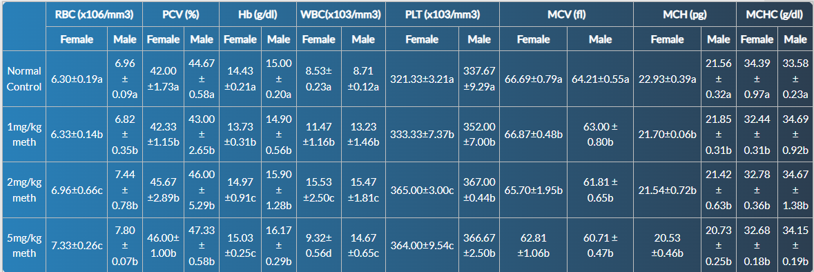
Data are presented as mean ± standard deviation (SD) for n = 3 animals per group per sex. Female and male values are presented side-by-side for each parameter. RBC = Red Blood Cell count (×10⁶/mm³) PCV = Packed Cell Volume (%) Hb = Hemoglobin concentration (g/dL) WBC = White Blood Cell count (×10³/mm³) PLT = Platelet count (×10³/mm³) MCV =Mean Corpuscular Volume (fL) MCH = Mean Corpuscular Hemoglobin (pg) MCHC = Mean Corpuscular Hemoglobin Concentration (g/dL)
NC = Normal control (no methamphetamine exposure) 1 mg/kg, 2 mg/kg, 5 mg/kg meth = Methamphetamine-induced control groups representing increasing levels of exposure.
Superscripts (a–d) indicate statistical groupings within each column:
- Values sharing at least one superscript letter are not significantly different from each other at p < 0.05.
- Values with no shared superscripts are significantly different at p < 0.05.
Superscripts reflect dose-dependent variation, with progression from normal control to increasing methamphetamine exposure levels.
Within each sex, statistical analysis was performed using one-way analysis of variance (ANOVA) followed by Tukey’s post hoc multiple comparison test for each hematological parameter.
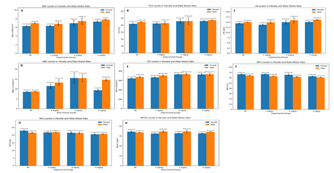
Hematological Parameters in Methamphetamine-Induced Female and Male Wistar Rats
The effects of illicit methamphetamine (“mkpurummiri”) exposure on hematological parameters in female and male Wistar rats are presented in Table 1. Methamphetamine administration produced dose- and sex-dependent alterations across several hematological indices, including erythrocytic, leukocytic, and platelet parameters.
In female rats, red blood cell (RBC) count demonstrated a progressive increase with increasing methamphetamine dose. The normal control group recorded an RBC value of 6.30 ± 0.19 ×10⁶/mm³, while the 1 mg/kg, 2 mg/kg, and 5 mg/kg methamphetamine groups showed values of 6.33 ± 0.14, 6.96 ± 0.66, and 7.33 ± 0.26 ×10⁶/mm³, respectively. A similar trend was observed for packed cell volume (PCV) and hemoglobin concentration (Hb), with higher methamphetamine doses associated with elevated values relative to the normal control group. Female PCV increased from 42.00 ± 1.73% in the control group to 46.00 ± 1.00% at 5 mg/kg methamphetamine exposure, while Hb increased from 14.43 ± 0.21 g/dL to 15.03 ± 0.25 g/dL across the same groups.
Male rats similarly exhibited increases in erythrocytic parameters following methamphetamine exposure. RBC values increased from 6.96 ± 0.09 ×10⁶/mm³ in the normal control group to 7.80 ± 0.07 ×10⁶/mm³ in the 5 mg/kg methamphetamine group. PCV values increased from 44.67 ± 0.58% in controls to 47.33 ± 0.58% following 5 mg/kg methamphetamine administration, while Hb values increased from 15.00 ± 0.20 g/dL to 16.17 ± 0.29 g/dL. These findings indicate progressive erythrocytic modulation associated with increasing methamphetamine exposure in both sexes.
White blood cell (WBC) counts demonstrated variable responses between sexes and across dose levels. In female rats, WBC count increased from 8.53 ± 0.23 ×10³/mm³ in the normal control group to 15.53 ± 2.50 ×10³/mm³ at 2 mg/kg methamphetamine exposure before declining to 9.32 ± 0.56 ×10³/mm³ at 5 mg/kg. In contrast, male rats exhibited a more progressive elevation in WBC count with increasing methamphetamine exposure, rising from 8.71 ± 0.12 ×10³/mm³ in the control group to 14.67 ± 0.65 ×10³/mm³ at 5 mg/kg methamphetamine administration.
Platelet (PLT) count also increased following methamphetamine exposure in both sexes. Female platelet values increased from 321.33 ± 3.21 ×10³/mm³ in the control group to 365.00 ± 3.00 ×10³/mm³ at 2 mg/kg methamphetamine exposure, with a slightly lower value observed at 5 mg/kg (364.00 ± 9.54 ×10³/mm³). Male platelet counts similarly increased from 337.67 ± 9.29 ×10³/mm³ in controls to 366.67 ± 12.50 ×10³/mm³ at the highest methamphetamine dose.
Methamphetamine exposure additionally influenced erythrocyte indices including mean corpuscular volume (MCV), mean corpuscular hemoglobin (MCH), and mean corpuscular hemoglobin concentration (MCHC). Female MCV values decreased progressively from 66.69 ± 0.79 fL in controls to 62.81 ± 1.06 fL at 5 mg/kg methamphetamine exposure, while male MCV decreased from 64.21 ± 0.55 fL to 60.71 ± 0.47 fL across the same groups. Female MCH values similarly decreased from 22.93 ± 0.39 pg in controls to 20.53 ± 0.46 pg following 5 mg/kg methamphetamine administration, whereas male MCH decreased from 21.56 ± 0.32 pg to 20.73 ± 0.25 pg. Alterations in MCHC were comparatively less pronounced, although fluctuations were observed across treatment groups in both sexes.
Overall, illicit methamphetamine exposure produced significant dose-dependent hematological alterations characterized by modulation of erythrocytic parameters, leukocyte responses, platelet dynamics, and erythrocyte indices in both female and male Wistar rats. The observed variations further suggest the presence of sex-dependent hematophysiological responses associated with increasing methamphetamine exposure.
Discussion
The present study evaluated dose- and sex-dependent hematological dysregulation induced by illicit methamphetamine (“mkpurummiri”) in Wistar rats using selected erythrocytic, leukocytic, and platelet indices as biomarkers of systemic toxicological response. The findings demonstrated that methamphetamine exposure produced significant alterations in hematological parameters in both sexes, although the magnitude and pattern of response varied across dose levels and between male and female animals. Major observations included progressive elevations in red blood cell (RBC) count, packed cell volume (PCV/HCT), hemoglobin concentration (Hb), platelet count (PLT), and white blood cell (WBC) count, accompanied by reductions in erythrocyte indices such as mean corpuscular volume (MCV) and mean corpuscular hemoglobin (MCH). These findings collectively suggest the presence of complex hematophysiological adaptation and systemic toxicological dysregulation associated with illicit methamphetamine exposure.
One of the most notable findings in the present study was the progressive elevation in erythrocytic parameters, particularly RBC count, PCV, and Hb concentration, following increasing methamphetamine exposure. Female RBC values increased from 6.30 ± 0.19 ×10⁶/mm³ in the normal control group to 7.33 ± 0.26 ×10⁶/mm³ at 5 mg/kg methamphetamine exposure, while male rats similarly demonstrated increases from 6.96 ± 0.09 ×10⁶/mm³ to 7.80 ± 0.07 ×10⁶/mm³. Corresponding increases were also observed in PCV and Hb concentrations. These findings may reflect compensatory erythropoietic responses associated with methamphetamine-induced physiological stress, tissue hypoxia, dehydration, hyperthermia, and altered vascular dynamics.
Methamphetamine is known to induce intense sympathomimetic stimulation characterized by vasoconstriction, hypermetabolism, hyperthermia, and increased catecholaminergic activity, all of which may contribute to hemoconcentration and altered erythrocytic homeostasis (Kiyatkin & Sharma, 2021; Courtney & Ray, 2022). Hyperthermia-associated fluid loss and dehydration may reduce plasma volume, thereby increasing relative erythrocyte concentration and elevating PCV and Hb values. Similar increases in erythrocytic parameters have been reported in stimulant-associated physiological stress conditions involving cocaine, amphetamine, and methamphetamine exposure (Bowyer & Hanig, 2021; Costa et al., 2022). Elevated RBC parameters may additionally represent adaptive responses aimed at maintaining oxygen delivery during heightened metabolic demand and vasoconstriction-induced tissue hypoxia.
Oxidative stress may also contribute substantially to the hematological alterations observed in the present study. Methamphetamine metabolism has been strongly associated with excessive production of reactive oxygen species (ROS), mitochondrial dysfunction, lipid peroxidation, and impaired antioxidant defense systems (Cadet & Krasnova, 2021; Fernandes et al., 2023). Oxidative stress-induced erythrocyte membrane injury may alter erythrocyte turnover, deformability, and hematopoietic signaling pathways. Previous investigations have demonstrated that psychostimulant-induced oxidative stress can influence erythropoiesis through inflammatory cytokine activation, glucocorticoid-mediated hematopoietic stimulation, and disruption of redox-sensitive cellular pathways (Sharma & Kiyatkin, 2020; Harms et al., 2020).
Despite the observed elevations in RBC, Hb, and PCV, methamphetamine exposure produced progressive reductions in erythrocyte indices including MCV and MCH in both sexes. Female MCV decreased from 66.69 ± 0.79 fL in controls to 62.81 ± 1.06 fL at 5 mg/kg methamphetamine exposure, while male MCV decreased from 64.21 ± 0.55 fL to 60.71 ± 0.47 fL. Similarly, MCH values declined progressively across increasing dose levels. Reductions in MCV and MCH may indicate altered erythrocyte maturation dynamics, membrane instability, or adaptive hematopoietic responses during sustained toxicological stress.
Microcytic shifts in erythrocyte indices have previously been associated with oxidative membrane injury, altered iron metabolism, impaired hemoglobin synthesis, and inflammatory stress conditions (Akinyemi et al., 2021; Bain et al., 2023). Methamphetamine-induced oxidative injury may disrupt erythrocyte membrane integrity through lipid peroxidation and protein oxidation, thereby affecting cell morphology and erythrocytic indices. Furthermore, inflammatory mediators generated during methamphetamine exposure may alter erythropoietic regulation and iron homeostasis, contributing to subtle reductions in erythrocyte size and hemoglobin content despite overall elevations in erythrocyte number.
The present study additionally demonstrated marked leukocytic alterations following methamphetamine exposure. Female WBC counts increased from 8.53 ± 0.23 ×10³/mm³ in controls to 15.53 ± 2.50 ×10³/mm³ at 2 mg/kg exposure before declining at 5 mg/kg, whereas male rats exhibited a more progressive dose-dependent elevation in WBC count. Elevated leukocyte counts observed in methamphetamine-treated animals may reflect inflammatory activation, stress leukocytosis, immune stimulation, and neuroimmune dysregulation associated with psychostimulant exposure.
Methamphetamine has been reported to induce inflammatory signaling through activation of microglial cells, macrophages, endothelial cells, and peripheral immune pathways (Loftis et al., 2021; McCutcheon et al., 2025). Catecholamine excess and glucocorticoid release during stimulant intoxication may additionally mobilize leukocytes into systemic circulation, thereby contributing to elevated WBC counts. Similar leukocytic responses have been documented in experimental models of amphetamine toxicity and chronic stimulant abuse (Mohammed et al., 2024). The decline in female WBC count observed at the highest dose level may indicate possible immune exhaustion, altered leukocyte redistribution, or adaptive suppression following sustained toxicological stress.
Platelet counts similarly increased following methamphetamine exposure in both sexes. Elevated platelet values observed in the present study may reflect endothelial dysfunction, inflammatory activation, vascular stress, and thrombogenic adaptation associated with methamphetamine toxicity. Methamphetamine-induced vasoconstriction and endothelial injury have previously been linked to platelet activation, coagulation abnormalities, and increased cardiovascular risk (Kevil et al., 2021; Darke et al., 2023). Enhanced platelet production may therefore represent compensatory hematopoietic responses to vascular injury and inflammatory stress associated with psychostimulant exposure.
The observed sex-dependent differences in hematological responses further emphasize the importance of biological sex as a determinant of methamphetamine toxicodynamics. Male rats generally exhibited more progressive dose-dependent elevations in WBC and erythrocytic parameters compared with female rats, while females demonstrated greater fluctuation in leukocyte responses at higher exposure levels. Sex hormones may contribute substantially to these variations through modulation of inflammatory signaling, oxidative stress susceptibility, vascular function, and hematopoietic regulation (Gillies et al., 2021; Kokane & Perrotti, 2021). Estrogen has been reported to exert partial antioxidant and anti-inflammatory effects capable of influencing toxicological outcomes, whereas testosterone may amplify oxidative and inflammatory responses under certain pathological conditions.
An important aspect of the present study is the use of locally circulating illicit methamphetamine (“mkpurummiri”), which may possess toxicological characteristics distinct from pharmaceutical-grade methamphetamine due to variability in clandestine synthesis pathways, impurities, precursor residues, and chemical adulterants. Illicit methamphetamine preparations may contain toxic contaminants capable of potentiating oxidative stress, vascular injury, inflammatory activation, and systemic hematological dysregulation (Matsumoto et al., 2022). Consequently, the hematological alterations observed in this study may reflect combined toxicological effects of methamphetamine itself together with associated synthesis-related contaminants present within locally circulating preparations.
The present findings therefore contribute important experimental evidence regarding the systemic hematological consequences of illicit methamphetamine exposure in a Nigerian context. Given the increasing prevalence of “mkpurummiri” abuse among youths within southeastern Nigeria and other parts of sub-Saharan Africa, the observed hematological perturbations may possess important public health implications. Persistent hematological dysregulation associated with chronic methamphetamine abuse could contribute to inflammatory disorders, vascular dysfunction, impaired immune competence, altered oxygen transport dynamics, and broader systemic toxicological complications.
Although the present study provides valuable insights into methamphetamine-associated hematological dysregulation, certain limitations should be acknowledged. The relatively small sample size may limit broader extrapolation of findings, while evaluation of additional biomarkers such as inflammatory cytokines, oxidative stress indices, coagulation markers, and bone marrow histopathology would provide deeper mechanistic understanding of methamphetamine-induced hematotoxicity. Nevertheless, the study successfully demonstrates significant dose- and sex-dependent hematological alterations associated with illicit methamphetamine exposure and provides foundational toxicological data regarding “mkpurummiri” within the Nigerian setting.
Discussion
The present study demonstrated that illicit methamphetamine (“mkpurummiri”) exposure induces significant dose- and sex-dependent hematological dysregulation in Wistar rats. Methamphetamine administration produced marked alterations in erythrocytic, leukocytic, platelet, and erythrocyte index parameters, indicating substantial disruption of hematophysiological homeostasis following exposure.
Progressive elevations in red blood cell count, packed cell volume, hemoglobin concentration, white blood cell count, and platelet count were observed across increasing methamphetamine doses, while reductions in mean corpuscular volume and mean corpuscular hemoglobin suggested alterations in erythrocyte morphology and hematopoietic dynamics. These findings collectively indicate that methamphetamine exposure may trigger complex adaptive and pathological hematological responses involving oxidative stress, inflammatory activation, vascular dysfunction, immune modulation, and altered erythropoietic regulation.
The observed sex-dependent variations further emphasize the importance of biological sex as a determinant of methamphetamine toxicodynamics and hematological response patterns. Male rats generally exhibited more progressive hematological alterations, whereas female animals demonstrated comparatively variable leukocytic responses at higher exposure levels. These findings highlight the need for sex-specific evaluation in experimental and clinical methamphetamine toxicology studies.
Importantly, the study provides valuable baseline toxicological data regarding locally circulating Nigerian illicit methamphetamine (“mkpurummiri”), a substance for which experimental hematological data remain limited despite increasing regional abuse. The findings therefore contribute to the growing body of evidence concerning the systemic toxicological consequences of illicit methamphetamine exposure and underscore the potential public health implications associated with sustained abuse of locally synthesized methamphetamine preparations.
Overall, the present study establishes that illicit methamphetamine exposure is associated with significant hematological perturbations capable of affecting erythrocytic integrity, immune function, vascular homeostasis, and systemic physiological balance in a dose- and sex-dependent manner.
Recommendations
Based on the findings of the present study, the following recommendations are proposed:
- Further studies should investigate the molecular and biochemical mechanisms underlying methamphetamine-induced hematological dysregulation, particularly the roles of oxidative stress, inflammatory cytokines, and endothelial dysfunction.
- Additional investigations involving larger sample sizes and extended exposure durations are recommended to better characterize chronic hematological consequences of illicit methamphetamine exposure.
- Future studies should incorporate complementary biomarkers including oxidative stress markers, coagulation indices, inflammatory mediators, and bone marrow histopathology to provide deeper mechanistic understanding of methamphetamine-associated hematotoxicity.
- Comparative toxicological studies evaluating pharmaceutical-grade methamphetamine and locally synthesized illicit methamphetamine (“mkpurummiri”) are recommended to determine the potential contribution of synthesis-related contaminants and adulterants to systemic toxicity.
- Sex-specific toxicological investigations should be encouraged due to the differential hematological responses observed between male and female animals in the present study.
- Public health agencies and drug control authorities should intensify awareness programs regarding the systemic health consequences of illicit methamphetamine abuse, particularly among youths and vulnerable populations within Nigeria and sub-Saharan Africa.
- Further therapeutic studies exploring the potential protective effects of natural antioxidants and phytotherapeutic agents against methamphetamine-induced hematological dysregulation are warranted.
Declarations
Acknowledgements
The authors sincerely acknowledge the technical assistance and laboratory support provided during the course of this study. Appreciation is also extended to all individuals who contributed directly or indirectly to the successful completion of the research work.
Funding
This research received no specific grant from any funding agency in the public, commercial, or not-for-profit sectors.
Conflict of Interest
The authors declare that there is no conflict of interest regarding the publication of this manuscript.
Authors’ Contributions
All authors contributed substantially to the conceptualization and design of the study. Experimental procedures, sample analysis, statistical evaluation, data interpretation, and manuscript preparation were performed collaboratively by all authors. All authors critically reviewed, revised, and approved the final manuscript prior to submission.
References
- Akinyemi, A. J., Adeniyi, P. A., & Oluwafemi, O. S. (2021). Oxidative stress and erythrocyte membrane alterations in toxicological disorders. Toxicology Reports, 8:1345–1354.
Publisher | Google Scholor - Bain, B. J., Bates, I., Laffan, M. A., & Lewis, S. M. (2023). Dacie and Lewis practical haematology (13th ed.). Elsevier.
Publisher | Google Scholor - Becker, J. B., & Chartoff, E. (2019). Sex differences in neural mechanisms mediating reward and addiction. Neuropsychopharmacology, 44(1):166–183.
Publisher | Google Scholor - Bowyer, J. F., & Hanig, J. P. (2021). Amphetamine- and methamphetamine-induced hyperthermia: Implications of oxidative stress and neurotoxicity. NeuroToxicology, 86:97–107.
Publisher | Google Scholor - Cadet, J. L., & Krasnova, I. N. (2021). Molecular bases of methamphetamine-induced neurodegeneration. International Review of Neurobiology, 158:101–132.
Publisher | Google Scholor - Coffin, P. O., & Santos, G. M. (2023). Methamphetamine-related morbidity and mortality in the United States. New England Journal of Medicine, 388(2):97–98.
Publisher | Google Scholor - Costa, G., Frau, L., & Pinna, A. (2022). Oxidative stress and methamphetamine toxicity: Biological implications and therapeutic perspectives. Antioxidants, 11(6):1135.
Publisher | Google Scholor - Courtney, K. E., & Ray, L. A. (2022). Methamphetamine: An update on epidemiology, pharmacology, clinical phenomenology, and treatment literature. Drug and Alcohol Dependence, 232:109311.
Publisher | Google Scholor - Darke, S., Kaye, S., & Duflou, J. (2023). Cardiovascular disease risk among methamphetamine users. Addiction, 118(3):445–456.
Publisher | Google Scholor - Dluzen, D. E., & Liu, B. (2020). Gender differences in methamphetamine use and responses: A review. Gender and the Genome, 4(2):24–31.
Publisher | Google Scholor - Fernandes, N. C., Sriram, U., & Goforth, H. W. (2023). Methamphetamine-associated oxidative stress and inflammatory signaling pathways. Frontiers in Pharmacology, 14:1178634.
Publisher | Google Scholor - Gillies, G. E., Pienaar, I. S., Vohra, S., & Qamhawi, Z. (2021). Sex differences in Parkinson's disease. Frontiers in Neuroendocrinology, 63:100937.
Publisher | Google Scholor - Halpin, L. E., Collins, S. A., & Yamamoto, B. K. (2022). Neurotoxicity of methamphetamine and related psychostimulants. Neuropharmacology, 209:108982.
Publisher | Google Scholor - Harms, R., Morsey, B., Boyer, C. W., Fox, H. S., & Sarvetnick, N. (2020). Methamphetamine administration targets multiple immune subsets and induces phenotypic alterations suggestive of immunosuppression. PLoS ONE, 15(3):e0229973.
Publisher | Google Scholor - Hoffbrand, A. V., & Moss, P. A. H. (2021). Essential haematology (8th ed.). Wiley-Blackwell.
Publisher | Google Scholor - Jones, C. M., Compton, W. M., & Mustaquim, D. (2020). Patterns and characteristics of methamphetamine use among adults—United States, 2015–2018. Morbidity and Mortality Weekly Report, 69(12):317–323.
Publisher | Google Scholor - Kevil, C. G., Goeders, N. E., Woolard, M. D., Bhuiyan, M. S., Dominic, P., Kolluru, G. K., & Traylor, J. G. (2021). Methamphetamine use and cardiovascular disease. Arteriosclerosis, Thrombosis, and Vascular Biology, 39(9):1739–1746.
Publisher | Google Scholor - Kiyatkin, E. A., & Sharma, H. S. (2021). Acute methamphetamine intoxication: Brain hyperthermia, blood–brain barrier, brain edema, and morphological cell abnormalities. International Review of Neurobiology, 158:65–100.
Publisher | Google Scholor - Kokane, S. S., & Perrotti, L. I. (2021). Sex differences and the role of estradiol in mesolimbic reward circuits and vulnerability to cocaine and opiate addiction. Frontiers in Behavioral Neuroscience, 14:74.
Publisher | Google Scholor - Loftis, J. M., & Janowsky, A. (2022). Neuroimmune basis of methamphetamine toxicity. International Review of Neurobiology, 162:211–240.
Publisher | Google Scholor - Loftis, J. M., Choi, D., Hoffman, W., & Huckans, M. S. (2021). Methamphetamine causes persistent immune dysregulation: A cross-species review. Neuroscience & Biobehavioral Reviews, 123:34–46.
Publisher | Google Scholor - Matsumoto, T., Kamijo, A., Miyakawa, T., Endo, K., Kishimoto, H., & Okudaira, K. (2022). Contaminants and toxicological implications of illicit methamphetamine production. Forensic Toxicology, 40(1):1–14.
Publisher | Google Scholor - McCutcheon, R. A., Bloomfield, M. A. P., & Marques, T. R. (2025). Methamphetamine-associated inflammation and systemic toxicity. Brain, Behavior, and Immunity, 123:120–132.
Publisher | Google Scholor - McDonnell-Dowling, K., & Kelly, J. P. (2017). The role of oxidative stress in methamphetamine-induced toxicity and sources of variation in the design of animal studies. Current Neuropharmacology, 15(2):300–314.
Publisher | Google Scholor - McKenzie, S. B., Williams, J. L., & Landis-Piwowar, K. R. (2022). Clinical laboratory hematology (4th ed.). Pearson.
Publisher | Google Scholor - Mohammed, M. A., Abd El-Aziz, T. A., & El-Hadidy, M. A. (2024). Hematological and inflammatory alterations among amphetamine abusers. Scientific Reports, 14:61182.
Publisher | Google Scholor - Moratalla, R., Khairnar, A., Simola, N., Granado, N., García-Montes, J. R., Porceddu, P. F., Tizabi, Y., Costa, G., & Morelli, M. (2021). Amphetamine-related drugs neurotoxicity in humans and in experimental animals: Main mechanisms. Progress in Neurobiology, 155: 149–170.
Publisher | Google Scholor - National Drug Law Enforcement Agency. (2024). National report on methamphetamine trafficking and abuse in Nigeria. NDLEA Press.
Publisher | Google Scholor - National Research Council. (2011). Guide for the care and use of laboratory animals (8th ed.). National Academies Press.
Publisher | Google Scholor - Ramli, F. F., Mohd Yusof, N. S., & Abdul Hamid, Z. (2025). A mechanistic review on toxicity effects of methamphetamine. International Journal of Medical Sciences, 22(2):482–501.
Publisher | Google Scholor - Saito, T., Takahashi, Y., & Hasegawa, M. (2022). Methamphetamine-associated immune activation and leukocyte dysregulation. Journal of NeuroImmune Pharmacology, 17(3):514–526.
Publisher | Google Scholor - Sharma, H. S., & Kiyatkin, E. A. (2020). Rapid morphological brain abnormalities during acute methamphetamine intoxication in the rat: An experimental study using light and electron microscopy. Journal of Chemical Neuroanatomy, 108:101807.
Publisher | Google Scholor - United Nations Office on Drugs and Crime. (2023). World drug report 2023. United Nations.
Publisher | Google Scholor - Volkow, N. D., & Morales, M. (2021). The brain on drugs: From reward to addiction. Cell, 184(6):1486–1501.
Publisher | Google Scholor - Yamamoto, B. K., Moszczynska, A., & Gudelsky, G. A. (2020). Amphetamine toxicities: Classical and emerging mechanisms. Annals of the New York Academy of Sciences, 1187(1):101–121.
Publisher | Google Scholor