

Case Report
Digital Impression for All-On-6 Implant Restoration in the Maxilla
1Reader, Department of Prosthodontics and Crown & Bridge, JSS Dental College and Hospital Mysore, Karnataka India.
2Assistant Professor, Department of Prosthodontics and Crown & Bridge, JSS Dental College and Hospital Mysore, Karnataka India.
3Postgraduate, Department of Prosthodontics and Crown & Bridge, JSS Dental College and Hospital Mysore, Karnataka India.
*Corresponding Author: Anupama Aradya, Assistant Professor, Department of Prosthodontics and Crown & Bridge, JSS Dental College and Hospital Mysore, Karnataka India.
Citation: Ravi M. B, Aradya A, Vinodkumar N. (2025). Digital Impression for All-On-6 Implant Restoration in the Maxilla. Dentistry and Oral Health Care, BioRes Scientia Publishers 4(2):1-4. DOI: 10.59657/2993-0863.brs.25.044
Copyright: © 2025 Anupama Aradya, this is an open-access article distributed under the terms of the Creative Commons Attribution License, which permits unrestricted use, distribution, and reproduction in any medium, provided the original author and source are credited.
Received: February 28, 2025 | Accepted: March 31, 2025 | Published: April 11, 2025
Abstract
Implant-supported full-arch prostheses have revolutionized the treatment of edentulous patients, offering improved function, aesthetics, and quality of life. The All-on-6 concept utilizes six strategically placed implants to support a fixed full-arch prosthesis, providing excellent biomechanical support while avoiding anatomical limitations such as the maxillary sinus. [1] Traditional impression techniques for full-arch implant cases have historically relied on conventional methods using elastomeric materials. These techniques, while effective, present challenges including patient discomfort, material distortion, and potential inaccuracies. Digital impression systems have emerged as an alternative approach, offering potential advantages in terms of accuracy, efficiency, and patient comfort. This case report documents the use of digital impression technology in the rehabilitation of an edentulous maxilla using the All-on-6 concept.
Keywords: all-on-6; peek implant scan bodies; intraoral scan; neodant implants
Case Report
A male patient in his 60s presented with an edentulous maxilla. The patient reported difficulty with his existing complete denture, citing poor retention, discomfort during function, and dissatisfaction with esthetics. After comprehensive examination and radiographic assessment using cone-beam computed tomography (CBCT), treatment planning was completed for an implant-supported fixed prosthesis. The patient was deemed suitable for the All-on-6 treatment concept using the Neodent implant system (Straumann Group). The surgical phase was completed with the placement of six Neodent implants in positions corresponding to #12, #14, #16, #21, #23, and #25 region. Following a healing period of four months, during which osseointegration was confirmed clinically and radiographically, the restorative phase was initiated. For the digital impression procedure, the existing healing abutments were first removed, and an initial scan of the soft tissue architecture and emergence profile was captured. (Figure 1) This preliminary scan provided valuable information about the soft tissue contours and the relationship between the implant platforms and surrounding tissues. (Figure 2).
Following this, PEEK scan bodies (Gm intraoral Scanbody, Straumann Group) were attached to each implant, and a second comprehensive scan was performed to accurately record the three-dimensional position, angulation, and orientation of each implant. (Figure 3) and (Figure 4) The intraoral scanning was performed using a chairside scanner (dfine intraoral scanner) following the manufacturer's recommended protocol. The scanning sequence began with the occlusal surfaces, followed by the buccal and palatal aspects. (Figure 5) Special attention was given to capturing the scan bodies from multiple angles to ensure accurate registration of implant positions. The opposing dentition and occlusal relationship were also scanned. The patient reported high satisfaction with the digital impression experience, noting that it was more comfortable than previous conventional impressions he had experienced with his mandibular implant treatment. (Figure 6).
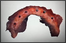
Figure1: Initial scan of the soft tissue architecture and emergence profile
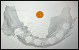
Figure 2: The implant platforms
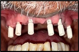
Figure 3: PEEK Scan Bodies attached
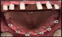
Figure 4: Occlusal view of PEEK Scan bodies
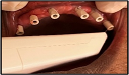
Figure 5: Scanning procedure
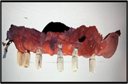
Figure 6: Digital impression of All on 6 implants
Discussion
Digital vs. Conventional Impression Techniques
Accuracy and Precision
Digital impressions have demonstrated comparable or superior accuracy to conventional methods in multiple studies. Research by Papaspyridakos et al. (2) showed that digital impressions for full-arch implant cases achieved accuracy within 15-30 μm, compared to 50-75 μm for conventional impressions. This increased precision is particularly crucial for implant-supported restorations, where passive fit is essential for long-term success.
In our case, the digital impression allowed for the fabrication of a framework with excellent passive fit, eliminating the need for sectioning and soldering often required with conventionally fabricated frameworks.
Time Efficiency
The digital workflow demonstrated significant time savings compared to conventional methods. The digital impression procedure was completed in approximately 20 minutes, whereas conventional procedures typically require 45-60 minutes including material setting time, multiple impressions, and verification jigs [3].
Additionally, the digital workflow eliminated the need for physical model fabrication, shipping delays, and potential remakes due to impression distortion or inaccuracies.
Patient Comfort
The patient reported greater comfort with the digital impression compared to his previous experience with conventional materials. This aligns with studies by Joda et al. (2019) [4] that showed significantly higher patient satisfaction scores for digital impressions compared to conventional methods, particularly for parameters including comfort, taste, and gag reflex [5].
The elimination of impression trays and materials is especially beneficial for patients with a pronounced gag reflex or those with limited mouth opening.
Workflow Efficiency
The digital workflow offered significant advantages in communication with the laboratory. The immediate visualization of the impression allowed for real-time assessment of capture quality, with the ability to rescan specific areas as needed rather than retaking the entire impression [6].
Furthermore, the digital files could be transmitted instantly to the laboratory, allowing for immediate design feedback and reducing the overall treatment time [7].
Limitations
Despite its advantages, digital impressions do present certain challenges. The scanning process can be technique-sensitive, particularly for capturing accurate data in the posterior regions or with deeply placed implants. Additionally, the initial investment cost for digital scanning equipment remains a barrier for some practitioners [8].
Conventional impressions still maintain advantages in certain situations, such as cases with subgingival margins where tissue retraction is required or in multi-unit cases where scanning angulation limitations may compromise accuracy [9].
Conclusion
This case report demonstrates the successful implementation of digital impression technology in the rehabilitation of an edentulous maxilla using the All-on-6 concept with the Neodent implant system. The digital workflow provided advantages in terms of accuracy, efficiency, and patient comfort compared to conventional impression techniques.
As digital technologies continue to evolve, their integration into implant prosthodontics will likely expand, potentially becoming the standard of care for complex implant cases. However, clinicians should maintain proficiency in both digital and conventional techniques to address the specific requirements of each clinical situation.
The successful outcome in this case supports the viability of digital impressions for full-arch implant rehabilitations, offering a predictable pathway to achieve functional and esthetic restorations while enhancing the patient experience throughout the treatment process.
Abbreviations
Cone-beam computed tomography (CBCT); Polyethertherketone (PEEK)
References
- Moriwaki H, Yamaguchi S, Nakano T, Yamanishi Y, Imazato S, Yatani H. (2016). Influence of implant length and diameter, bicortical anchorage, and sinus augmentation on bone stress distribution: Three-dimensional finite element analysis. Int J Oral Maxillofac Implants, 31(4):e84-e91.
Publisher | Google Scholor - Chochlidakis K, Papaspyridakos P, Tsigarida A, Romeo D, Chen YW, Natto Z, et al. (2020). Digital versus conventional full-arch implant impressions: A prospective study on 16 edentulous maxillae. J Prosthodont, 29(4):281-286.
Publisher | Google Scholor - Mangano F. (2022). Digital versus conventional workflows for implant rehabilitations: Accuracy and efficiency. Journal of Prosthodontics, 31(5):376-384.
Publisher | Google Scholor - Joda T, Bragger U, Zitzmann NU. (2019). CAD/CAM implant crowns in a digital workflow: Five-year follow-up of a prospective clinical trial. Clin Implant Dent Relat Res, 21(1):169–74.
Publisher | Google Scholor - Londono J, Tadros M, Salgueiro M, Baker PS. (2018). Digital design and 3D printing of an implant-supported prosthetic stent for protecting complete arch soft tissue grafts around dental implants: A dental technique. J Prosthet Dent, 120(6):801-804.
Publisher | Google Scholor - Alkindi S, Hamdoon Z, Aziz AM. Effect of different impression coping and scan body designs on the accuracy of conventional versus digital implant impressions: An in vitro study. J Dent, 146(105045):105045.
Publisher | Google Scholor - Lawand G, Ismail Y, Revilla-León M, Tohme H. (2024). Effect of implant scan body geometric modifications on the trueness and scanning time of complete arch intraoral implant digital scans: An in vitro study. J Prosthet Dent, 131(6):1189-1197.
Publisher | Google Scholor - Buda M, Bratos M, Sorensen JA. (2018). Accuracy of 3-dimensional computer-aided manufactured single-tooth implant definitive casts. J Prosthet Dent, 120(6):913-918.
Publisher | Google Scholor - Sanda M, Miyoshi K, Baba K. (2016). Trueness and precision of digital implant impressions by intraoral scanners: a literature review. Int J Implant Dent, 7(1):97.
Publisher | Google Scholor


