

Case Report
An Anatomical Variation in the Flexor Digitorum Brevis Muscle
- Alka Aggarwal *
Department of Anatomy, HIMS, SRHU, Jolly Grant, Dehradun, Uttarakhand, India.
*Corresponding Author: Alka Aggarwal, Department of Anatomy, HIMS, SRHU, Jolly Grant, Dehradun, Uttarakhand, India.
Citation: Aggarwal A. (2025). An Anatomical Variation in the Flexor Digitorum Brevis Muscle, Clinical Case Reports and Studies, BioRes Scientia Publishers. 10(5):1-4. DOI: 10.59657/2837-2565.brs.25.272
Copyright: © 2025 Alka Aggarwal, this is an open-access article distributed under the terms of the Creative Commons Attribution License, which permits unrestricted use, distribution, and reproduction in any medium, provided the original author and source are credited.
Received: August 01, 2025 | Accepted: August 15, 2025 | Published: August 22, 2025
Abstract
The 1st layer of the foot sole contains 3 muscles: Abductor hallucis, Flexor digitorum brevis (FDB), and Abductor digiti minimi. The FDB lies lateral to abductor hallucis. The central portion of plantar aponeurosis (PA) lies below FDB. FDB belly arises from medial process of calcanean tuberosity, the PA, and intermuscular septa (medial and Lateral).
Keywords: metatarsophalangeal; napalese cadavers; FDB
Introduction
After origin FDB muscle belly divided into 4 parts, each part continues as a slender tendon. At the proximal phalanx base, each tendon bifurcates into 2 slips and the corresponding FDL tendon passes through this bifurcation to enter the fibrous flexor sheath of the toe. These two slips curve over the sides of the FDL tendon; reunite and partially decussate to form a tunnel. The tendon of FDL passes through this tunnel up to the distal phalanx base and attaches there. FDB tendon divides again and attaches to sides of middle phalanx shaft. The medial plantar nerve innervates the FDB muscle. FDB act on the metatarsophalangeal (MP) joints and proximal interphalangeal (IP) joints and also helps to reinforce the foot arches [1, 2].
FDB muscle develops from the somatic mesoderm. Various studies reported variation in FDB especially the absence of its 4th tendon (for little toe) and the variation frequencies vary in different populations. Reported variations vary in cadaveric studies from reports of radiological or surgical studies. Ilayperuma studied morphological features of FDB muscle on 270 soles of 135 (81 males and 54 females) healthy adult embalmed Sri Lankan cadavers and found the bilateral absence of the 4th tendon of FDB in 134 limbs of 67 cadavers (Incidence - 71.85%)
[3]. Lobo SW and colleagues reported the absence of the 4th tendon (tendon for little toe) of FDB in all 60 soles of 30 (20 males and 10 females) adult embalmed Napalese cadavers [4].
Clinically, the mayo-cutaneous flap of FDB is used to reconstruct heel soft tissue due to its nearby position, good recovery of skin sensation, and weight-bearing function [5]. For the claw toe/ hammer toe deformity correction, FDB tendon transfer via the intramedullary approach is possible [6].
FDB paralysis leads to distortion of the arches of the foot [7]. FDB variation can lead to altered foot biomechanics, foot pain, stiffness, mobility-related dysfunction, and hammertoe or claw- toe-like toe deformities. Understanding foot muscle architecture and their variations may improve surgical outcomes as well as foot diagnostic imaging techniques, and assist in prosthesis designing and foot function analysis [3].
The present case report reports anatomical variation of FDB of muscle, observed during routine cadaveric dissection at the Anatomy dissection hall.
Methods
Present case report was observed during a routine cadaver dissection for undergraduate medical students at the Anatomy Department, Himalayan Institute of Medical Sciences (HIMS), Swami Rama Himalayan University (SRHU), Dehradun, Uttarakhand. Because this was a routine dissection so exempted from permission from Institutional Ethical Committee. Written informed consent was also waived. Two feet (1 Right and 1 Left feet) soles of embalmed (with 10% formalin) adult male cadaver were studied. The cadaver was about 60 years old. Cause of death, Clinical history, and family history was not available.
Dissection of the feet was performed as follows: A longitudinal incision was made on the sole extending from the heel to the middle toe. The planter skin and superficial fascia of the sole were carefully stripped from the plantar aponeurosis (deep fascia) with a knife. The plantar aponeurosis was cut across 2-3 cm in front of the heel. Reflect plantar aponeurosis away from the underlying FDB, abductor hallucis, and abductor digiti minimi. Cut the fibrous flexor sheath longitudinally and follow the tendons of FDB and FDL up to its insertion. 2nd layer of sole muscles of both feet of cadaver also examined after detaching and reflecting FDB.
Results
In the present case report three variations in FDB muscle were found in the right foot sole. These variations are-1st variation - After origin, from the medial process of the calcaneal tuberosity, PA, and intermuscular septum (medial and Lateral) FDB muscle belly divides into 3 parts. There was the absence of the 4th part which continues as the tendon for the 5th (little) toe. The medial part continues as thin fascia for the second toe that was attached to the nearby fascial tissue. The remaining two lateral parts continue as well-formed slender tendons for the 3rd and 4th toes. Each of these tendons bifurcates and enters into the fibrous flexor sheath of the 3rd & 4th toe with the FDL tendon (Figure 1).
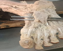
Figure 1: Flexor Digitorum Brevis (FDB) of right Foot showing three parts from its distal end. From 1st part only a fascia arises. From 2nd and 3rd part tendon arise for the 3rd and 4th toe.
2nd variation – For the 2nd toe there was the presence of a well-developed long tendon arising from a separate well-developed small muscle belly attached to the medial tubercle of calcanean tuberosity just beneath the muscle belly of FDB. This tendon was about two times longer than the tendons of the FDB muscle. This tendon bifurcates into two slips at 2nd toe proximal phalanx base. The tendon of FDL passes through these two slips and then enters into the fibrous flexor sheath of the 2nd toe. These slips further unite and split and attach to both sides of the middle phalanx of the second toe. (Figure 2).
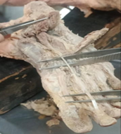
Figure 2: Right foot sole after reflecting the FDB showing the muscle and its long tendon which replace FDB tendon for the 2nd toe.
3rd variation-There was the presence of a muscle arising from the lateral intermuscular septum and fascial tissue; compensating the 4th part and 4th tendon of FDB muscle. This tendon bifurcates into 2 slips through which the corresponding tendon of FDL passes and then enters into the fibrous flexor sheath of the little toe and inserts on both sides of the middle phalanx of the little toe (Figure 3).
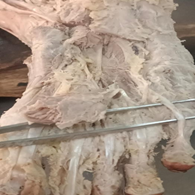
Figure 3: Right foot sole showing the muscle belly with tendon which replace the 4th part and tendon of FDB muscle for little (5th) toe.
The FDB muscle of the left foot sole of the same cadaver follow the normal description as given in anatomy textbooks.
Discussion
In the present case report, the variation was found only in the right foot. The left foot had normal FDB. The found variation was - there was a very thin fascia arising from the first part of the FDB muscle for the second toe which did not reach up to the proximal phalanx base of 2nd toe but ended after attaching to the nearby fascial tissue. The FDB tendon for the 2nd toe was compensated by the well-developed small muscle belly with a long tendon. This muscle arises from the medial tubercle of the calcaneum and lies in the central part of the foot sole in between the 1st and 2nd muscle layer of the sole. There was an absence of 4th part & 4th tendon of FDB meant for the little (5th) toe. This tendon was also compensated by a separate muscle & its tendon. This muscle appeared to take origin from the lateral intermuscular septum, and fascial tissue.
Previous studies also reported anatomical variations in the form of absent FDB tendon that is replaced or compensated by separate muscle. Bergmann reported that a small tendon replaces the missing fifth toe tendon of FDB (8). A study reported an atrophied 4th tendon of FDB in the left foot and an absent 4th tendon of FDB in the right foot of a 75-year-old male cadaver [9].
Bulent Yalcin found that out of 33 feet, the FDB muscle of 18 feet differed from the classical description. In 12 feet there was small muscle belly for the 5th toe and in 6 feet it was missing. In 1 cadaver (female), left foot FDB was formed by superficial & deep head and has 3 muscle bellies and 4 tendons. In right foot, a separate very thin muscle belly for 5th toe originated from intermuscular septum as a flat fascia under FDB [10].
Tendon variation can occur genetically, developmental anomaly, or an evolutionary impact. FDB tendon variations are diagnosed by the help of CT and MRI. FDB variations are clinically important because FDB is used in the heel pad, tendoachillis, medial malleolus, and lateral malleolus soft tissue reconstruction. FDB tendon are used in the correction of congenital deformities [11].
Due to bipedal walking, in humans, the FDB muscle along with intrinsic muscles of sole help in support of longitudinal arch and postural control, contribute to stiffening the MP joints during walking or running. FDB muscle helps FDL muscle in plantarflexion of 2nd to 5th toe. Studies showed that toe plantarflexion and foot stability during gait were not affected by removing FDB tendon for corrective clawtoe/hammer toe deformities. Most authors said that variations of the 4th tendon of FDB may be due to degenerative change or an evolutionary adaptation as being more peripheral and no need for opposition action the muscles of the little toe of the foot regress because of less function, less use the muscles [12]. However, the present case report showed that a separate muscle with its tendon compensates for the absent fourth part and fourth tendon of the FDB muscle. This indicates that the present variation may be a developmental deficiency of the fourth part of FDB. However, this deficiency is compensated by the development of separate muscle and tendon.
For an anatomist, variations of the sole muscles is important for enhanced education of foot anatomy. Variation documentation promotes a deeper understanding of foot anatomy, updates the online resources, anatomy textbooks, and atlases; helps radiologists in their reporting; may assist in foot function analysis, accurate diagnosis, and effective surgical management of tendon transfer, prosthesis designing, and biomechanical foot modelling. Physical medicine and rehabilitation specialists, and podiatrists can develop effective treatment plans for patients with foot disorders or injuries which reduce complications and improve patient outcomes. It also facilitates collaboration among educators, researchers, clinicians, and educators [12].
Conclusion
The present case report gave knowledge about the variations found in the structure of the FDB muscle. In this case report, FDB in the right foot sole of one cadaver although there was a less developed tendon for the second toe and an absent tendon for the little toe but these were compensated by separate muscles and their tendons which follow the normal description as given in anatomical textbooks for FDB.
Declarations
Funding: None
Conflicts of Interest: No conflictof interest.
References
- Romanes, G. J. (2020). Leg and foot. In R. Koshi (Ed.):Cunningham’s manual of practical anatomy, Vol. 1: Upper and lower limbs Oxford University Press, (17th ed.):231–278.
Publisher | Google Scholor - Standring, S. (Ed.). (2020). Plantar muscles of the foot – First layer. In Gray’s anatomy: The anatomical basis of medicine and surgery Churchill Livingstone, Elsevier, (42nd ed.,):1555.
Publisher | Google Scholor - Ilayperuma, I. (2012). On the variations of the muscles flexor digitorum brevis: Anatomical insight. International Journal of Morphology, 30(1):337–340.
Publisher | Google Scholor - Lobo, S. W., Menezes, R. G., Mamata, S., Baral, P., Hunnargi, S. A., Kanchan, T., et al. (2008). Phylogenetic variation in flexor digitorum brevis: A Nepalese cadaveric study. Nepal Medical College Journal, 10(4):230–232.
Publisher | Google Scholor - Yang, L., Zhang, Z., & Fang, S. (1999). [Repair of soft tissue defect of heel with myocutaneous flap of flexor digitorum brevis]. Zhongguo Xiu Fu Chong Jian Wai Ke Za Zhi, 13(1):16–17.
Publisher | Google Scholor - Cosin-Matamoros, J., Becerro-de-Bengoa-Vallejo, R., Losa-Iglesias, M. E., Casado-Hernandez, I., Benito-de-Pedro, M., Calvo-Lobo, C., et al. (2021). Intramedular transfer of the flexor digitorum brevis tendon for the correction of claw toe deformity: A cross-sectional study. Annals of Anatomy – Anatomischer Anzeiger, 234:151646.
Publisher | Google Scholor - Rosse, C., & Gaddum-Rosse, P. (1997). Hollinshead’s textbook of anatomy (6th ed.). Lippincott-Raven.
Publisher | Google Scholor - Bergman, R. A., Thompson, S. A., Afifi, A. K., & Saadeh, F. A. (1988). Compendium of human anatomic variations, Urban & Schwarzenberg, 27–28.
Publisher | Google Scholor - Claassen, H., & Wree, A. (2003). Isolated flexor muscles of the little toe in the feet of an individual with atrophied or lacking 4th head of the M. extensor digitorum brevis and lacking the 4th tendon of the M. extensor digitorum longus. Annals of Anatomy, 185(1):81–84.
Publisher | Google Scholor - Yalcin, B., & Ozan, H. (2005). Some variations of the musculus flexor digitorum brevis. Anatomical Science International, 80(4):189–192.
Publisher | Google Scholor - Chuang, G., & Craig, D. P. M. (2020). Intrinsic muscle flaps for coverage of small defects in the foot. Clinics in Podiatric Medicine and Surgery, 37(4):789–802.
Publisher | Google Scholor - Jallenne, I. Q., Cristian, M., Felix, R., Catalina, I. V., Juan, F., Maryvi, G., et al. (2022). A missing flexor digitorum brevis tendon and its relationship to sex and ancestry: Evaluation in Hispanic population. Anatomia, 1(2):210–216.
Publisher | Google Scholor


