
Research Article
Acute Toxicity, Phytochemical Profile, and Antioxidant Activity of Mangifera Indica Leaf Extract in Mice
- Getahun Tsegaye Dibaba 2*
- Abebaye Aragaw Leminie 2
- Wossene Habtu Tadesse 3
- Worku Gemechu Lemmi 4
- Samuel Woldekidan Hirpesa 4
- Rekik Ashebir 4
- Abiy Abebe Gelagle 4
- Sofia Yimam Hussen 5
- Moti Sori Reje 2
- Tsegay Beyene Weldemariam 1
- Beza Tasew Degefu 5
- Mekoya Mengistu 2
- Memberework Chanyalew 5
- Tesfaye Tolessa Dugul 2
1School of Medicine, College of Health Sciences, Aksum University, Aksum, Ethiopia.
2School of Medicine, College of Health Sciences, Addis Ababa University, Ethiopia.
3National Clinical Chemistry Reference Laboratories, Public Health Institute, Addis Ababa, Ethiopia.
4Traditional and Modern Medicine Directorates, Armauer Hansen Research Institute, Addis Ababa, Ethiopia.
5Non communicable Disease Division, Armauer Hansen Research Institute, Addis Ababa, Ethiopia.
*Corresponding Author: Getahun Tsegaye Dibaba, School of Medicine, College of Health Sciences, Addis Ababa University, Ethiopia.
Citation: Dibaba GT, Leminie AA, Tadesse WH, Lemmi WG, Hirpesa SW, et al. (2026). Acute Toxicity, Phytochemical Profile, and Antioxidant Activity of Mangifera Indica Leaf Extract in Mice, International Journal of Biomedical and Clinical Research, BioRes Scientia Publishers. 7(2):1-10. DOI: 10.59657/2997-6103.brs.26.144
Copyright: © 2026 Getahun Tsegaye Dibaba, this is an open-access article distributed under the terms of the Creative Commons Attribution License, which permits unrestricted use, distribution, and reproduction in any medium, provided the original author and source are credited.
Received: June 02, 2026 | Accepted: July 16, 2026 | Published: July 27, 2026
Abstract
Background: Mangifera indica (mango) leaf aqueous extract is widely used in traditional medicine for diabetes, asthma, and inflammatory conditions. However, comprehensive safety data are limited. This study evaluated the acute oral toxicity, phytochemical composition, and in vitro antioxidant activity of the aqueous leaf extract in Swiss albino mice.
Materials and Methods: Acute oral toxicity was assessed following OECD Guideline 425 (limit test at 2000 mg/kg). Ten mice (five per group) received a single oral dose of 2000 mg/kg body weight of lyophilized aqueous extract or distilled water (control). Clinical signs, body weight, and mortality were monitored for 14 days. Haematological (Hb, RBC, WBC, differential counts, platelets) and serum biochemical parameters (electrolytes, urea, creatinine, liver enzymes, proteins) were measured. Vital organs (liver, kidney, heart) were examined histopathologically. Phytochemical screening was performed using standard qualitative tests. Antioxidant activity was evaluated by DPPH radical scavenging assay.
Results: No mortality or clinical signs of toxicity were observed at 2000 mg/kg. Body weight gain was normal and comparable to controls. Haematological parameters showed no significant differences between groups except for a physiological neutrophilia (80.1% in treated vs. 26.4% in control, p < 0.001) and relative lymphopenia (14.3% vs. 67.5%), which are consistent with a stress response to gavage rather than toxicity. All other parameters (RBC, platelets, Hb, etc.) remained unchanged. Serum biochemical markers (electrolytes, urea, creatinine, ALT, AST, ALP, total protein, albumin) showed no significant alterations (p > 0.05). Histopathological examination revealed normal architecture of the liver, kidney, and heart, with only minor, nonspecific inflammatory foci in two treated mice. Phytochemical analysis revealed the presence of alkaloids, flavonoids, tannins, saponins, terpenoids, and phenols. The extract exhibited concentration dependent DPPH radical scavenging activity with an IC₅₀ of 145.7 µg/mL (95% CI: 130.5-160.9 µg/mL).
Conclusion: The aqueous leaf extract of M. indica is well tolerated in mice at a single oral dose of 2000 mg/kg, with no adverse effects on clinical signs, body weight, major organ histology, or standard haematological and biochemical parameters. The neutrophilia observed is a physiological stress response, not a toxic effect. The no observed adverse effect level (NOAEL) is 2000 mg/kg. The extract contains multiple bioactive phytochemicals and demonstrates moderate antioxidant activity, supporting its traditional use and safety for further pharmacological studies.
Keywords: mangifera indica; acute toxicity; phytochemical screening; antioxidant activity; DPPH; haematology; histopathology; mice
Introduction
The mango tree (Mangifera indica L., family Anacardiaceae) is one of the most widely cultivated fruit trees in tropical and subtropical regions. Beyond its nutritional value, various parts of the plant, especially the leaves, have been used in traditional medicine for centuries. In Ethiopian traditional medicine, as well as in other cultures (e.g., Ayurveda, traditional Chinese medicine), mango leaf decoctions are used to manage diabetes mellitus, asthma, dysentery, and inflammatory conditions [1,2].
Pharmacological studies have identified a wide range of bioactive compounds in mango leaves, including polyphenols (gallic acid, caffeic acid), flavonoids (quercetin, kaempferol), benzophenone C glucosides (mangiferin), terpenoids, and volatile compounds [3-5]. These constituents are responsible for multiple biological activities: antioxidant, anti-inflammatory, antidiabetic, antimicrobial, anthelmintic, and antiallergic [6,7]. Notably, mangiferin has been shown to inhibit triglyceride accumulation in adipocytes and improve glucose and lipid homeostasis via PI3K/AKT and AMPK signaling pathways in KK Ay mice [8].
Despite the long history of ethno medicinal use and promising pharmacological data, comprehensive toxicological evaluations of M. indica leaf extracts are relatively sparse. Regulatory agencies such as the World Health Organization (WHO) and the U.S. Food and Drug Administration (FDA) require rigorous safety data before herbal products can be recommended for widespread use or incorporated into standardized formulations [9,10]. Acute toxicity testing is the first step in this safety assessment, providing information on the lethal dose, target organ effects, and the no observed adverse effect level (NOAEL).
Therefore, the present study aimed to: (i) evaluate the acute oral toxicity of an aqueous extract of M. indica leaves in Swiss albino mice following OECD Guideline 425, (ii) perform qualitative phytochemical screening to identify major secondary metabolites, and (iii) determine the in vitro antioxidant activity using the DPPH radical scavenging assay. The findings will contribute to the safety profile of this widely used medicinal plant and support its rational use in traditional medicine and future clinical studies.
Materials and Methods
Plant Material Collection and Authentication
Fresh, healthy leaves of Mangifera indica were collected in June 2024 from Seka, Jimma Zone, Oromia Region, Ethiopia. The collection was carried out following the WHO guidelines on good agricultural and collection practices (GACP) for medicinal plants [11]. A botanist at the National Herbarium, College of Natural and Computational Sciences, Addis Ababa University authenticated the plant. A voucher specimen (GT001) was deposited in the herbarium for future reference.
Preparation of Aqueous Extract
The leaves were thoroughly washed with tap water followed by distilled water to remove soil and debris. They were shade dried at room temperature (25 ± 2°C) with adequate ventilation for 10-14 days until constant weight was achieved. The dried leaves were ground into a fine powder using an electric grinder and passed through a 40-mesh sieve.
Two hundred grams of powdered leaves were macerated in 3.2 L of distilled water (1:16 w/v) and boiled for 30 minutes with occasional stirring, then allowed to cool to room temperature. The mixture was placed on a mechanical shaker for 2 hours to ensure thorough extraction. After settling for 30 minutes, the supernatant was decanted and allowed to stand for an additional 2 hours to separate fine sediments. The clear supernatant was filtered through sterile muslin cloth, followed by Whatman No. 1 filter paper to obtain a particle free filtrate. The filtrate was frozen at -20°C and lyophilized (freeze dried) for 2-3 weeks using a laboratory freeze dryer (Christ Alpha 1 4 LDplus). The lyophilized powder (yield: approximately 8.5% w/w) was stored in airtight containers at -20°C until use.
Experimental Animals
Healthy Swiss albino mice (10 weeks old, weight 30-40 g) of both sexes were obtained from the animal breeding facility of the College of Health Sciences, Addis Ababa University. Animals were housed in polypropylene cages (five per cage) under standard laboratory conditions: temperature 22 ± 2°C, relative humidity 55 ± 10%, 12 h light/dark cycle. They had free access to standard rodent pellets and tap water ad libitum. Mice were acclimatized for one week before the start of the experiment. All procedures were approved by the Institutional Review Board of the College of Health Sciences, Addis Ababa University (Protocol No. 044/23/physio) and followed the guidelines of the Canadian Council on Animal Care (CCAC) and the Ethiopian National Research Ethics Committee.
Acute Oral Toxicity Study (OECD 425)
The acute oral toxicity was evaluated using the up and down procedure (UDP) as described in OECD Test Guideline 425 [12]. Because the extract was expected to have low toxicity, a limit test at 2000 mg/kg was performed.
Dose preparation and administration: The lyophilized extract was suspended in distilled water to achieve a concentration of 200 mg/mL. The dose volume was 10 mL/kg body weight, delivering 2000 mg/kg. Mice were fasted for 3-4 hours (water ad libitum) prior to dosing. A single dose was administered orally by gavage using a stainless-steel feeding needle.
Grouping: Ten mice were randomly assigned to two groups (n=5 per group, mixed sexes):
Control Group: Received distilled water (10 mL/kg).
Treated Group: Received 2000 mg/kg of M. indica leaf extract.
Observation: After administration, mice were observed individually for signs of toxicity at 30 min, 1, 2, 3, 4, 6, 8, and 24 hours, and then daily for 14 days. Observations included changes in skin and fur, eyes, mucous membranes, respiratory pattern, circulation, autonomic effects (salivation, lacrimation, diarrhoea), and central nervous system effects (tremors, convulsions, sedation, gait abnormalities). Body weight was recorded on days 0, 1, 3, 7, and 14. Food and water consumption were measured daily. Any mortality was recorded.
Euthanasia and Sample Collection
At the end of the 14-day observation period, all mice were fasted overnight but allowed free access to water. On day 15, animals were anaesthetized by inhalation of isoflurane (3% induction, 1.5% maintenance in 100% oxygen). Blood samples were collected via cardiac puncture using a 1 mL syringe with a 23-gauge needle [13]. Approximately 1 mL of blood was transferred into EDTA coated tubes for haematological analysis, and another 1 mL was placed into plain tubes without anticoagulant for serum biochemistry. After blood collection, all mice were euthanized by cervical dislocation under deep anesthesia, following the American Veterinary Medical Association (AVMA) guidelines for euthanasia [14]. Death was confirmed by cessation of heartbeat and respiration. The liver, kidneys, and heart were immediately dissected out, rinsed with ice cold normal saline, blotted dry, and weighed for relative organ weight calculation. Tissues were then fixed in 10% neutral buffered formalin for histopathological examination [15].
Haematological Analysis
Blood samples collected in EDTA tubes were analyzed using a fully automated hematology analyzer (KX 21, Sysmex, Japan). The following parameters were measured: hemoglobin (Hb), red blood cell count (RBC), packed cell volume (PCV), mean corpuscular volume (MCV), mean corpuscular hemoglobin (MCH), mean corpuscular hemoglobin concentration (MCHC), total white blood cell count (WBC), differential WBC counts (neutrophils, lymphocytes, monocytes, eosinophil, basophils), and platelet count. All analyses were performed within 4 hours of collection.
Serum Biochemical Analysis
Blood samples in plain tubes were allowed to clot at room temperature for 30 minutes and centrifuged at 3000 rpm for 10 minutes to obtain serum. Serum was stored at -80°C until analysis. Biochemical parameters were measured using a semi-automatic biochemical analyser (BioSystems BTS 350, Spain) with commercially available kits (Randox Laboratories, UK). The parameters included:
Electrolytes: sodium (Na⁺), potassium (K⁺), chloride (Cl⁻) by ion selective electrodes.
Renal Function: blood urea nitrogen (BUN), creatinine, uric acid.
Liver Function: total protein, albumin, globulin (calculated by difference), alkaline phosphatase (ALP), aspartate aminotransferase (AST), alanine aminotransferase (ALT).
All assays were performed in duplicate according to the manufacturer’s instructions.
Histopathological Examination
The liver, kidneys, and heart were fixed in 10% neutral buffered formalin for 48 hours, dehydrated through graded alcohols, cleared in xylene, and embedded in paraffin wax. Sections of 5 μm thickness were cut using a rotary microtome, stained with haematoxylin and eosin (H&E), and examined under a light microscope (Olympus BX43) by a pathologist who was blinded to the treatment groups. Any histopathological changes were recorded and photographed.
Phytochemical Screening
The aqueous extract was subjected to qualitative phytochemical tests using standard procedures [16]:
Alkaloids: 0.1 mL of extract was treated with 2-3 drops of Dragendorff’s reagent (potassium bismuth iodide). Formation of an orange red precipitate indicated alkaloids.
Flavonoids: To 1 mL of extract, a piece of magnesium ribbon and 3 drops of concentrated HCl were added. A red to crimson colour indicated flavonoids.
Saponins: 0.5 g of extract was shaken vigorously with 5 mL of distilled water for 2 minutes. Persistent foam for 15 minutes indicated saponins.
Tannins: 2 mL of extract was diluted with distilled water, and 2-3 drops of 5 percentage ferric chloride solution were added. A green black or blue colour indicated tannins.
Terpenoids: 0.5 g of extract was dissolved in 2 mL of chloroform, then 2 mL of concentrated H₂SO₄ was added carefully. A reddish brown colour at the interface indicated terpenoids.
Phenols: 1 mL of extract was treated with a few drops of 5 percentage ferric chloride. A bluish black colour indicated phenols.
DPPH Radical Scavenging Assay
The antioxidant activity was determined using the stable free radical DPPH (2,2 diphenyl 1 picrylhydrazyl) as described by Blois [17] with minor modifications. Stock solutions of the extract and ascorbic acid (positive control) were prepared in methanol at 1 mg/mL. Working solutions were diluted to final concentrations of 50, 100, 150, 200, 250, 300, and 350 µg/mL in methanol.
Procedure: In a 96 well micro plate, 100 µL of each concentration was mixed with 100 µL of 0.1 mM DPPH in methanol. The mixture was incubated in the dark at room temperature for 30 minutes. Absorbance was measured at 517 nm using a microplate reader (Multiskan GO, Thermo Fisher Scientific). Methanol with DPPH served as the control (100% radical activity). All tests were performed in triplicate. The percentage of radical scavenging activity (% RSA) was calculated as:
% RSA = [(A_control - A_sample) / A_control] × 100
Where A- control is the absorbance of DPPH solution without extract, and A_sample is the absorbance of the extract DPPH mixture.
The concentration required to scavenge 50% of DPPH radicals (IC₅₀) was calculated by nonlinear regression (log dose vs. response) using GraphPad Prism 9.0 (GraphPad Software, San Diego, CA, USA).
Statistical Analysis
All data are expressed as mean ± standard error of the mean (SEM). Comparisons between control and treated groups were performed using unpaired Student’s t test for normally distributed data. Normality was assessed by Shapiro Wilk test. A p value less than 0.05 was considered statistically significant. Analyses were conducted using SPSS version 26 (IBM Corp., Armonk, NY, USA).
Results
Acute Oral Toxicity - Clinical Observations
No mortality was recorded in either group during the 14-day observation period. In the treated group, no signs of acute toxicity were observed: behavior was normal, no tremors, convulsions, salivation, diarrhea, or respiratory distress occurred. The mice showed normal exploratory activity, grooming, and feeding behavior. The extract was well tolerated, and the limit dose of 2000 mg/kg produced no observable adverse effects. According to OECD 425, the LD₅₀ is >2000 mg/kg and the NOAEL is 2000 mg/kg.
Body Weight Changes: There was no significant difference in body weight between control and treated groups at any time point (Table 1). Both groups showed a gradual, normal weight gain over 14 days, indicating no impairment of general health.
Table 1: Body weight (g) of mice during the acute toxicity study (mean ± SEM, n=5).
| Day | Control group | Treated group (2000 mg/kg) | p‑value* |
| 0 | 34.2 ± 1.2 | 34.5 ± 1.3 | 0.87 |
| 1 | 34.8 ± 1.1 | 35.0 ± 1.2 | 0.90 |
| 3 | 35.6 ± 1.0 | 35.9 ± 1.1 | 0.84 |
| 7 | 36.9 ± 1.0 | 37.2 ± 1.0 | 0.83 |
| 14 | 38.5 ± 1.1 | 38.9 ± 1.2 | 0.81 |
*Unpaired t test, not significant (p > 0.05) for all days.
Relative Organ Weights: The relative organ weights (liver, kidneys, heart) are presented in Table 2. No significant differences were observed between control and treated groups (p > 0.05), indicating that the extract did not cause organ enlargement or atrophy.
Table 2: Relative organ weights (g/100 g body weight) after acute treatment (mean ± SEM, n=5).
| Organ | Control Group | Treated Group (2000 mg/kg) | p‑value |
| Liver | 2.20 ± 0.14 | 2.00 ± 0.12 | 0.29 |
| Kidney | 0.50 ± 0.02 | 0.48 ± 0.02 | 0.48 |
| Heart | 0.44 ± 0.02 | 0.41 ± 0.02 | 0.33 |
Hematological Parameters
The hematological profile is shown in Table 3. Most parameters (Hb, RBC, PCV, MCV, MCH, MCHC, WBC, platelets) showed no significant differences between control and treated groups (p > 0.05). The differential white blood cell count revealed a higher percentage of neutrophils in the treated group (80.1%) compared to controls (26.4%), with a corresponding lower lymphocyte percentage (14.3% vs. 67.5%). These differences were statistically significant (p less than 0.001) but are within the range of physiological stress responses commonly observed in mice following oral gavage and handling [18,19]. No clinical signs of infection or inflammation were present, and all other parameters remained normal, indicating this is a physiological adjustment rather than a toxic effect.
Table 3: Hematological parameters after acute oral administration (mean ± SEM, n=5).
| Parameter | Unit | Control Group | Treated Group (2000 mg/kg) | p‑value |
| Hb | g/dL | 14.12 ± 3.11 | 14.67 ± 2.02 | 0.88 |
| RBC | ×10¹²/L | 6.52 ± 0.23 | 6.31 ± 0.31 | 0.60 |
| PCV | L/L | 0.43 ± 0.01 | 0.46 ± 0.00 | 0.09 |
| MCV | fL | 54.30 ± 0.00 | 54.21 ± 0.77 | 0.92 |
| MCH | pg | 18.55 ± 0.66 | 18.01 ± 0.42 | 0.51 |
| MCHC | g/L | 328.0 ± 4.7 | 327.0 ± 4.1 | 0.88 |
| WBC | ×10⁹/L | 7.52 ± 0.22 | 9.00 ± 1.30 | 0.30 |
| Neutrophils (%) | % | 26.4 ± 1.9 | 80.1 ± 2.5 | less than 0.001* |
| Lymphocytes (%) | % | 67.5 ± 2.0 | 14.3 ± 1.8 | less than 0.001* |
| Monocytes (%) | % | 4.3 ± 0.5 | 3.9 ± 0.6 | 0.60 |
| Eosinophils (%) | % | 1.2 ± 0.2 | 1.2 ± 0.3 | 0.95 |
| Basophils (%) | % | 0.6 ± 0.1 | 0.5 ± 0.1 | 0.48 |
| Platelet count | ×10⁹/L | 870 ± 10.7 | 865 ± 17.1 | 0.81 |
*Significant difference (p less than 0.001), but attributed to physiological stress from gavage, not toxicity.
Serum Biochemical Parameters
Table 4 summarises the serum biochemistry results. No significant differences were found between control and treated groups for any parameter (p > 0.05). Electrolytes (Na⁺, K⁺, Cl⁻) remained within normal ranges. Renal function markers (urea, creatinine, uric acid) showed no evidence of nephrotoxicity; the numerically higher urea in the treated group (26.0 vs. 15.0 mg/dL) was not statistically significant (p=0.07) and remained within the normal reference range for mice (10-30 mg/dL). Liver function tests (total protein, albumin, globulin, ALP, AST, ALT) were not significantly altered, indicating no hepatocellular damage or cholestasis.
Table 4: Serum biochemical parameters after acute oral administration (mean ± SEM, n=5).
| Parameter | Unit | Control Group | Treated Group (2000 mg/kg) | p‑value |
| Sodium | mmol/L | 138.0 ± 0.20 | 138.2 ± 0.08 | 0.38 |
| Potassium | mmol/L | 6.12 ± 0.32 | 6.33 ± 0.35 | 0.66 |
| Chloride | mmol/L | 104.0 ± 0.27 | 104.3 ± 0.01 | 0.24 |
| Urea | mg/dL | 15.0 ± 0.34 | 26.0 ± 0.20 | 0.07 |
| Creatinine | mg/dL | 1.07 ± 0.33 | 1.12 ± 0.48 | 0.93 |
| Uric acid | mmol/L | 0.17 ± 0.03 | 0.19 ± 0.01 | 0.55 |
| Total protein | g/L | 69.37 ± 0.21 | 69.53 ± 0.43 | 0.75 |
| Albumin | g/L | 36.73 ± 0.67 | 37.13 ± 0.71 | 0.69 |
| Globulin | g/L | 32.64 ± 0.47 | 32.40 ± 0.51 | 0.73 |
| ALP | U/L | 135.0 ± 8.3 | 135.0 ± 9.7 | 0.99 |
| AST | U/L | 74.31 ± 4.31 | 74.03 ± 3.39 | 0.96 |
| ALT | U/L | 45.41 ± 0.71 | 45.81 ± 0.52 | 0.65 |
Histopathological Findings
Liver: Microscopic examination of liver sections from the control group showed normal hepatic architecture: well defined hepatocytes arranged in cords around central veins, with normal sinusoids and portal triads. In the treated group, the majority of liver sections appeared normal. Occasional, scattered, minimal lymphocytic infiltration was observed in two of five treated mice (Figure 1), but no necrosis, steatosis, or fibrosis was present. Such minor inflammatory foci are commonly seen in untreated laboratory animals and are not considered treatment related or toxicologically significant [20].
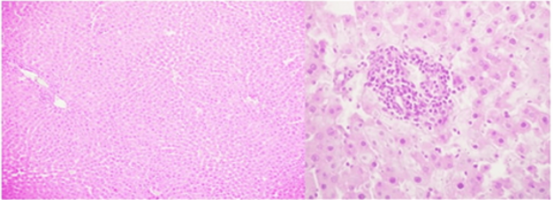
Figure 1: H&E stain, 100x: [Liver], Inset (200x): mononuclear inflammatory infiltrate G1, 2 and 3.
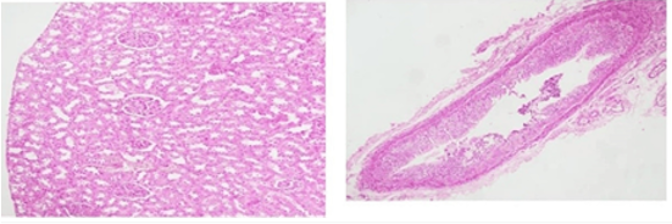
Kidney: Both control and treated groups exhibited normal renal histology. Glomeruli showed normal cellularity and capillary loops. Tubules (proximal and distal) were intact without vacuolation, necrosis, or casts. No interstitial inflammation or fibrosis was observed (Figure 2).
Heart: Myocardial fibers were well organized with normal striations. No evidence of myocyte necrosis, inflammatory infiltrate, or fibrosis was seen in either group (Figure 3).
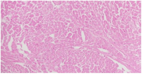
Figure 3: H&E stain, a) 200x: [Heart].
Overall, histopathological examination confirmed that a single oral dose of 2000 mg/kg M. indica leaf extract did not induce any deleterious structural changes in the liver, kidney, or heart.
Phytochemical Screening
The qualitative analysis revealed the presence of several bioactive phytochemicals, as summarized in Table 5.
Table 5: Phytochemical constituents of M. indica leaf aqueous extract.
| Phytochemical Class | Result |
| Alkaloids | + |
| Flavonoids | + |
| Saponins | + |
| Tannins | + |
| Terpenoids | + |
| Phenols | + |
(+) = present
DPPH Radical Scavenging Activity
The aqueous extract of Mangifera indica leaves scavenged DPPH radicals in a concentration-dependent manner (Figure 4). The percentage of radical scavenging activity (% RSA) increased from 40.2% at 50 µg/mL to 93.9% at 350 µg/mL, with intermediate values as follows: 46.1% (100 µg/mL), 58.8% (150 µg/mL), 68.6% (200 µg/mL), 78.4% (250 µg/mL), and 86.1% (300 µg/mL). Nonlinear regression analysis yielded an IC₅₀ value of 145.7 µg/mL (95% CI: 130.5-160.9 µg/mL). Under identical conditions, the positive control (ascorbic acid) exhibited an IC₅₀ of 12.5 µg/mL.
Figure 4 Concentration-dependent DPPH radical scavenging activity of Mangifera indica leaf aqueous extract. Various concentrations (50-350 µg/mL) of the extract were incubated with 0.1 mM DPPH for 30 min at room temperature. The % RSA was calculated from absorbance at 517 nm. Data are presented as mean ± SEM (n = 3). The extract showed a clear concentration-response relationship, with % RSA increasing from 40.2% (50 µg/mL) to 93.9% (350 µg/mL). The calculated IC₅₀ (concentration required to scavenge 50% of DPPH radicals) was 145.7 µg/mL (95% CI: 130.5-160.9 µg/mL). Under the same conditions, the positive control (ascorbic acid) gave an IC₅₀ of 12.5 µg/mL.
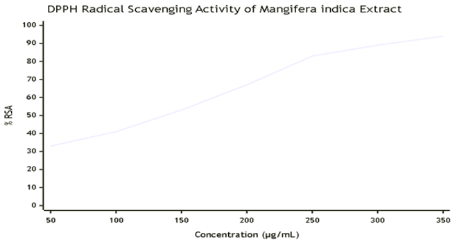
Figure 4: DPPH radical scavenging activity of M. indica leaf aqueous extract.
Concentration-response curve (50-350 µg/mL). Values are mean ± SEM (n=3). IC₅₀ = 145.7 µg/mL (95% CI: 130.5-160.9). Ascorbic acid IC₅₀ = 12.5 µg/mL.
Discussion
The present study provides a comprehensive acute toxicity profile, phytochemical characterization, and antioxidant activity of the aqueous leaf extract of Mangifera indica in a murine model. The increasing global reliance on herbal medicines necessitates rigorous safety evaluations to protect public health [9,10,21]. Our findings demonstrate that a single oral dose of 2000 mg/kg of the extract is safe in mice, with no mortality, clinical signs of toxicity, or significant adverse effects on major organ histology or standard biochemical markers. These results are consistent with earlier acute toxicity studies on M. indica leaf extracts, which reported an LD₅₀ > 5000 mg/kg in rodents [22,23].
Clinical Observations and Body Weight: The absence of behavioral or autonomic changes during the 14-day observation period suggests that the extract does not cause neurotoxicity or acute organ failure. Normal body weight gain is a sensitive indicator of overall health and metabolic function; the comparable weight gain between control and treated groups indicates that the extract did not impair food intake, digestion, or nutrient absorption [24].
Hematology: Hematological parameters are valuable biomarkers of systemic toxicity, as the bone marrow and circulating blood cells are highly sensitive to xenobiotic [25]. In our study, RBC, Hb, PCV, and red cell indices remained unchanged, indicating no effect on erythropoiesis or red cell survival. The WBC count was not significantly elevated, but the differential count showed a marked increase in neutrophils (80.1% vs. 26.4%) and a decrease in lymphocytes (14.3% vs. 67.5%). This pattern (neutrophilia with lymphopenia) is a classic stress response in rodents, often induced by handling, gavage, or the stress of a novel environment [18,19]. It is not indicative of infection or inflammation because there were no clinical signs of illness, no fever, and no histopathological evidence of tissue damage. Importantly, the platelet count was normal, ruling out bleeding or thrombotic disorders. Therefore, the hematological changes are considered physiological and not toxicologically significant.
Biochemistry: Serum biochemistry reflects the functional integrity of the liver, kidneys, and other organs. The liver enzymes ALT, AST, and ALP are sensitive markers of hepatocellular injury and cholestasis [26]. In our study, these enzymes remained stable, indicating that the extract did not cause hepatocyte membrane damage or bile duct obstruction. Total protein and albumin were unchanged, suggesting normal synthetic function of the liver. Renal function markers (urea, Creatinine, uric acid) showed no significant elevation. Although urea in the treated group was numerically higher (26.0 vs. 15.0 mg/dL), this difference was not statistically significant (p=0.07) and remained within the normal reference range for mice (10-30 mg/dL). Moreover, Creatinine, a more specific marker of glomerular filtration, was unchanged. Thus, no nephrotoxicity was evident. Electrolyte balance was preserved, indicating normal tubular function.
Histopathology: Gross and microscopic examination of the liver, kidneys, and heart revealed no treatment related abnormalities. The minor, scattered lymphocytic foci in the liver of two treated mice are commonly observed in laboratory rodents and are not considered evidence of toxicity, as they also occur in control animals at low frequency [20]. The absence of necrosis, fibrosis, or structural disorganisation supports the biochemical findings of organ safety.
Phytochemical Profile: The presence of alkaloids, flavonoids, tannins, saponins, terpenoids, and phenols aligns with previous reports on M. indica leaves [27,28]. These secondary metabolites are known to exert a wide range of pharmacological effects, including antioxidant, anti-inflammatory, hypoglycemic, and antimicrobial activities. Flavonoids and phenolic, in particular, are potent radical scavengers due to their ability to donate hydrogen atoms or electrons [29]. Tannins and saponins may contribute to antidiarrheal and immunomodulatory effects, respectively. The rich phytochemical composition justifies the traditional use of mango leaves for various ailments.
Antioxidant Activity: The DPPH assay demonstrated that the extract neutralises free radicals in a concentration dependent manner, with an IC₅₀ of 145.7 µg/mL. This activity is moderate compared to ascorbic acid (IC₅₀ 12.5 µg/mL) but is comparable to other medicinal plant extracts [30]. The antioxidant capacity is likely due to the high content of phenolic compounds and flavonoids, which can scavenge reactive oxygen species (ROS) and prevent oxidative stress related diseases [31]. This finding supports the ethno medicinal use of mango leaf decoctions as a health promoting beverage.
Comparison with previous studies: Our acute toxicity results are consistent with a study by Zhang et al. [22], who found no adverse effects in rats and mice at doses up to 2000 mg/kg of mango leaf extract. Similarly, Ojo et al. [23] reported an LD₅₀ > 5000 mg/kg for the aqueous extract in Wistar rats. The NOAEL of 2000 mg/kg established in our study provides a safe starting dose for future sub chronic and efficacy studies. The phytochemical and antioxidant findings corroborate those of Madunagu et al. [27] and other researchers, confirming that mango leaves are a rich source of natural antioxidants.
Limitations of The Study
While this study followed OECD guidelines for acute toxicity, it did not evaluate sub-acute (28 day) or chronic toxicity, which would be necessary to assess cumulative effects, target organ toxicity after repeated dosing, and potential carcinogenicity. Additionally, the histopathological evaluation was limited to the liver, kidneys, and heart; other organs (e.g., spleen, lungs, and reproductive organs) were not examined. The DPPH assay is an in vitro test; in vivo antioxidant activity (e.g., by measuring malondialdehyde levels, superoxide dismutase activity) was not performed. Future studies should address these gaps.
Conclusion
The aqueous leaf extract of Mangifera indica administered orally at a single dose of 2000 mg/kg to Swiss albino mice produced no mortality, no clinical signs of toxicity, and no significant alterations in body weight, relative organ weights, serum biochemical markers, or histopathology of the liver, kidneys, and heart. The observed neutrophilia and lymphopenia are consistent with a physiological stress response to gavage and handling and are not indicative of toxicity. The no observed adverse effect level (NOAEL) is 2000 mg/kg. Qualitative phytochemical screening revealed the presence of alkaloids, flavonoids, tannins, saponins, terpenoids, and phenols. The extract exhibited moderate concentration dependent DPPH radical scavenging activity (IC₅₀ = 145.7 µg/mL). These findings support the traditional use of mango leaves as a safe herbal remedy and provide a basis for further pharmacological and toxicological studies, including repeated dose toxicity and efficacy assessments in disease models.
What is Already Known on This Topic
- Mangifera indica leaves have been traditionally used for diabetes, asthma, and inflammatory conditions.
- The leaves contain polyphenols, flavonoids, and mangiferin with antioxidant and antidiabetic properties.
- Limited acute toxicity studies in rodents suggest a high safety margin.
What This Study Adds
- First comprehensive acute toxicity evaluation of the aqueous leaf extract of Ethiopian M. indica following OECD 425 guidelines, including haematology, serum biochemistry, and histopathology.
- Confirmation of NOAEL at 2000 mg/kg in Swiss albino mice.
- Detailed qualitative phytochemical profiling and DPPH antioxidant activity data for the Ethiopian variety.
- Evidence that the extract does not adversely affect vital organs, supporting its safe use in traditional medicine.
Declarations
Competing Interests
The authors declare that they have no competing financial or personal interests that could have influenced the work reported in this paper.
Author Contributions
Getahun Tsegaye Dibaba: Conceptualisation, methodology, investigation, formal analysis, writing - original draft, funding acquisition, project administration. Abebaye Aragaw Leminie, Worku Gemechu Lemmi: Supervision, validation, resources, writing - review & editing. Wossene Habtu Tadesse, Samuel Woldekidan Hirpesa, Rekik Ashebir, Abiy Abebe Gelagle, Sofia Yimam Hussen, Moti Sori Reje, Tsegay Beyene Weldemariam, Beza Tasew Degefu, Mekoya Mengistu, Memberework Chanyalew, Tesfaye Tolessa Dugul: Investigation, data curation, formal analysis, writing - review & editing. All authors have read and approved the final version of the manuscript.
Acknowledgments
The authors are grateful to Addis Ababa University, Department of Physiology, for providing laboratory facilities and administrative support. We thank the Armauer Hansen Research Institute for collaborative assistance and access to equipment. Special thanks go to Dr. Abebaye Aragaw Leminie and Mr. Worku Gemechu Lemmi for their continuous guidance. We also acknowledge the contribution of the animal house staff for their care of the experimental animals. This research received no specific grant from any funding agency in the public, commercial, or not for profit sectors.
References
- Shah KA, Patel MB, Patel RJ, Parmar PK. (2010). Mangifera Indica (Mango). Pharmacogn Rev. 4(7):42-48.
Publisher | Google Scholor - Ediriweera MK, Tennekoon KH, Samarakoon SR. (2017). A Review on Ethnopharmacological Applications, Pharmacological Activities, and Bioactive Compounds of Mangifera Indica (Mango). Evid Based Complement Alternat Med. 6949835.
Publisher | Google Scholor - Barreto, J. C., Trevisan, M. T., Hull, W. E., Erben, G., De Brito, E. S., et al. (2008). Characterization and Quantitation of Polyphenolic Compounds in Bark, Kernel, Leaves, and Peel of Mango (Mangifera Indica L.). Journal of Agricultural and Food Chemistry, 56(14):5599-5610.
Publisher | Google Scholor - Gebara, S. S., de Oliveira Ferreira, W., Ré-Poppi, N., Simionatto, E., Carasek, E. (2011). Volatile Compounds of Leaves and Fruits of Mangifera Indica Var. Coquinho (Anacardiaceae) Obtained Using Solid Phase Microextraction and Hydrodistillation. Food Chemistry, 127(2):689-693.
Publisher | Google Scholor - Masibo, M., He, Q. (2008). Major Mango Polyphenols and Their Potential Significance to Human Health. Comprehensive Reviews in Food Science and Food Safety, 7(4):309-319.
Publisher | Google Scholor - Hannan, A., Asghar, S., Naeem, T., Ullah, M. I., Ahmed, I., et al. (2013). Antibacterial Effect of Mango (Mangifera Indica Linn.) Leaf Extract Against Antibiotic Sensitive and Multi-Drug-Resistant Salmonella Typhi. Pak. J. Pharm. Sci, 26(4):715-719.
Publisher | Google Scholor - Selles AJ, Alvarez ME, Rocha M, et al. (2015). Antioxidant and Antidiabetic Potential of Mangifera Indica L. Leaf Extract. J Med Plants Res. 9(9):301-309.
Publisher | Google Scholor - Zhang, Y., Han, L., Ge, D., Liu, X., Liu, E., et al. (2013). Isolation, Structural Elucidation, MS Profiling, and Evaluation of Triglyceride Accumulation Inhibitory Effects of Benzophenone C-Glucosides from Leaves of Mangifera Indica L. Journal of Agricultural and Food Chemistry, 61(8):1884-1895.
Publisher | Google Scholor - World Health Organization. (2004). WHO Guidelines on Safety Monitoring of Herbal Medicines in Pharmacovigilance Systems. Geneva: WHO.
Publisher | Google Scholor - U.S. Food and Drug Administration. (2016). Guidance for Industry: Botanical Drug Products. FDA.
Publisher | Google Scholor - World Health Organization. (2003). WHO Guidelines on Good Agricultural and Collection Practices (GACP) for Medicinal Plants. Geneva: WHO.
Publisher | Google Scholor - OECD. (2008). Test No. 425: Acute Oral Toxicity - Up and Down Procedure. OECD Guidelines for the Testing of Chemicals, Section 4. OECD Publishing.
Publisher | Google Scholor - Hoff J. (2000). Methods of Blood Collection in The Mouse. Lab Animal. 29(10):47-53.
Publisher | Google Scholor - American Veterinary Medical Association. (2020). AVMA Guidelines for The Euthanasia of Animals: 2020 Edition. Schaumburg, IL: AVMA.
Publisher | Google Scholor - Bancroft JD, Gamble M. (2013). Theory and Practice of Histological Techniques. 7th ed. Churchill Livingstone.
Publisher | Google Scholor - Shaikh JR, Patil M. (2020). Qualitative Tests for Preliminary Phytochemical Screening: An Overview. Int J Chem Stud. 8(2):603-608.
Publisher | Google Scholor - Blois MS. (1958). Antioxidant Determinations by The Use of a Stable Free Radical. Nature. 181(4617):1199-1200.
Publisher | Google Scholor - Tuli, J. S., Smith, J. A., Morton, D. B. (1995). Stress Measurements in Mice after Transportation. Laboratory Animals, 29(2):132-138.
Publisher | Google Scholor - Balcombe JP, Barnard ND, Sandusky C. (2004). Laboratory Routines Cause Animal Stress. Contemp Top Lab Anim Sci. 43(6):42-51.
Publisher | Google Scholor - Abebe, M. S. (2023). Acute and Subacute Toxicity of Rhamnus Prinoides Leaves on Histopathology of Liver, Kidney, and Brain Tissues, and Biochemical Profile of Rats. Journal of Toxicology, 1:3105615.
Publisher | Google Scholor - Moreira, D. D. L., Teixeira, S. S., Monteiro, M. H. D., De-Oliveira, A. C. A., Paumgartten, F. J. (2014). Traditional Use and Safety of Herbal Medicines. Revista Brasileira de Farmacognosia, 24(2):248-257.
Publisher | Google Scholor - Zhang, Y., Li, J., Wu, Z., Liu, E., Shi, P., et al. (2014). Acute and Long‐Term Toxicity of Mango Leaves Extract in Mice and Rats. Evidence‐Based Complementary and Alternative Medicine, 1:691574.
Publisher | Google Scholor - Ojo OA, Ojo AB, Ajiboye BO, et al. (2019). Acute and Sub-Acute Toxicity Studies of Aqueous Leaf Extract of Mangifera Indica in Wistar Rats. J Basic Clin Physiol Pharmacol. 30(3):20180100.
Publisher | Google Scholor - National Research Council. (1984). Toxicity Testing: Strategies to Determine Needs and Priorities. Washington, DC: National Academies Press.
Publisher | Google Scholor - Muthuraman A, Singh N. (2012). Neuroprotective Effect of Saponin Rich Extract of Acorus Calamus L. in Rat Model of Chronic Constriction Injury of Sciatic Nerve Induced Neuropathic Pain. J Ethnopharmacol. 142(3):723-731.
Publisher | Google Scholor - Dybing, E., Doe, J., Groten, J., Kleiner, J., O'brien, J., et al. (2002). Hazard Characterisation of Chemicals in Food and Diet: Dose Response, Mechanisms and Extrapolation Issues. Food and Chemical Toxicology, 40(2-3):237-282.
Publisher | Google Scholor - Madunagu KE, et al. (2020). Phytochemical and Antioxidant Analysis of Mangifera Indica Leaf Extracts. J Med Plants Res. 14(5):230-236.
Publisher | Google Scholor - Doughari JH. (2012). Phytochemical, Antimicrobial and Radical Scavenging Studies of Mangifera Indica (L.) Stem Bark. J Med Plants Res. 6(22):3886-3893.
Publisher | Google Scholor - Rice-Evans, C. A., Miller, N. J., Paganga, G. (1996). Structure-Antioxidant Activity Relationships of Flavonoids and Phenolic Acids. Free Radical Biology and Medicine, 20(7):933-956.
Publisher | Google Scholor - Sanna, D., Delogu, G., Mulas, M., Schirra, M., Fadda, A. (2012). Determination of Free Radical Scavenging Activity of Plant Extracts Through DPPH Assay: An EPR and UV-Vis Study. Food Analytical Methods, 5(4):759-766.
Publisher | Google Scholor - Nimse, S. B., Pal, D. (2015). Free Radicals, Natural Antioxidants, and Their Reaction Mechanisms. RSC Advances, 5(35):27986-28006.
Publisher | Google Scholor